- Appendicular skeleton the scull

Содержание
- 2. Clavicle | Collar Bone It is a modified long bone having two curves. Medial 2/3 is
- 3. Scapula | Shoulder Blade It is a flat triangular bone. It has three borders; Vertebral (medial)
- 4. Humerus Humerus has three main parts: the proximal end, the shaft, the distal end. The proximal
- 5. The distal end of the humerus is furnished with two articular surfaces. The lateral of these
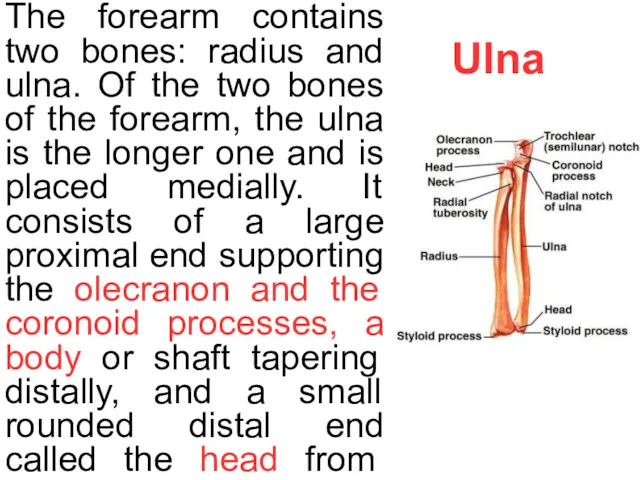
- 6. Ulna The forearm contains two bones: radius and ulna. Of the two bones of the forearm,
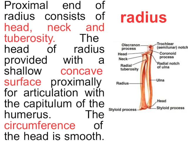
- 7. radius Proximal end of radius consists of head, neck and tuberosity. The head of radius provided
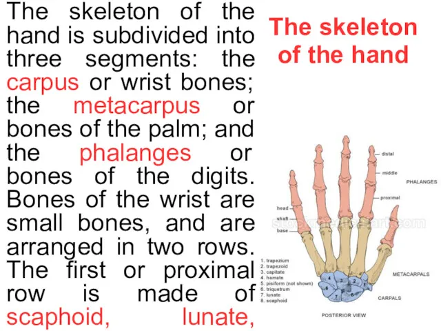
- 8. The skeleton of the hand The skeleton of the hand is subdivided into three segments: the
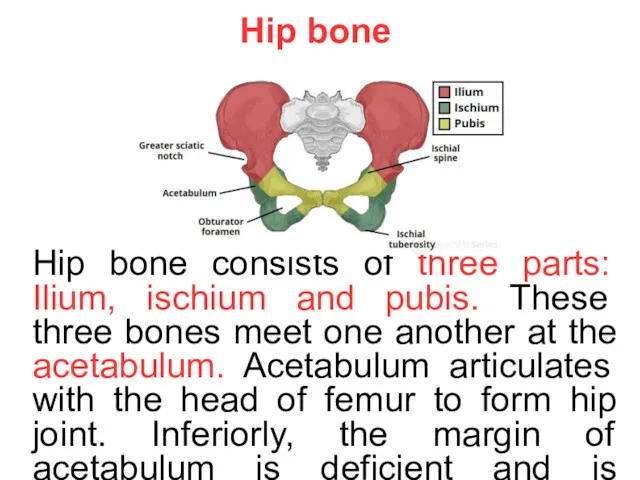
- 9. Hip bone Hip bone consists of three parts: Ilium, ischium and pubis. These three bones meet
- 10. ilium The ilium possesses a iliac crest.The crest ends in the front at the anterior superior
- 11. Ischium Ischium possesses the body of ischium and the ramus of ischium. The ischial spine intervenes
- 12. Pubis Pubis consists of three parts: the body, superior ramus and inferior ramus. Upper part of
- 13. Femur Femur is the longest bone of human body. Upper end of the femur has a
- 14. The shaft of femur has a ridge for many muscles of thigh known as linea aspera;
- 15. Tibia | Shinbone The proximal end of tibia has massive medial and lateral condyles and an
- 16. Fibula Fibula consists of a proximal end, a long shaft and a distal end forming lateral
- 17. foot The human foot is a complex structure containing 26 bones. The foot can be subdivided
- 18. human skull The human skull supports the structures of the face and forms a cavity for
- 19. frontal bone The frontal bone consists of three portions.These are the squamous part, the orbital part,
- 20. * The internal surface has small furrows for the anterior branches of the middle meningeal vessels,
- 21. occipital bone The occipital bone is the main bone of the occiput. The occipital bone, like
- 22. * Near the middle of the outer surface of the squamous part of the occipital (the
- 23. parietal bones The parietal bones form the sides and roof of the cranium. Each bone is
- 24. * The internal surface is concave; it presents depressions corresponding to the cerebral convolutions, and numerous
- 25. ethmoid bone The ethmoid bone (from Greek ethmos, "sieve") is an unpaired bone in the skull
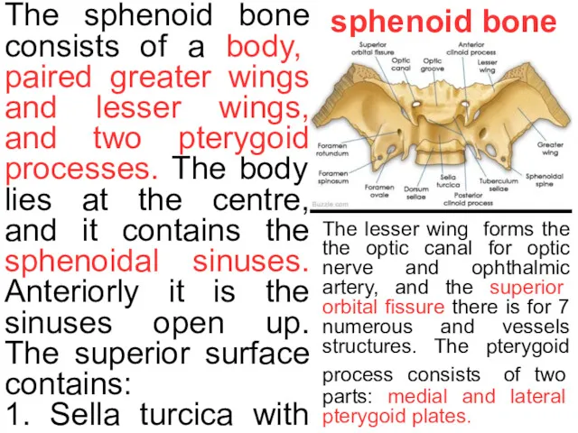
- 26. sphenoid bone The sphenoid bone consists of a body, paired greater wings and lesser wings, and
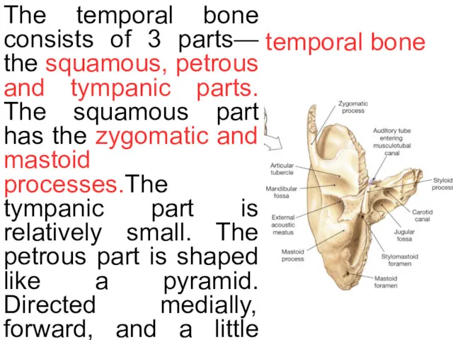
- 27. temporal bone The temporal bone consists of 3 parts— the squamous, petrous and tympanic parts. The
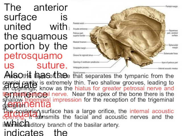
- 28. * The anterior surface is united with the squamous portion by the petrosquamous suture. Also it
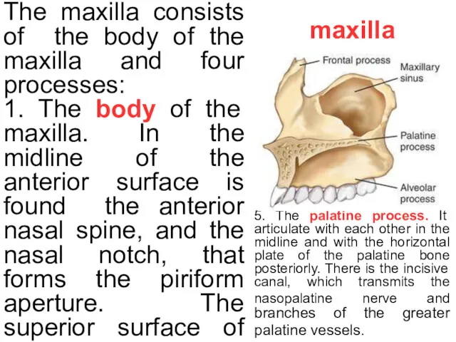
- 29. maxilla The maxilla consists of the body of the maxilla and four processes: 1. The body
- 31. Скачать презентацию
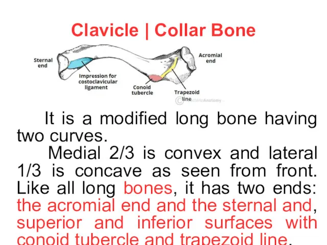
Clavicle | Collar Bone
It is a modified long bone having
Clavicle | Collar Bone
It is a modified long bone having
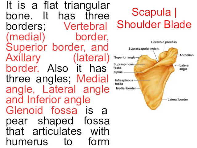
Scapula | Shoulder Blade
It is a flat triangular bone. It has
Scapula | Shoulder Blade
It is a flat triangular bone. It has
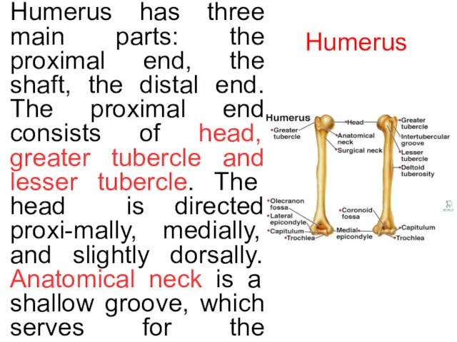
Humerus
Humerus has three main parts: the proximal end, the shaft,
Humerus
Humerus has three main parts: the proximal end, the shaft,
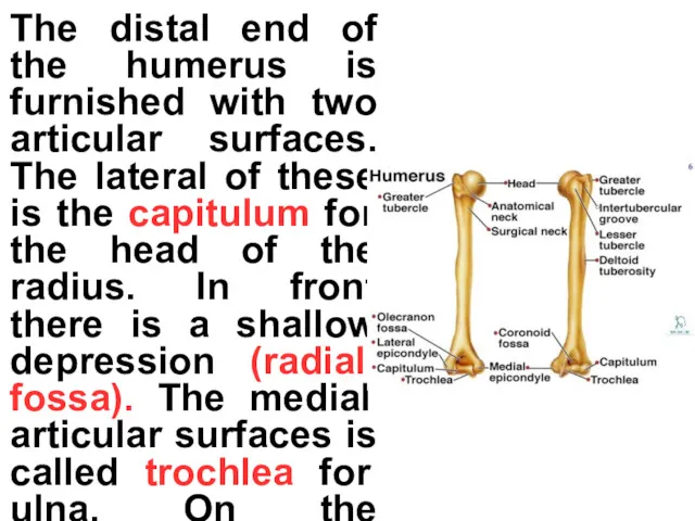
The distal end of the humerus is furnished with two articular
The distal end of the humerus is furnished with two articular
Ulna
The forearm contains two bones: radius and ulna. Of the two
Ulna
The forearm contains two bones: radius and ulna. Of the two
radius
Proximal end of radius consists of head, neck and tuberosity. The
radius
Proximal end of radius consists of head, neck and tuberosity. The
The skeleton of the hand
The skeleton of the hand is subdivided
The skeleton of the hand
The skeleton of the hand is subdivided
Hip bone
Hip bone consists of three parts: Ilium, ischium and pubis.
Hip bone
Hip bone consists of three parts: Ilium, ischium and pubis.
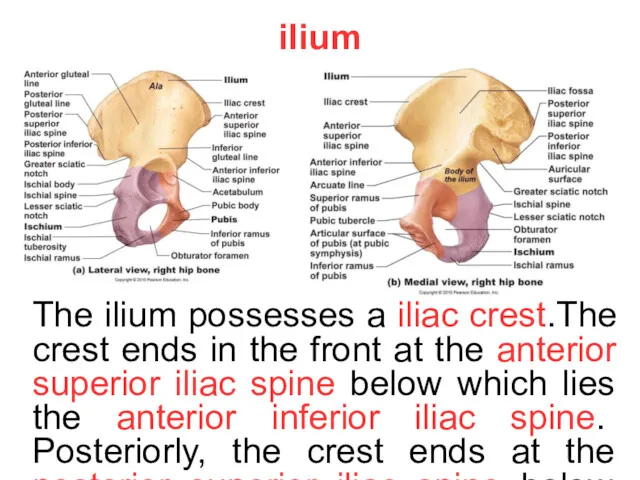
ilium
The ilium possesses a iliac crest.The crest ends in the front
ilium
The ilium possesses a iliac crest.The crest ends in the front
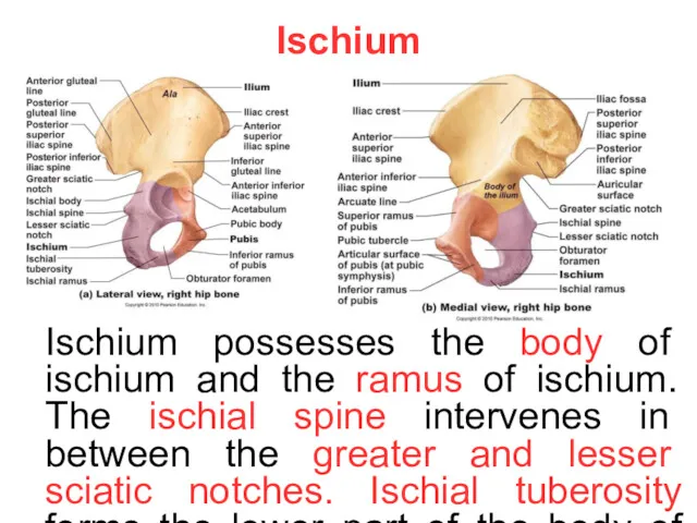
Ischium
Ischium possesses the body of ischium and the ramus of ischium.
Ischium
Ischium possesses the body of ischium and the ramus of ischium.
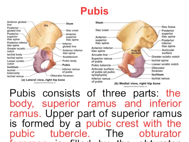
Pubis
Pubis consists of three parts: the body, superior ramus and inferior
Pubis
Pubis consists of three parts: the body, superior ramus and inferior
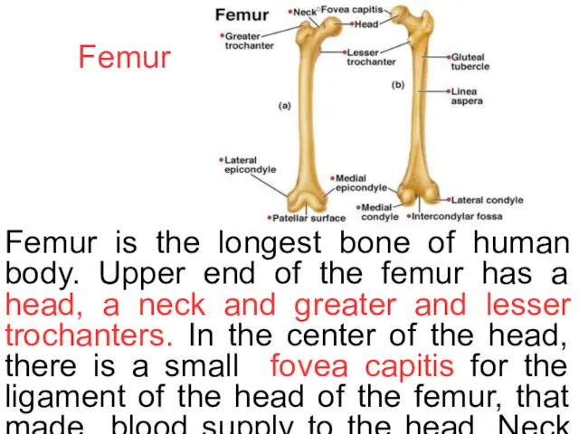
Femur
Femur is the longest bone of human body. Upper end of
Femur
Femur is the longest bone of human body. Upper end of
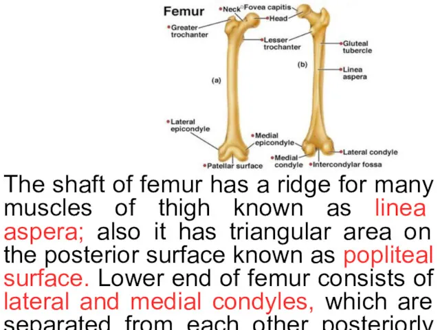
The shaft of femur has a ridge for many muscles of
The shaft of femur has a ridge for many muscles of
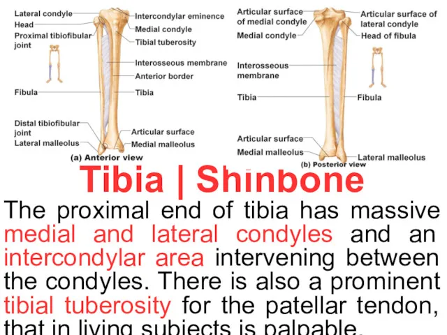
Tibia | Shinbone
The proximal end of tibia has massive medial and
Tibia | Shinbone
The proximal end of tibia has massive medial and
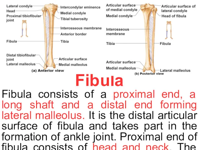
Fibula
Fibula consists of a proximal end, a long shaft and a
Fibula
Fibula consists of a proximal end, a long shaft and a
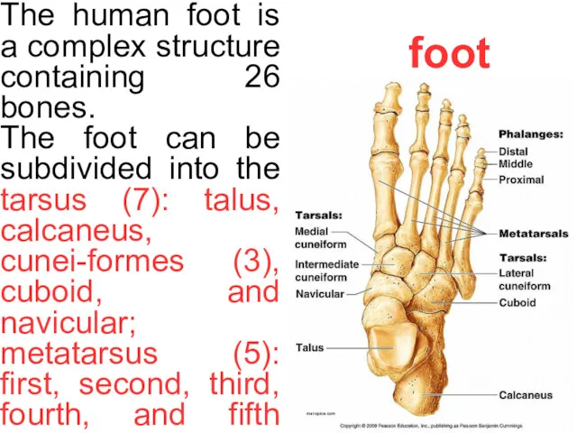
foot
The human foot is a complex structure containing 26 bones.
The foot
foot
The human foot is a complex structure containing 26 bones.
The foot
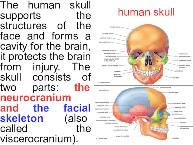
human skull
The human skull supports the structures of the face and
human skull
The human skull supports the structures of the face and
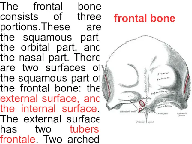
frontal bone
The frontal bone consists of three portions.These are the squamous
frontal bone
The frontal bone consists of three portions.These are the squamous
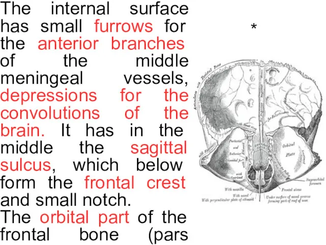
*
The internal surface has small furrows for the anterior branches of
*
The internal surface has small furrows for the anterior branches of
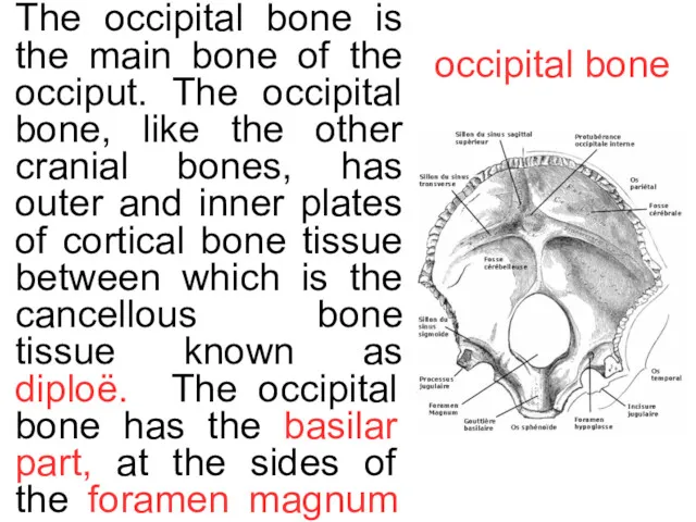
occipital bone
The occipital bone is the main bone of the occiput.
occipital bone
The occipital bone is the main bone of the occiput.
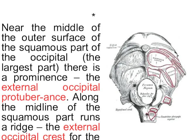
*
Near the middle of the outer surface of the squamous part
*
Near the middle of the outer surface of the squamous part
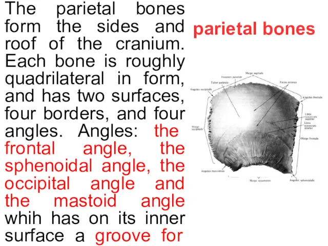
parietal bones
The parietal bones form the sides and roof of the
parietal bones
The parietal bones form the sides and roof of the
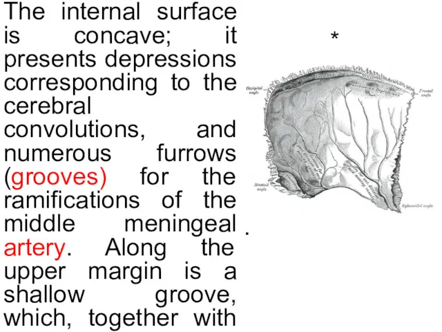
*
The internal surface is concave; it presents depressions corresponding to the
*
The internal surface is concave; it presents depressions corresponding to the
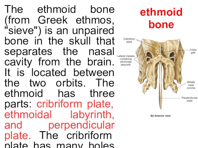
ethmoid bone
The ethmoid bone (from Greek ethmos, "sieve") is an unpaired
ethmoid bone
The ethmoid bone (from Greek ethmos, "sieve") is an unpaired
sphenoid bone
The sphenoid bone consists of a body, paired greater wings
sphenoid bone
The sphenoid bone consists of a body, paired greater wings
temporal bone
The temporal bone consists of 3 parts— the squamous, petrous
temporal bone
The temporal bone consists of 3 parts— the squamous, petrous
*
The anterior surface is united with the squamous portion by the
*
The anterior surface is united with the squamous portion by the
maxilla
The maxilla consists of the body of the maxilla and four
maxilla
The maxilla consists of the body of the maxilla and four
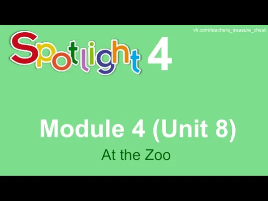 Spotlight 4. Module 4 (Unit 8). At the Zoo
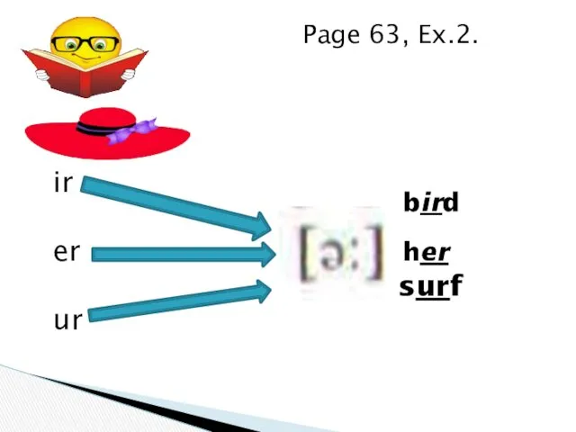 Ir, er, ou, ow, ur, wor, aw
 Conditionals. Типы условных предложений
 Pre-translation analysis
 National dishes of Germany
 Krasnoyarsk regional museum of local lore
 Body Parts Hidden Pictures Game
 Can-can
 Past Simple Tense
 Going shopping
 The State Hermitage Museum
 Friends and relationship
 100 invitations to write. Writing prompts for grades 4–8
 Origami animals
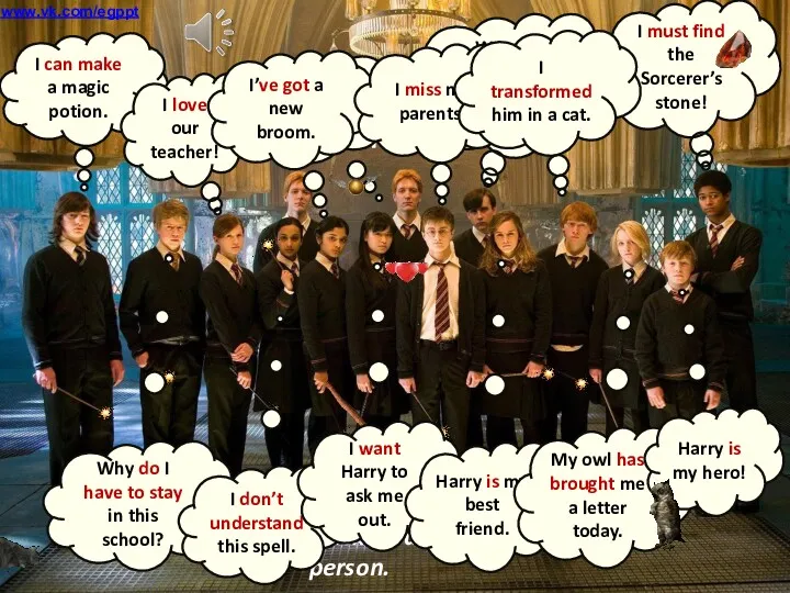 Reported speech Harry Potter
 Let me introduce myself…
 Singular and Plural. One and More. 6 класс
 Spotlight 3 Unit 13. Present Continuous
 Rules for writing a personal letter
 The weather is
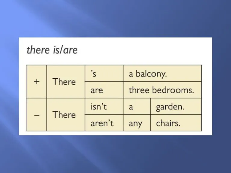 There is/are
 Иностранный язык (английский)
 Fernand Magellan
 Live and learn
 Virtual Assistants
 Going Shopping. Shopping Vocabulary
 Внеклассное интегрированное мероприятие Полиглот (русский и английский языки)
 What are they doing