- Skeleton

Содержание
- 2. SKELETON The skeleton (from Greek σκελετός, skeletós "dried up" is the body part that forms the
- 3. ENDOSKELETON Exoskeletons are external, and are found in many invertebrates; they enclose and protect the soft
- 4. An external skeleton can be quite heavy in relation to the overall mass of an animal,
- 5. The cytoskeleton (gr. kytos = cell) is used to stabilize and preserve the form of the
- 6. A hydrostatic skeleton is a semi-rigid, soft tissue structure filled with liquid under pressure, surrounded by
- 7. The endoskeletons of echinoderms and some other soft-bodied invertebrates such as jellyfish and earthworms are also
- 8. The skeleton of the echinoderms, which include, among other things, the starfish, is composed of calcite
- 9. The skeleton of the echinoderms, which include, among other things, the starfish, is composed of calcite
- 10. VERTEBRATES In most vertebrates, the main skeletal component is referred to as bone. Another important component
- 11. The skeleton, which forms the support structure inside the fish is either made of cartilage as
- 12. HUMAN The human skeleton consists of both fused and individual bones supported and supplemented by ligaments,
- 13. There exist several general differences between the male and female skeletons. The male skeleton, for example,
- 15. Скачать презентацию
SKELETON
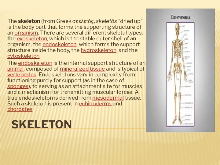
The skeleton (from Greek σκελετός, skeletós "dried up" is the body
SKELETON
The skeleton (from Greek σκελετός, skeletós "dried up" is the body
ENDOSKELETON
Exoskeletons are external, and are found in many invertebrates;
ENDOSKELETON
Exoskeletons are external, and are found in many invertebrates;
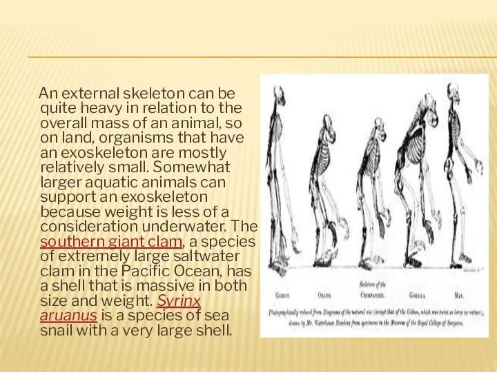
An external skeleton can be quite heavy in relation to
An external skeleton can be quite heavy in relation to
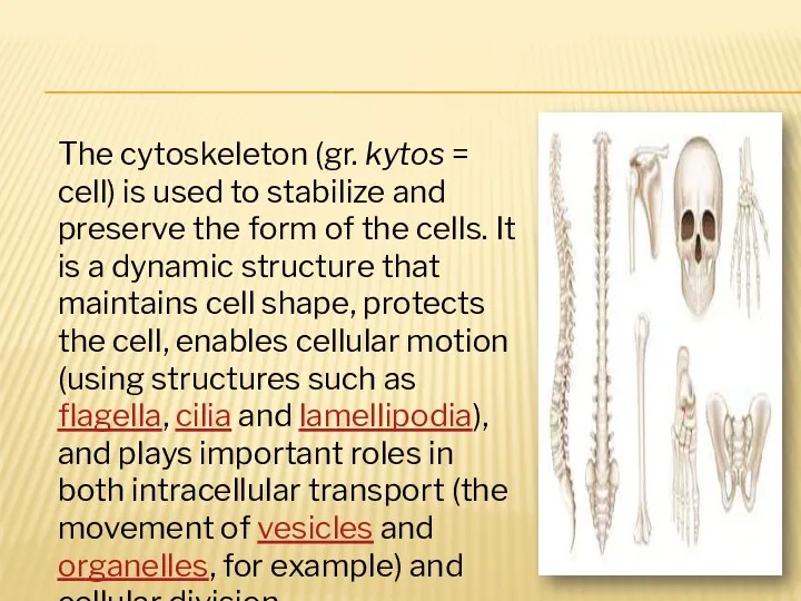
The cytoskeleton (gr. kytos = cell) is used to stabilize and
The cytoskeleton (gr. kytos = cell) is used to stabilize and
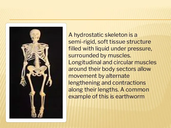
A hydrostatic skeleton is a semi-rigid, soft tissue structure filled with
A hydrostatic skeleton is a semi-rigid, soft tissue structure filled with
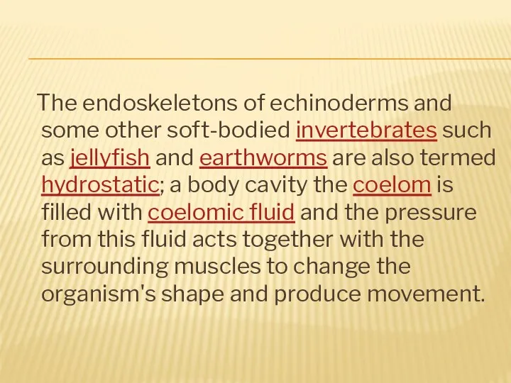
The endoskeletons of echinoderms and some other soft-bodied invertebrates such
The endoskeletons of echinoderms and some other soft-bodied invertebrates such
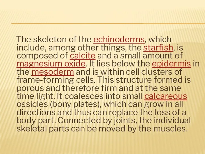
The skeleton of the echinoderms, which include, among other things,
The skeleton of the echinoderms, which include, among other things,
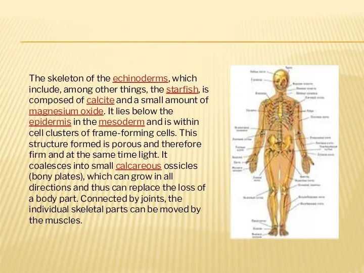
The skeleton of the echinoderms, which include, among other things, the
The skeleton of the echinoderms, which include, among other things, the
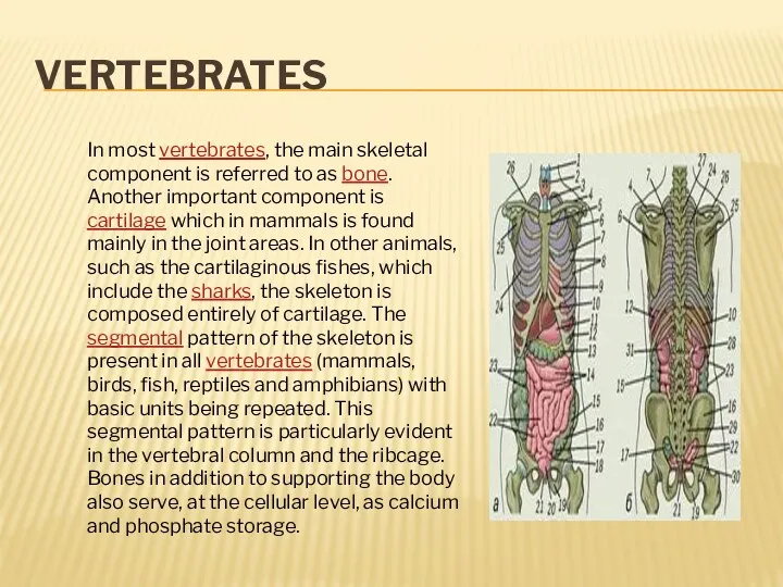
VERTEBRATES
In most vertebrates, the main skeletal component is referred to as
VERTEBRATES
In most vertebrates, the main skeletal component is referred to as
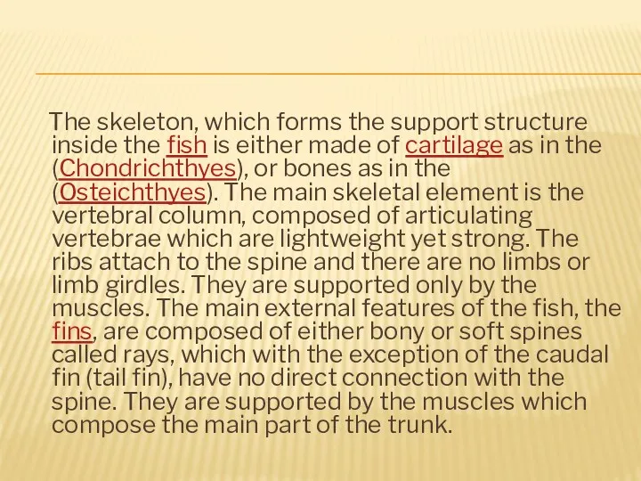
The skeleton, which forms the support structure inside the fish
The skeleton, which forms the support structure inside the fish
HUMAN
The human skeleton consists of both fused and individual
HUMAN
The human skeleton consists of both fused and individual
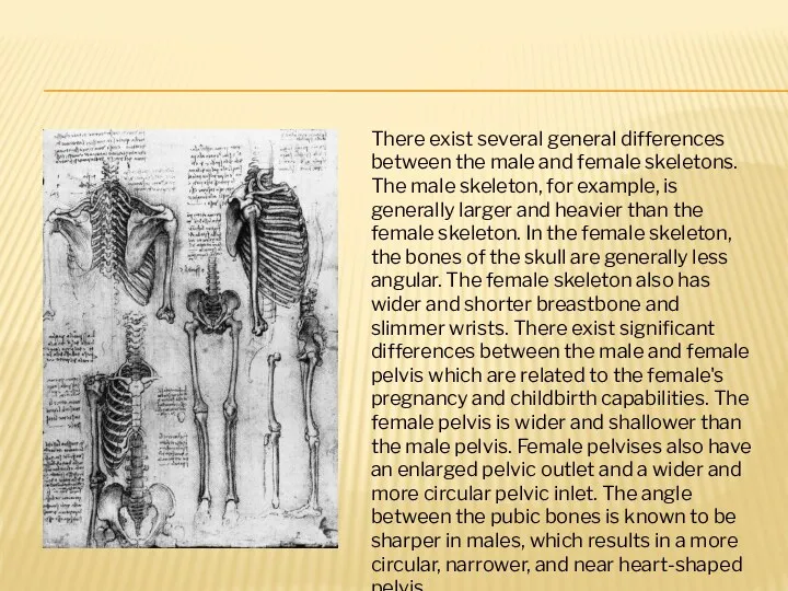
There exist several general differences between the male and female skeletons.
There exist several general differences between the male and female skeletons.
 “Who Wants to Be a Millionaire?”
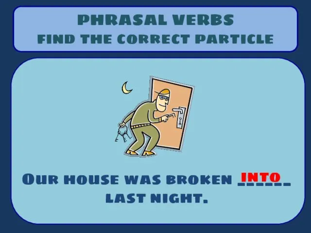 Phrasal verbs find the correct particle into
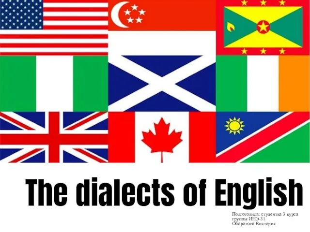 The dialects of English
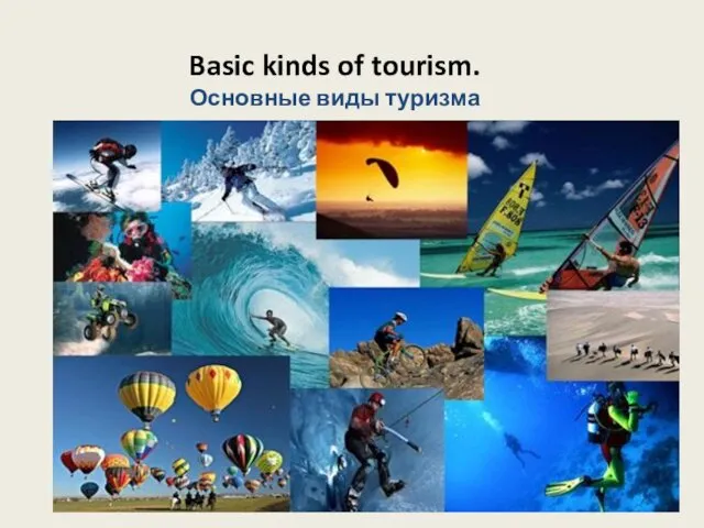 Basic kinds of tourism. Основные виды туризма
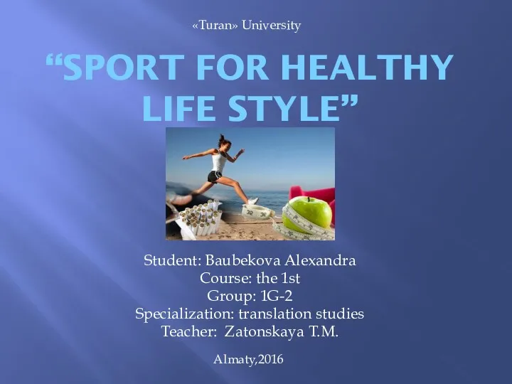 Sport for Healthy Life Style
 A trip to London
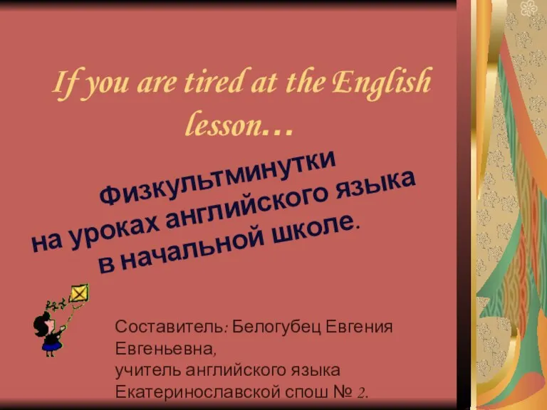 If you are tired at the english lesson. Физминутки на уроках английского языка в начальной школе
 В пути. On the move
 The presentation is made
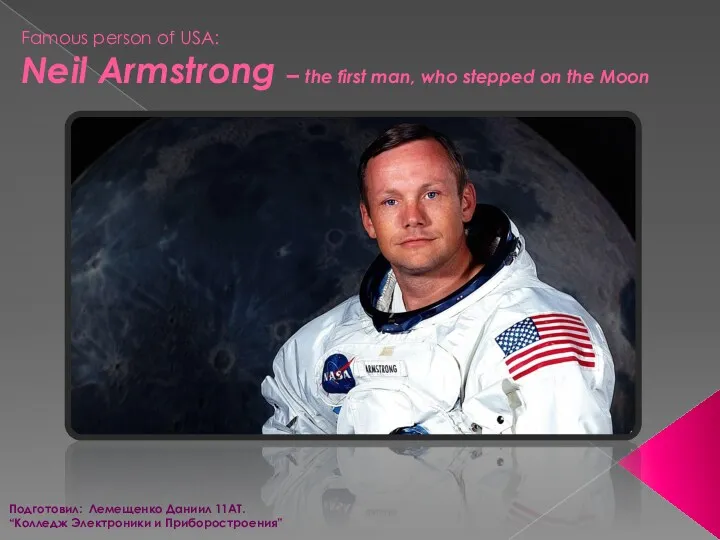 Famous person of USA: Neil Armstrong – the first man, who stepped on the Moon
 The importance of carbohydrates in animal nutrition
 What has changed
 schools in Brirain
 Dangerous hobbies
 Family relationships
 Physical appearance
 Places of interest of London
 Symbols of the USA
 Fizminutki for English lessons
 Диалектные особенности английского языка
 My family. What are the most important things in people’s life
 English for Academic Purposes
 Methods of using songs in developing English speaking skill in primary school pupils
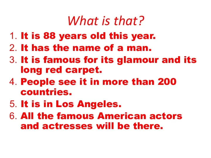 Oscars
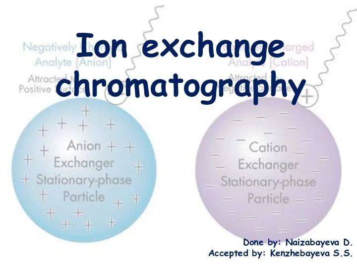 Ion exchange chromatography
 Time
Time Fruit, drink, eat, teatime,
 Lexicology as the science of the Vocabulary