- Sylvian Fissure

Содержание
- 2. Sylviun fissure, to whom we owe, in this part, everything that the brain has the most,
- 3. Definition The sylvian fissure ,is the most distinct & consistent landmark on the lateral surface, that
- 4. Parts Superficial Deep
- 5. Superficial part
- 6. Deep Part (Sylvian Cistern) Sphenoidal Operculoinsular compartment
- 7. Sphenoidal Compartment It extends laterally from the cistern around the internal carotid artery, between the frontal
- 8. Sphenoidal Compartment Roof is formed by: Post. orbital surface of the frontal lobe Anterior perforated substance.
- 9. Roof of Sphenoidal Compartment
- 11. Basal Ganglia
- 12. Floor: anterior part of the planum polare, an area free of gyri on the upper temporal
- 13. The operculoinsular compartment Opercular Insular
- 14. Opercular Cleft This is situated where the sylvian surfaces of the F lobe, & the P
- 15. Operculum
- 16. Lower Lip Of Opercular cleft from post to ant: by the planum temporale, composed of the
- 17. Insula The insular lobe (linked to emotion & self-perceptione is not visible from the outside of
- 18. Insular Clefts
- 19. Picture slide M1: Sphenoidal M2: Insular M3: Opercular M4: Cortical
- 20. Drainage Area M1:head +body of caudate, globus pallidus, putamen & posterior limb of internal capsule. M2:temporal
- 21. Radiographic Classification M1: before bifurcation M2: after bifurcation
- 22. Variations
- 23. Dominant superior Division (Early Bifurcation 187
- 24. MCA Trifurcation
- 25. Dominant inferior division
- 26. The superior division (red) can be traced to the frontal lobe (purple). The inferior division (yellow)
- 27. Short M1 segment (red) with smaller superior division (yellow) supplying the frontal convexity, & larger inferior
- 28. Acessory & Duplicated MCA aMCA configuration:both branches (purple) appear to originate proximal to the A1 complex
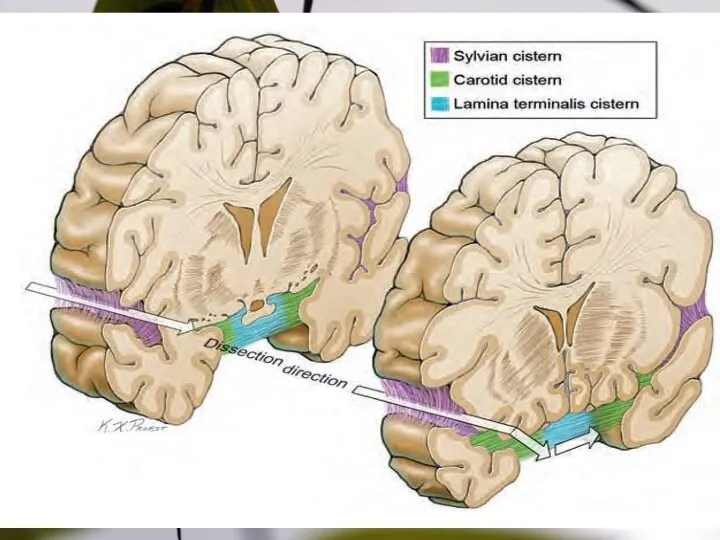
- 29. SYLVIAN FISSURE Splitting
- 30. Sylvian Vein Variations
- 31. Step 1, cortical arachnoid incision; Step 2, temporal mobilization of the sylvian veins. Dissection steps in
- 32. Venous systems draining the sylvian fissure
- 34. Steps in splitting the sylvian fissure (arteries & deep dissection). Step 3: following the cortical MCA
- 35. Types of sylvian fissures
- 36. Arteries branch temporally or frontally, but never to both lobes. Consequently, arteries in the sylvian fissure
- 37. MCA aneurysm dome projections Coronal views: lateral (A), inferior (B), and superior (C) projection. Axial views:
- 38. MCA aneurysm dissection strategy, distal-to-proximal dissection Step 1: following the superior trunk (outer surface); Step 2:
- 39. MCA aneurysm dissection strategy, proximal-to-distal dissection Step 1, dissecting the supraclinoid ICA; Step 2, dissecting the
- 40. Simple clipping technique for MCA aneurysms.
- 41. Draining Areas
- 42. Thank You
- 43. Colour scheme
- 44. Sample Graph (3 colours)
- 45. Process Flow Bullet 1 Bullet 2 Bullet 3 Bullet 1 Bullet 2 Bullet 3 Bullet 1
- 46. Example of a table Note: PowerPoint does not allow you to have nice default tables -
- 47. Examples of default styles Text and lines are like this Hyperlinks like this Visited hyperlinks like
- 49. Скачать презентацию

Sylviun fissure, to whom we owe, in this part, everything that
Sylviun fissure, to whom we owe, in this part, everything that
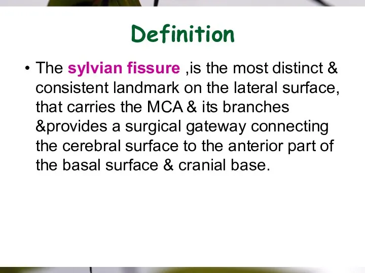
Definition
The sylvian fissure ,is the most distinct & consistent landmark on
Definition
The sylvian fissure ,is the most distinct & consistent landmark on
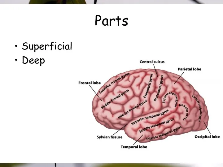
Parts
Superficial
Deep
Parts
Superficial
Deep
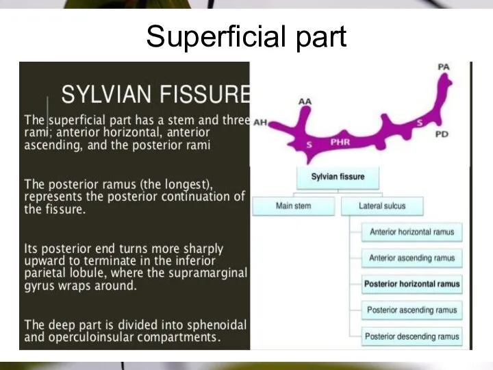
Superficial part
Superficial part
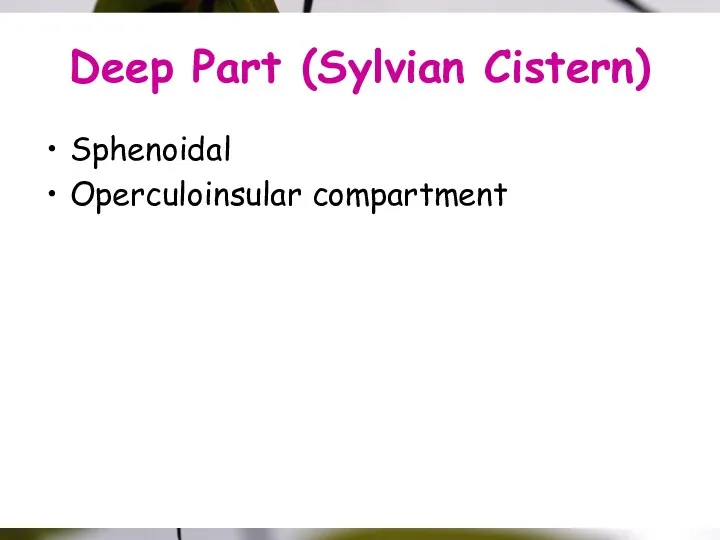
Deep Part (Sylvian Cistern)
Sphenoidal
Operculoinsular compartment
Deep Part (Sylvian Cistern)
Sphenoidal
Operculoinsular compartment
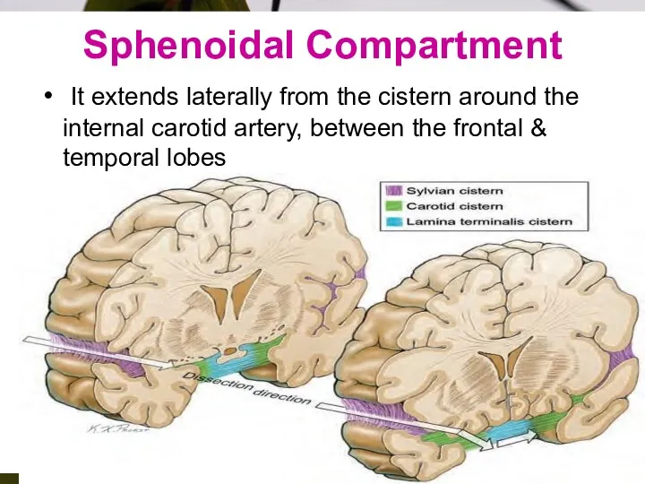
Sphenoidal Compartment
It extends laterally from the cistern around the internal
Sphenoidal Compartment
It extends laterally from the cistern around the internal
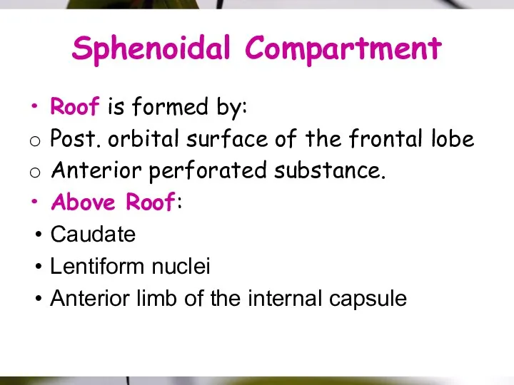
Sphenoidal Compartment
Roof is formed by:
Post. orbital surface of the frontal
Sphenoidal Compartment
Roof is formed by:
Post. orbital surface of the frontal
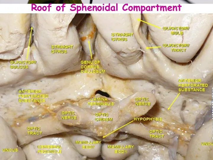
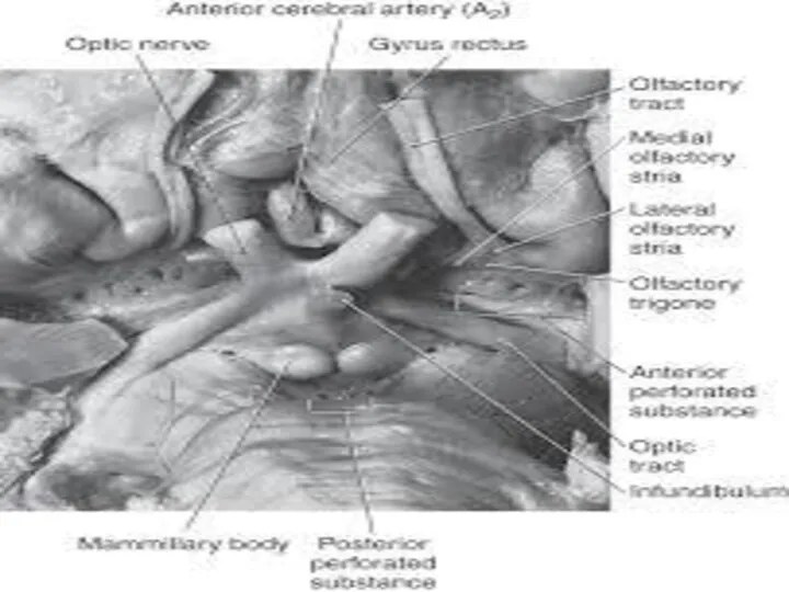
Roof of Sphenoidal Compartment
Roof of Sphenoidal Compartment
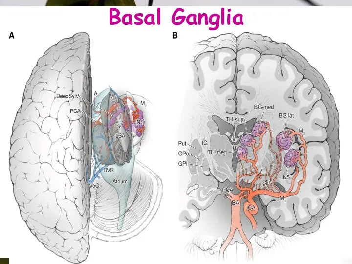
Basal Ganglia
Basal Ganglia
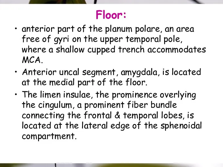
Floor:
anterior part of the planum polare, an area free of gyri
Floor:
anterior part of the planum polare, an area free of gyri
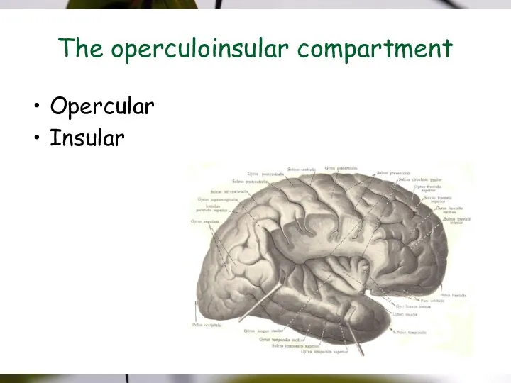
The operculoinsular compartment
Opercular
Insular
The operculoinsular compartment
Opercular
Insular
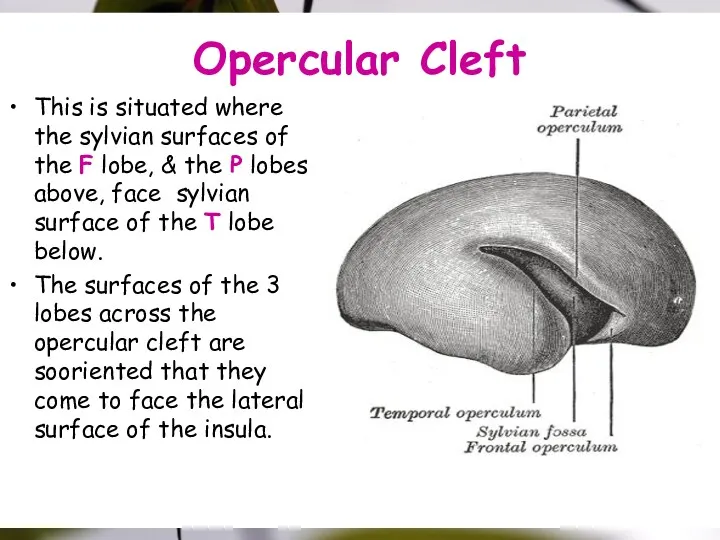
Opercular Cleft
This is situated where the sylvian surfaces of the F
Opercular Cleft
This is situated where the sylvian surfaces of the F
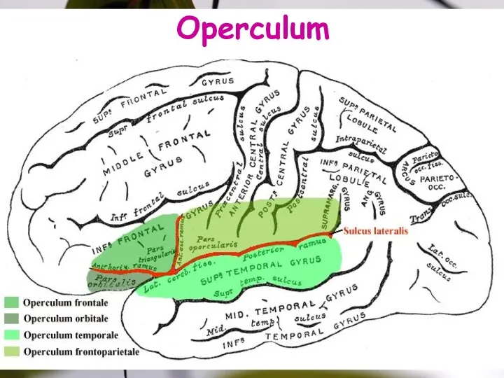
Operculum
Operculum
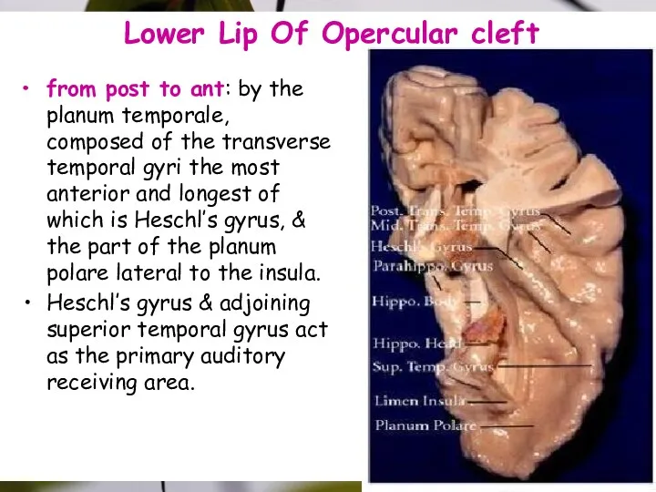
Lower Lip Of Opercular cleft
from post to ant: by the planum
Lower Lip Of Opercular cleft
from post to ant: by the planum

Insula
The insular lobe (linked to emotion & self-perceptione is not visible
Insula
The insular lobe (linked to emotion & self-perceptione is not visible
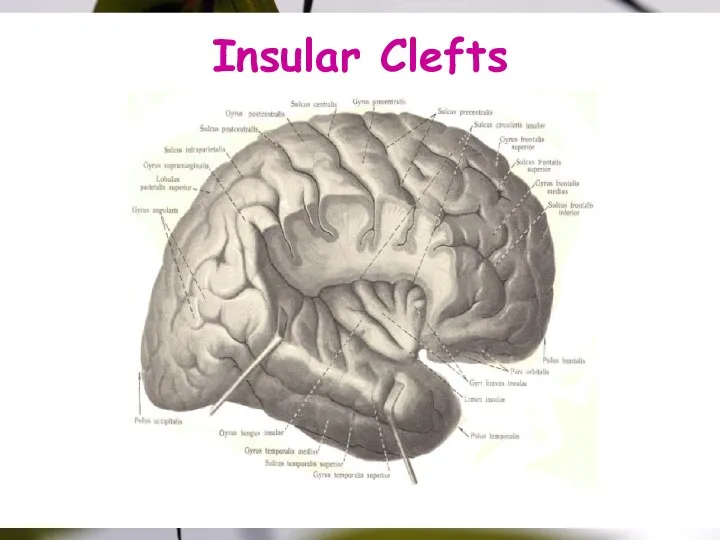
Insular Clefts
Insular Clefts
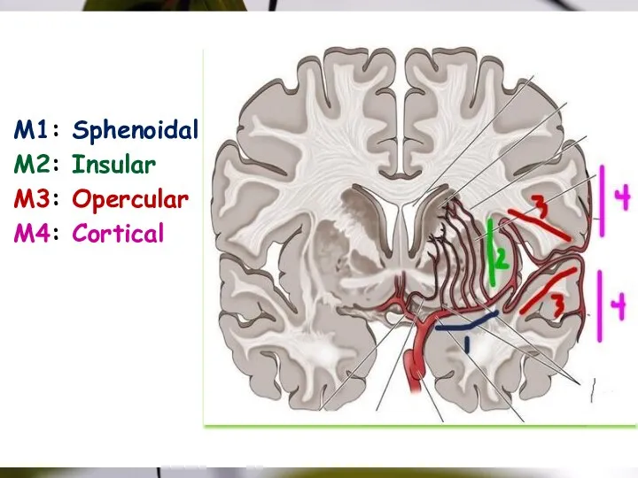
Picture slide
M1: Sphenoidal
M2: Insular
M3: Opercular
M4: Cortical
Picture slide
M1: Sphenoidal
M2: Insular
M3: Opercular
M4: Cortical
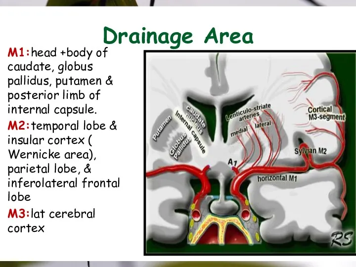
Drainage Area
M1:head +body of caudate, globus pallidus, putamen & posterior limb
Drainage Area
M1:head +body of caudate, globus pallidus, putamen & posterior limb
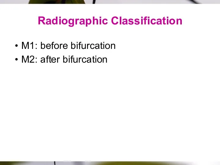
Radiographic Classification
M1: before bifurcation
M2: after bifurcation
Radiographic Classification
M1: before bifurcation
M2: after bifurcation
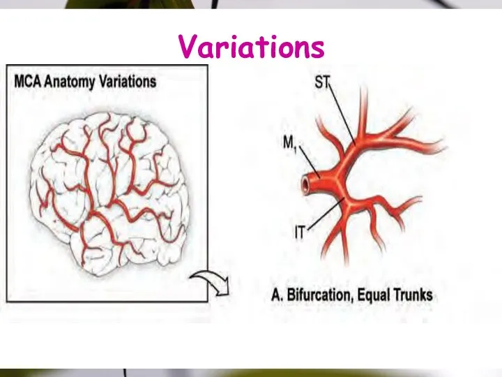
Variations
Variations
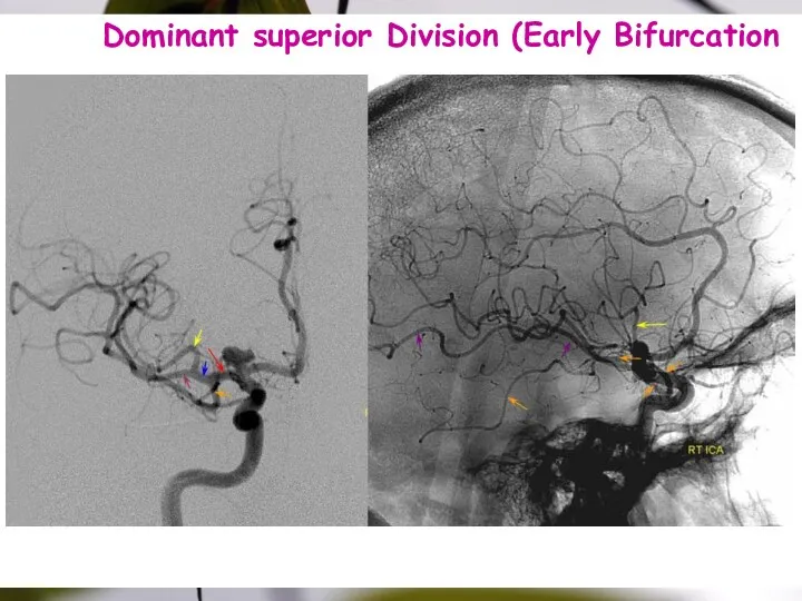
Dominant superior Division (Early Bifurcation
187
Dominant superior Division (Early Bifurcation
187
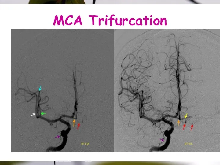
MCA Trifurcation
MCA Trifurcation
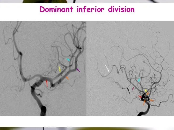
Dominant inferior division
Dominant inferior division
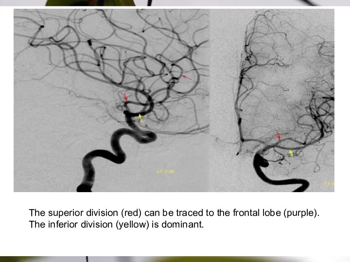
The superior division (red) can be traced to the frontal lobe
The superior division (red) can be traced to the frontal lobe
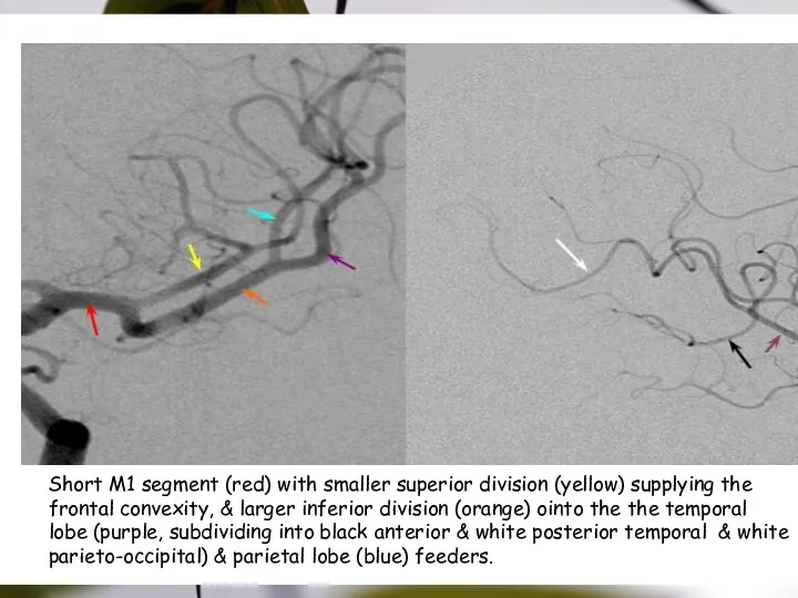
Short M1 segment (red) with smaller superior division (yellow) supplying the
Short M1 segment (red) with smaller superior division (yellow) supplying the
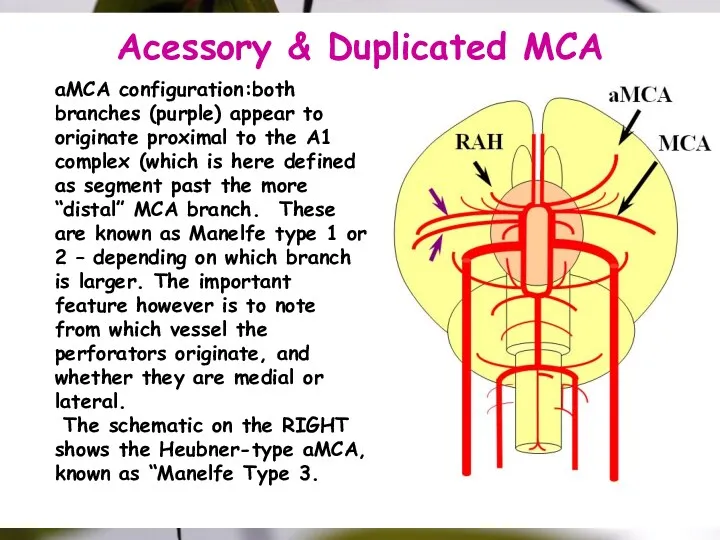
Acessory & Duplicated MCA
aMCA configuration:both branches (purple) appear to originate proximal
Acessory & Duplicated MCA
aMCA configuration:both branches (purple) appear to originate proximal

SYLVIAN FISSURE
Splitting
SYLVIAN FISSURE
Splitting
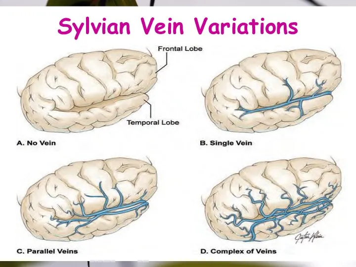
Sylvian Vein Variations
Sylvian Vein Variations
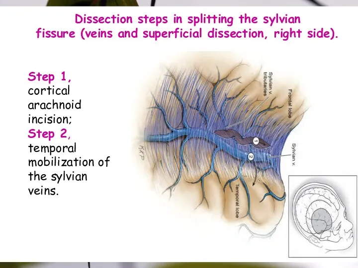
Step 1, cortical arachnoid incision;
Step 2, temporal
mobilization of the sylvian
Step 1, cortical arachnoid incision;
Step 2, temporal
mobilization of the sylvian
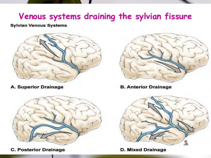
Venous systems draining the sylvian fissure
Venous systems draining the sylvian fissure
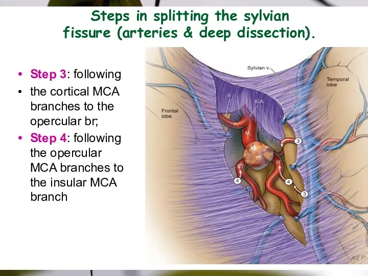
Steps in splitting the sylvian
fissure (arteries & deep dissection).
Step 3: following
the
Steps in splitting the sylvian
fissure (arteries & deep dissection).
Step 3: following
the
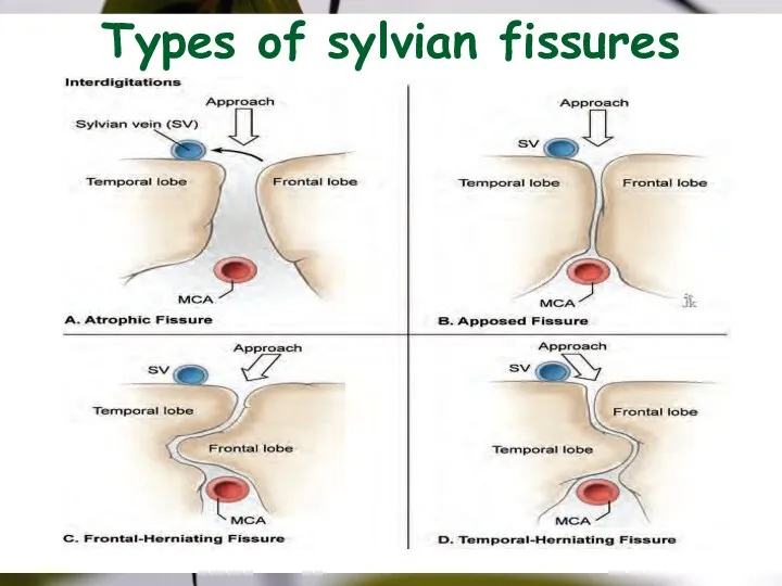
Types of sylvian fissures
Types of sylvian fissures
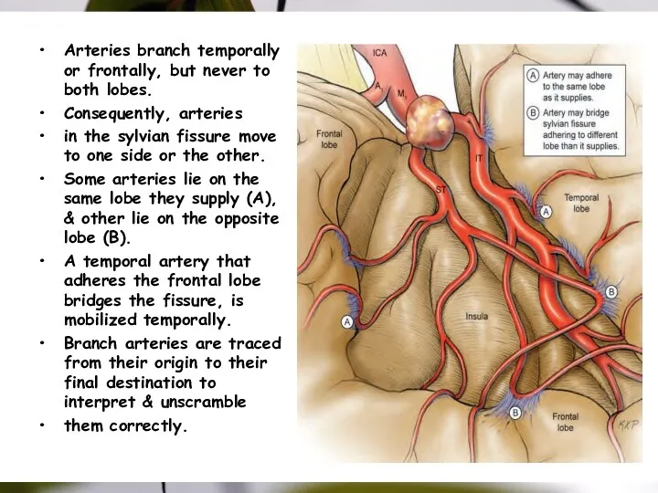
Arteries branch temporally or frontally, but never to both lobes.
Consequently,
Arteries branch temporally or frontally, but never to both lobes.
Consequently,
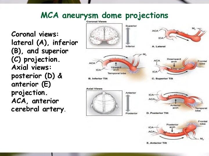
MCA aneurysm dome projections
Coronal views: lateral (A), inferior (B), and superior
MCA aneurysm dome projections
Coronal views: lateral (A), inferior (B), and superior
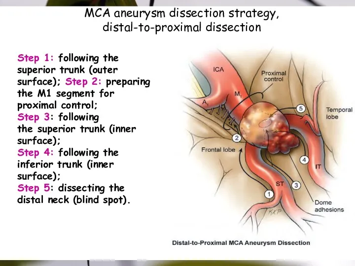
MCA aneurysm dissection strategy,
distal-to-proximal dissection
Step 1: following the
superior trunk (outer surface);
MCA aneurysm dissection strategy,
distal-to-proximal dissection
Step 1: following the
superior trunk (outer surface);
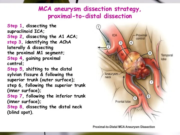
MCA aneurysm dissection strategy,
proximal-to-distal dissection
Step 1, dissecting the
supraclinoid ICA;
Step 2, dissecting
MCA aneurysm dissection strategy,
proximal-to-distal dissection
Step 1, dissecting the
supraclinoid ICA;
Step 2, dissecting
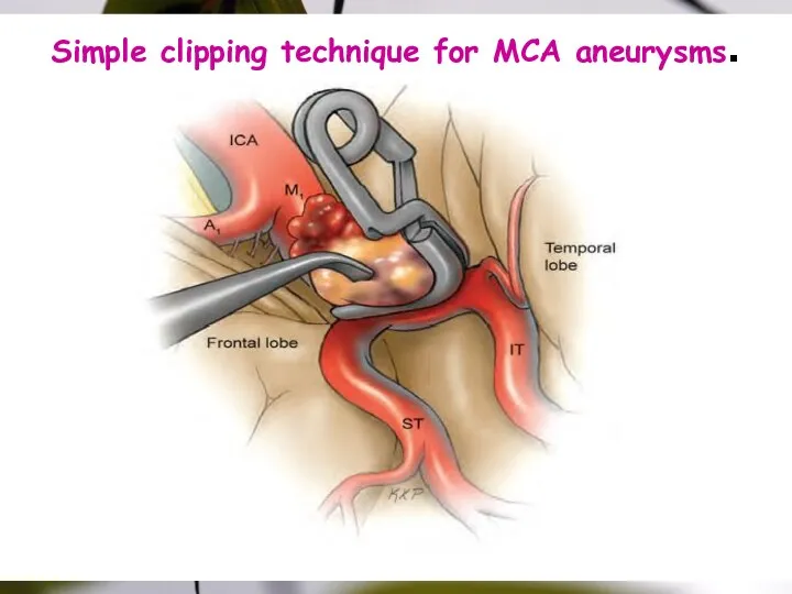
Simple clipping technique for MCA aneurysms.
Simple clipping technique for MCA aneurysms.
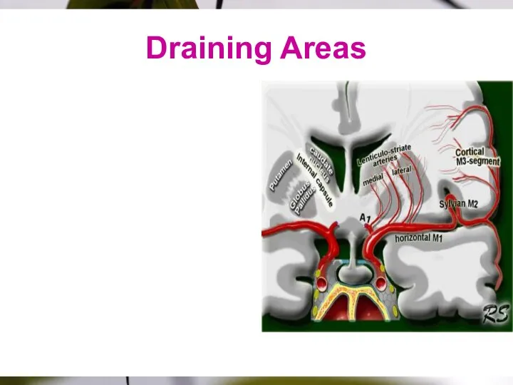
Draining Areas
Draining Areas

Thank You
Thank You
Colour scheme
Colour scheme

Sample Graph (3 colours)
Sample Graph (3 colours)
Process Flow
Bullet 1
Bullet 2
Bullet 3
Bullet 1
Bullet 2
Bullet 3
Bullet 1
Bullet 2
Bullet 3
Bullet
Process Flow
Bullet 1
Bullet 2
Bullet 3
Bullet 1
Bullet 2
Bullet 3
Bullet 1
Bullet 2
Bullet 3
Bullet

Example of a table
Note: PowerPoint does not allow you to have
Example of a table
Note: PowerPoint does not allow you to have

Examples of default styles
Text and lines are like this
Hyperlinks like this
Visited
Examples of default styles
Text and lines are like this
Hyperlinks like this
Visited
 I like school
 An Englishman`s House Is His Castle
 Philippines
 My native town Arzamas
 Цвета. Сolours
 My summer holidays
 Существительное в притяжательном падеже
 The Adjective. The Pronoun. Lecture 8
 Past simple tense
 What number is this
 Job Interview in English. Guestions
 Present Cont
 Emirates society. (Сhapter 3)
 Science: insects
 Global warming
 Раздел грамматики синтаксис. (Лекция 10)
 Etymology
 Gerund Infinitive
 My family
 Проблемы больших гоодов
 Устная часть ЕГЭ. Тренировочные картинки для описания и сравнения. Задания 3,4
 Relative pronouns and adverbs
 Ecological problems
 Прошедшее совершенное время
 To say, to tell, to speak, to talk
 Can and Can’t
 Look at the pictures and be ready to compare them
 Adjective or Adverb