- Anatomy of nervous system

Содержание
- 2. MAJOR CHAPTER OBJECTIVES Relate the developmental processes of the embryonic nervous system to the adult structures
- 3. 13.1 THE EMBRYOLOGIC PERSPECTIVE MAJOR SECTION OBJECTIVES Describe the growth and differentiation of the neural tube
- 4. FIGURE 13.2 Early Embryonic Development of Nervous System (at about 16 days) The neuroectoderm begins to
- 5. FIGURE 13.3 Primary and Secondary Vesicle Stages of Development The embryonic brain develops complexity through enlargements
- 6. FIGURE 13.4 Human Neuraxis The mammalian nervous system is arranged with the neural tube running along
- 7. TABLE 13.1 Stages of Embryonic Development N.B. A synonym for “cerebral aqueduct” is “aqueduct of Sylvius”.
- 8. 13.2 THE CENTRAL NERVOUS SYSTEM MAJOR SECTION OBJECTIVES Name the major regions of the adult brain
- 9. FIGURE 13.6 The Cerebrum The cerebrum is a large component of the CNS in humans, and
- 10. FIGURE 13.7 Lobes of the Cerebral Cortex The cerebral cortex is divided into four five lobes.
- 11. FIGURE 13.8 Brodmann’s Areas of the Cerebral Cortex Brodmann mapping of functionally distinct regions of the
- 12. FIGURE 13.9 Frontal Section of Cerebral Cortex and Basal Nuclei The major components of the basal
- 13. FIGURE 13.11 The Diencephalon The diencephalon is composed primarily of the thalamus and hypothalamus, which together
- 14. FIGURE 13.12 The Brain Stem The brain stem comprises three regions: the midbrain, the pons, and
- 15. FIGURE 13.13 The Cerebellum The cerebellum is situated on the posterior surface of the brain stem.
- 16. FIGURE 13.14 Cross-section of Spinal Cord The cross-section of a thoracic spinal cord segment shows the
- 17. 13.3 CIRCULATION IN THE CENTRAL NERVOUS SYSTEM MAJOR SECTION OBJECTIVES Describe the vessels that supply the
- 18. FIGURE 13.15 Circle of Willis The blood supply to the brain enters through the internal carotid
- 19. FIGURE 13.16 Dural Sinuses and Veins Blood drains from the brain through a series of sinuses
- 20. FIGURE 13.17 Meningeal Layers of Superior Sagittal Sinus The layers of the meninges in the longitudinal
- 21. FIGURE 13.18 Cerebrospinal Fluid Circulation The choroid plexus in the four ventricles produce CSF, which is
- 22. MODIFIED TABLE 13.2 Components of CSF Circulation
- 23. 13.4 THE PERIPHERAL NERVOUS SYSTEM MAJOR SECTION OBJECTIVES Describe the structures found in the PNS Distinguish
- 24. FIGURE 13.19 Dorsal Root Ganglion The cell bodies of sensory neurons, which are unipolar neurons by
- 25. FIGURE 13.20 Spinal Cord and Root Ganglion The slide includes both a cross-section of the lumbar
- 26. FIGURE 13.21 Nerve Structure The structure of a nerve is organized by the layers of connective
- 27. FIGURE 13.22 Close-Up of Nerve Trunk Zoom in on this slide of a nerve trunk to
- 28. FIGURE 13.23 The Cranial Nerves The anatomical arrangement of the roots of the cranial nerves observed
- 29. MODIFIED TABLE 13.3 Cranial Nerves
- 30. FIGURE 13.24 Nerve Plexuses of the Body There are four main nerve plexuses in the human
- 31. TYPOS N.B. Nerves do not enervate targets! To enervate means to make weak, tired… Nerves innervate
- 32. DEVELOPMENT Embryonic Germ Layers - Review To begin, a sperm cell and an egg cell fuse
- 33. DEVELOPMENT Neural Tube Formation Early formation of the nervous system depends on the formation of the
- 34. AGING Anosmia The sensory neurons of the olfactory epithelium have a limited lifespan of approximately one
- 35. EVERYDAY CONNECTIONS Left Brain/Right Brain To say that people are “right-brained” or “left-brained” is an oversimplification
- 36. DISORDERS & HOMEOSTATIC IMBALANCES Spina Bifida Results from a failure of the neural tube to close.Two
- 37. FIGURE 13.5 Spinal Bifida Spina bifida is a birth defect of the spinal cord caused when
- 38. DISORDERS & HOMEOSTATIC IMBALANCES Basal Nuclei and Parkinson’s Disease Parkinson’s disease is a disorder of the
- 39. DISORDERS & HOMEOSTATIC IMBALANCES Meningitis Meningitis is an inflammation of the meninges, the three layers of
- 40. DISORDERS & HOMEOSTATIC IMBALANCES CNS Perfusion Disorders Without a steady supply of oxygen, and to a
- 41. INTERACTIVE LINKS Watch this animation http://openstaxcollege.org/l/braindevel to examine the development of the brain, starting with the
- 42. INTERACTIVE LINKS Watch this animation http://openstaxcollege.org/l/bloodflow1 to see how blood flows to the brain and passes
- 43. INTERACTIVE LINKS Visit this site http://openstaxcollege.org/l/NYTmeningitis to read about a man who wakes with a headache
- 45. Скачать презентацию
 Методы биологических исследований
 Вред и польза сахара
 Биология как наука, её история. Методы исследования в биологии
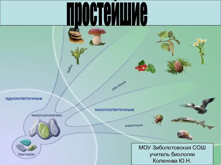 Подцарство простейшие
 Презентация Достижения и основные направления селекции
 Миазы. Классификация
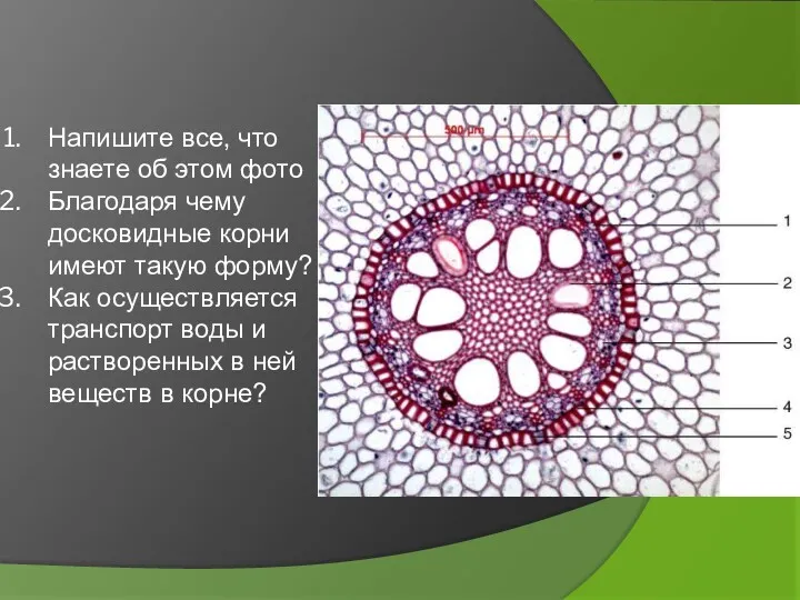 Основные вегетативные органы растений. Лекция 6
 Сүт және сүт тағамдары микрофлорасы
 Биологияны оқытудың инновациялық модульдердің қолдану ерекшеліктері
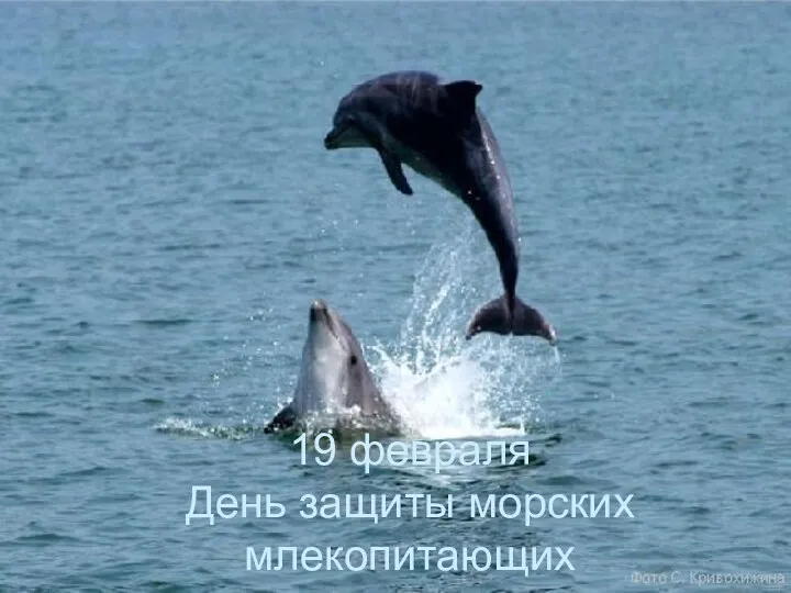 День защиты морских млекопитающих
 Отдел Мохообразные. Размножение и значение
 Ядовитые растения
 Красная книга Челябинской области
 эволюция кровеносной системы
 Пищеварение в ротовой полости
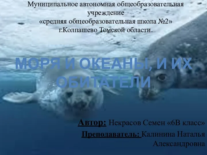 Моря и океаны и их обитатели
 10 цікавих фактів про рептилій
 Цветок. Плод. Семя
 Zarys Fizjologii - Układu Krążenia cz. I
 Потенциал покоя и потенциал действия клетки
 Многолетние бобовые травы. Клевер луговой, гибридный, ползучий
 Устройство увеличительных приборов и правила работы с ними
 20230419_belki
 Жизнь в архейскую эру
 Определение чистоты воздуха по лишайникам (лихеноиндикация)
 Обобщающий урок – КВН Общее знакомство с цветковыми растениями Клеточное строение растительного организма
 Методы исследования и симптомы поражения органов чувств – зрения, слуха, обоняния, вкуса
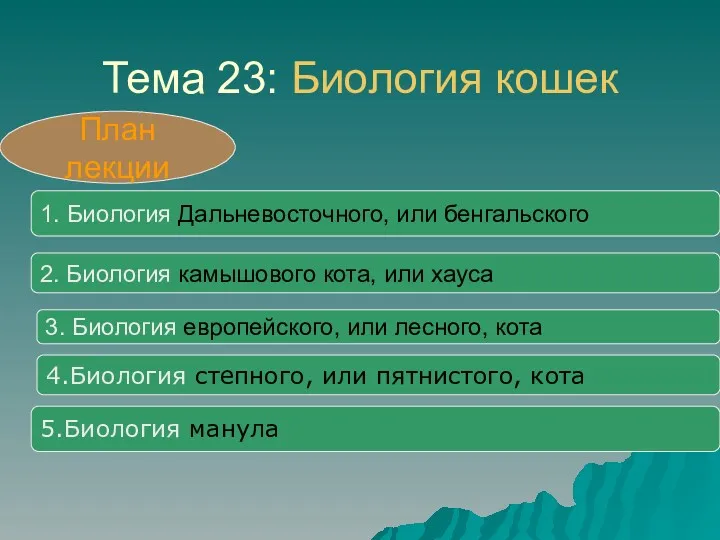 Биология кошек