- Биохимия клеточной поверхности микроорганизмов

Содержание
- 3. Функции клеточной оболочки Защита Транспорт Проведение сигналов Рост и деление клеток Межклеточные коммуникации
- 4. Цитоплазматическая мембрана ( ЦПМ ) – липидный бислой со встроенными в него белками Клеточная стенка -
- 6. Клеточные стенки бактерий состоят из пептидогликана (муреина) и бывают двух типов: грамположительного и грамотрицательного Клеточные стенки
- 7. a | Schematic representation of a cross-section of the cell envelope of Sulfolobus solfataricus showing the
- 8. Electron micrographs of ultra-thin sections of the euryarchaeote Methanocaldococcus villosus (a) and the crenarchaeote Metallosphaera prunae
- 9. M.lysodeicticus Gram+ (monoderma) Bacteroides melaninogenicus Gram- (diderma) Клеточная оболочка бактерий
- 10. Фотография цианобактерии Phormidium uncinatum, полученная с помощью трансмиссионного электронного микроскопа. Клеточная оболочка (CW) состоит из, цитоплазматической
- 11. A: A whole yeast cell (Сandida albicans) B: Section of a yeast cell; C: Schematic diagram
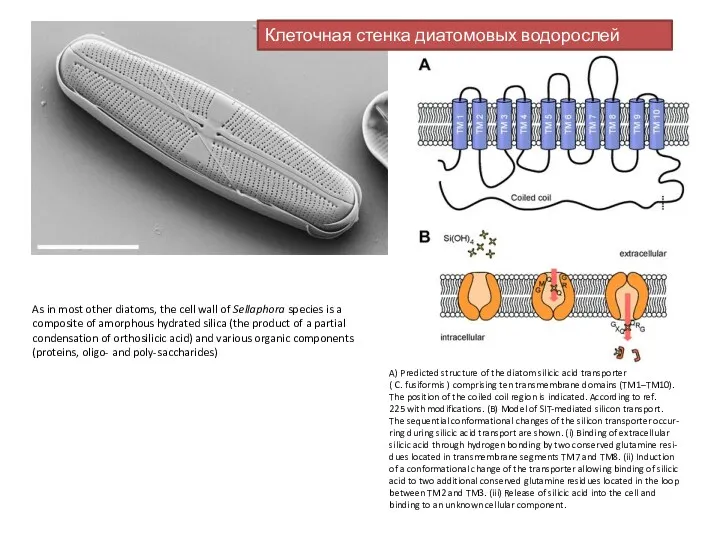
- 12. Клеточная стенка диатомовых водорослей As in most other diatoms, the cell wall of Sellaphora species is
- 14. Скачать презентацию
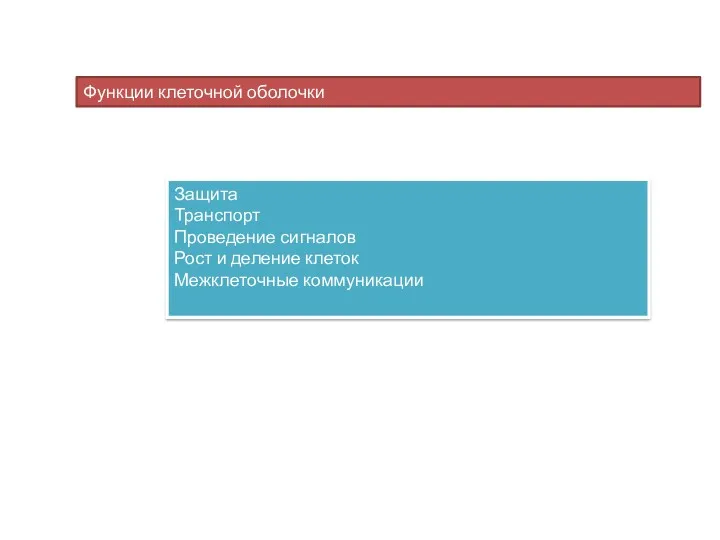
Функции клеточной оболочки
Защита
Транспорт
Проведение сигналов
Рост и деление клеток
Межклеточные коммуникации
Функции клеточной оболочки
Защита
Транспорт
Проведение сигналов
Рост и деление клеток
Межклеточные коммуникации
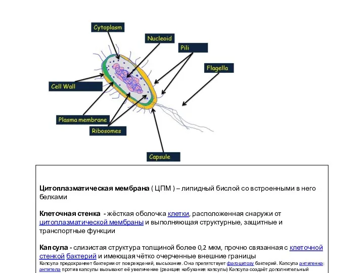
Цитоплазматическая мембрана ( ЦПМ ) – липидный бислой со встроенными в
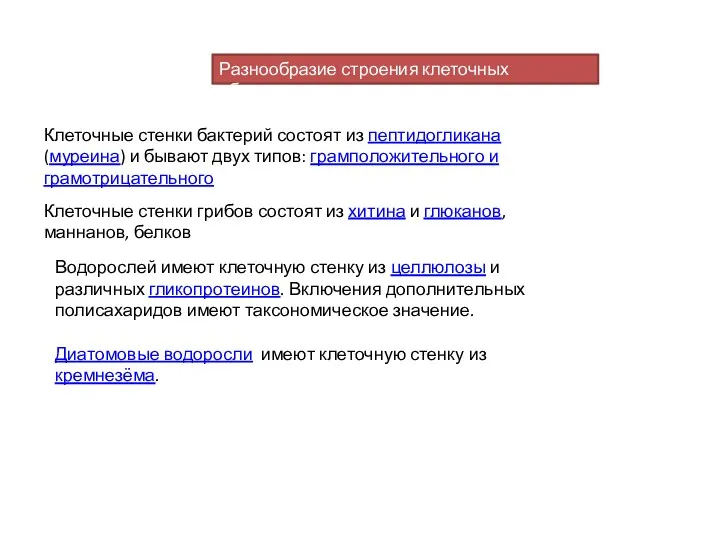
Клеточные стенки бактерий состоят из пептидогликана (муреина) и бывают двух типов:
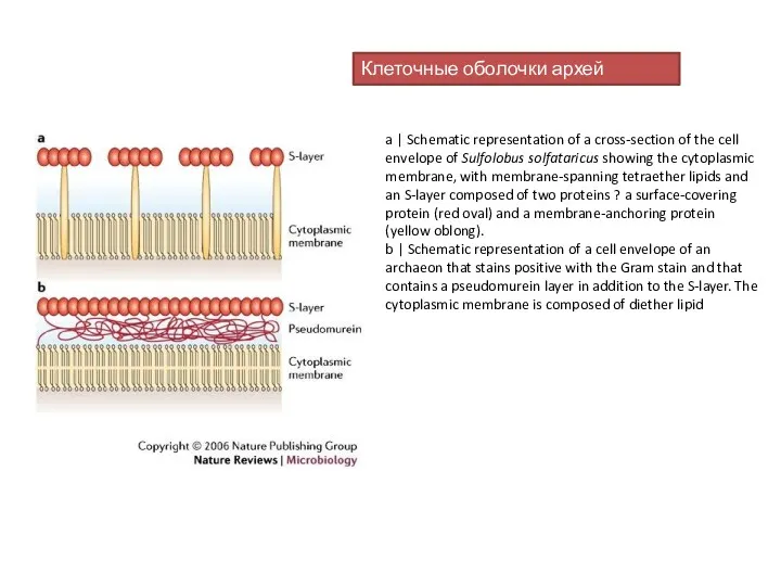
a | Schematic representation of a cross-section of the cell envelope
a | Schematic representation of a cross-section of the cell envelope
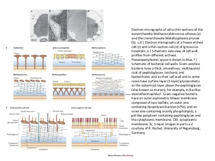
Electron micrographs of ultra-thin sections of the euryarchaeote Methanocaldococcus villosus (a)
Electron micrographs of ultra-thin sections of the euryarchaeote Methanocaldococcus villosus (a)
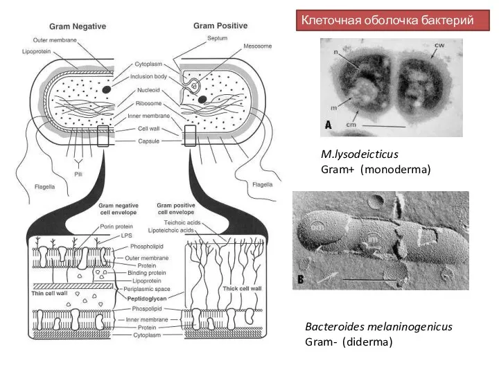
M.lysodeicticus
Gram+ (monoderma)
Bacteroides melaninogenicus Gram- (diderma)
Клеточная оболочка бактерий
M.lysodeicticus
Gram+ (monoderma)
Bacteroides melaninogenicus Gram- (diderma)
Клеточная оболочка бактерий
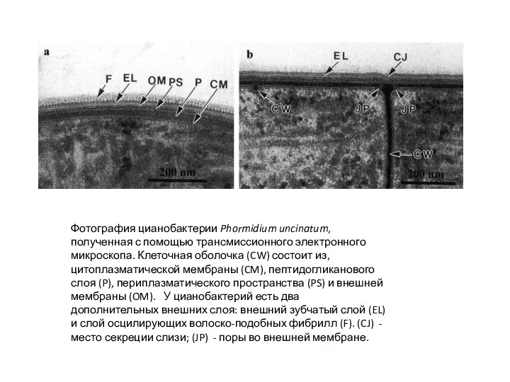
Фотография цианобактерии Phormidium uncinatum, полученная с помощью трансмиссионного электронного микроскопа. Клеточная
Фотография цианобактерии Phormidium uncinatum, полученная с помощью трансмиссионного электронного микроскопа. Клеточная
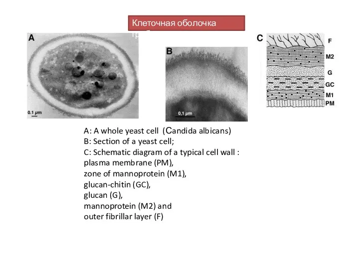
A: A whole yeast cell (Сandida albicans)
B: Section of a yeast
A: A whole yeast cell (Сandida albicans) B: Section of a yeast
Клеточная стенка диатомовых водорослей
As in most other diatoms, the cell wall
Клеточная стенка диатомовых водорослей
As in most other diatoms, the cell wall
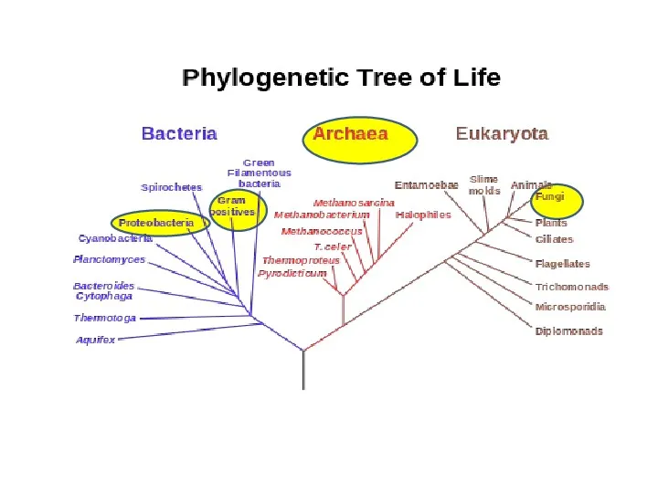
 Современная биосистематика
 Анализаторы. Органы чувств
 Многообразие млекопитающих. 7 класс
 Мои домашние животные
 Углеводы. Классификация. Роль углеводов в природе. Углеводы в жизни человека
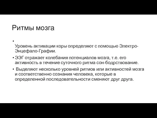 Ритмы мозга
 Развитие агрохимии как науки о взаимодействия почвы, растений и удобрений
 Digestion
 Растительный мир Донбасса
 Основные понятия генетики
 Praktyczne wykorzystanie wiedzy z zakresu genetyki cech jakościowych i ilościowych u koni
 Общие принципы организации тканей. Эпителиальные ткани
 Вегетативное размножение растений
 Птицы нашего края
 Компоненты опорно-двигательной системы. Кровеносная система
 20231217_stseplennoe_s_polom_nasledovanie
 Значення птахів у природі та житті людини. Птахівництво. Охорона птахів
 Рентгеноанатомия черепа. Обозначьте кости мозгового черепа
 Прокариотическая клетка
 Древесина и её топливные свойства
 Растительный и животный мир океана
 Лечебные свойства яда жабы обыкновенной
 Бесполое размножение
 Систематика растений
 Закономерности наследственной изменчивости
 Ствол мозга: продолговатый, задний, средний и промежуточный мозг. Занятие № 15
 Вид. Критерии вида. Популяция
 Тірі ағзалардағы қоректену типтері