- Biology of the Cell

Содержание
- 2. Understanding the Cell All body processes dependent upon cells for their activities Cells known as “the
- 3. Introduction to Cells: How Cells Are Studied Cells Studied through the discipline of cytology Discovered after
- 4. Microscopy
- 5. TEM vs. SEM
- 6. Figure 4.1 Copyright © The McGraw-Hill Companies, Inc. Permission required for reproduction or display. TEM 50,000x
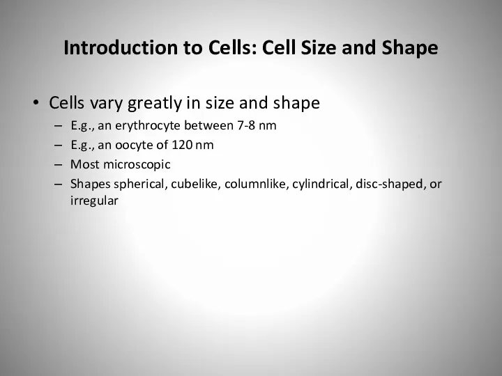
- 7. Introduction to Cells: Cell Size and Shape Cells vary greatly in size and shape E.g., an
- 8. Figure 4.2 Copyright © The McGraw-Hill Companies, Inc. Permission required for reproduction or display. Electron microscope
- 9. Figure 4.3 Copyright © The McGraw-Hill Companies, Inc. Permission required for reproduction or display. Irregular: Nerve
- 10. Introduction to Cells: Common Features and General Functions Overview of Cellular Components Plasma membrane Forms the
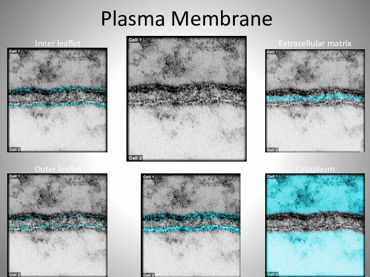
- 11. Plasma Membrane
- 12. Introduction to Cells: Common Features and General Functions Overview of Cellular Components (continued) Nucleus Largest structure
- 13. Nucleus
- 14. Cytoplasm Cytoplasm Nucleus Mitochondria Peroxisomes Vesicles
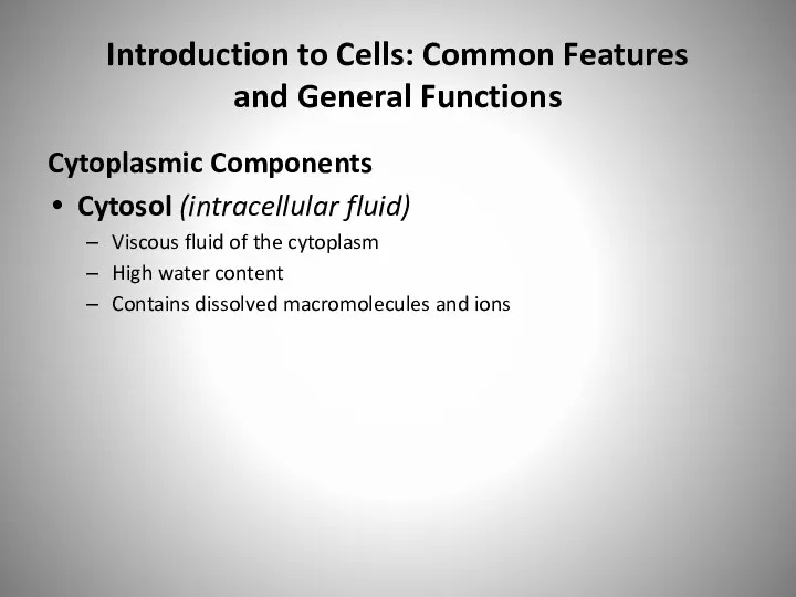
- 15. Introduction to Cells: Common Features and General Functions Cytoplasmic Components Cytosol (intracellular fluid) Viscous fluid of
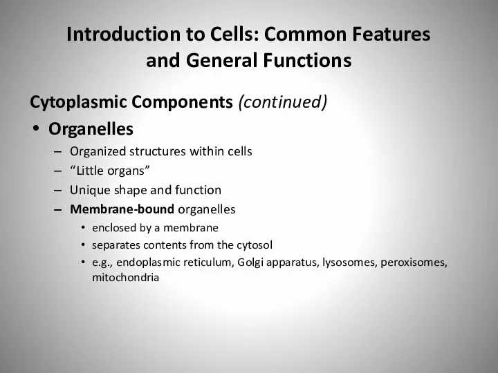
- 16. Introduction to Cells: Common Features and General Functions Cytoplasmic Components (continued) Organelles Organized structures within cells
- 17. Introduction to Cells: Common Features and General Functions Cytoplasmic Components Organelles (continued) Non-membrane-bound organelles not enclosed
- 18. Figure 4.4 Copyright © The McGraw-Hill Companies, Inc. Permission required for reproduction or display. Flagellum Non-membrane-
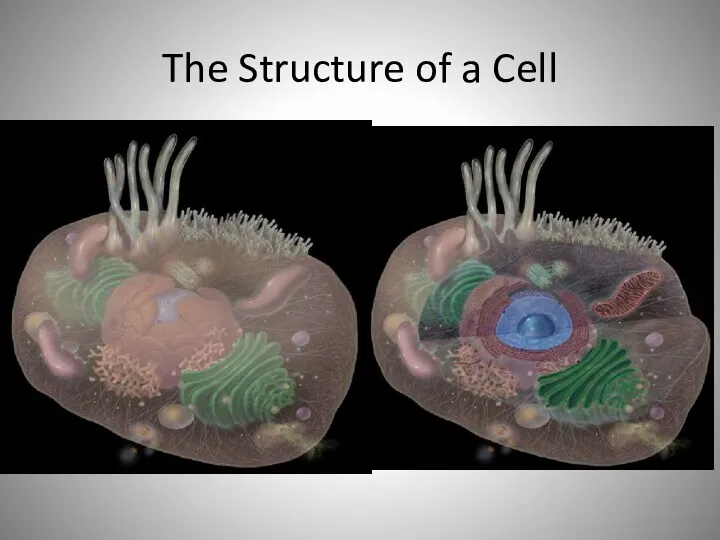
- 19. The Structure of a Cell
- 20. Introduction to Cells: Common Features and General Functions General Cell Functions Performed by most cells Maintain
- 21. Introduction to Cells: Common Features and General Functions General Cell Functions (continued) Performed by some cells
- 22. Plasma Membrane Inner leaflet Outer leaflet Plasma membranes Cytoplasm Extracellular matrix
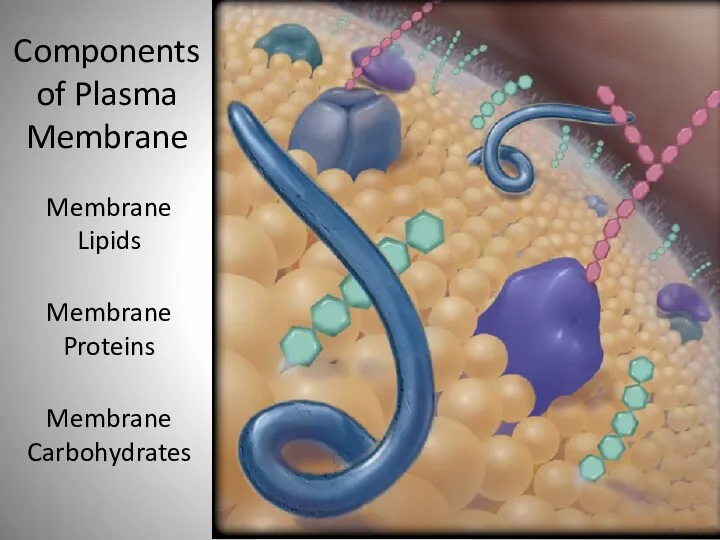
- 23. Membrane Lipids Membrane Proteins Membrane Carbohydrates Components of Plasma Membrane
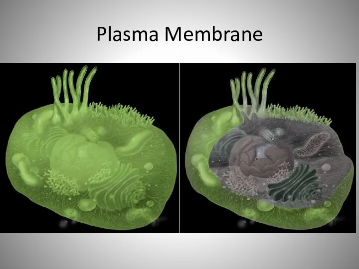
- 24. Plasma Membrane
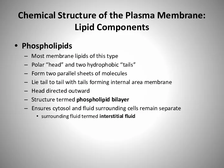
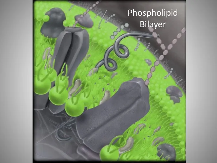
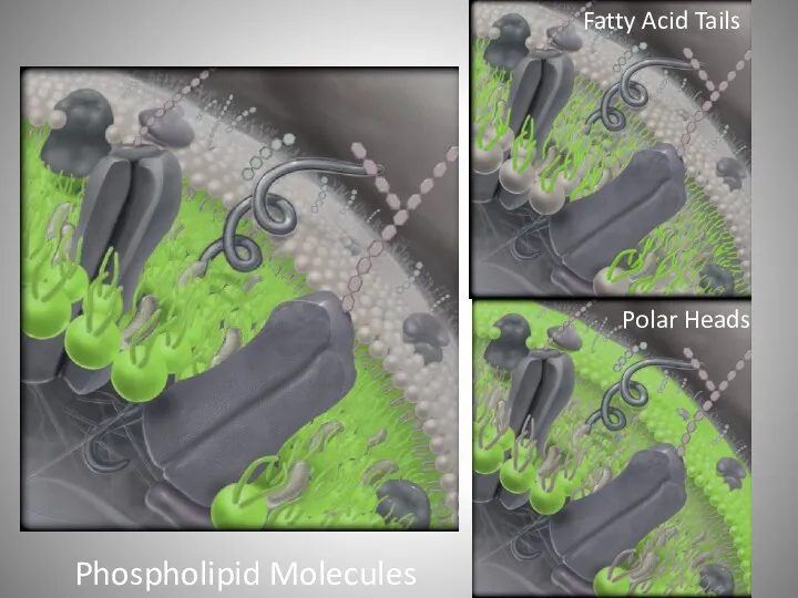
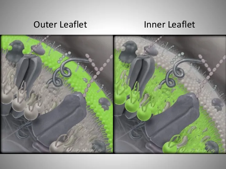
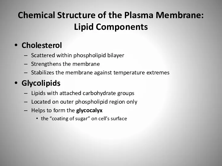
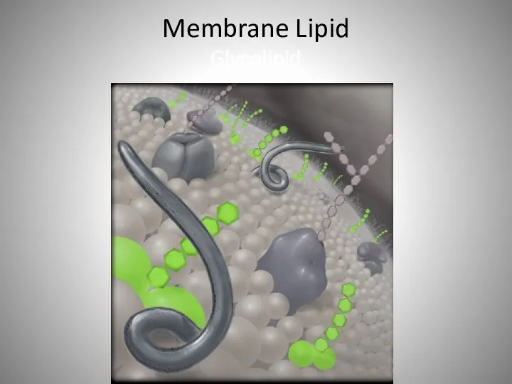
- 25. Chemical Structure of the Plasma Membrane: Lipid Components Phospholipids Most membrane lipids of this type Polar
- 26. Phospholipid Bilayer
- 27. Phospholipid Molecules Fatty Acid Tails Polar Heads
- 28. Outer Leaflet Inner Leaflet
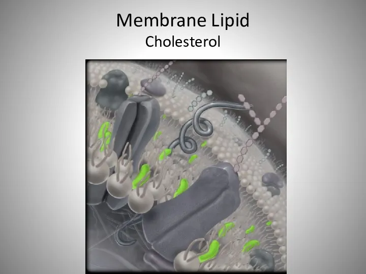
- 29. Chemical Structure of the Plasma Membrane: Lipid Components Cholesterol Scattered within phospholipid bilayer Strengthens the membrane
- 30. Figure 4.5 b: © Don W. Fawcett/Photo Researchers, Inc. Copyright © The McGraw-Hill Companies, Inc. Permission
- 31. Membrane Lipid Cholesterol
- 32. Membrane Lipid Glycolipid
- 33. Membrane Carbohydrates Glycocalyx
- 34. Chemical Structure of the Plasma Membrane: Membrane Proteins Membrane proteins Compose half of plasma membrane by
- 35. Membrane Protein
- 36. Transmembrane Proteins
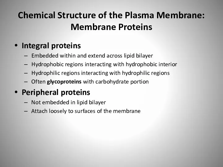
- 37. Chemical Structure of the Plasma Membrane: Membrane Proteins Integral proteins Embedded within and extend across lipid
- 38. Channel Pore
- 39. Peripheral Protein
- 40. Glycoprotein
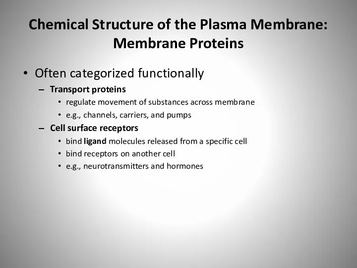
- 41. Chemical Structure of the Plasma Membrane: Membrane Proteins Often categorized functionally Transport proteins regulate movement of
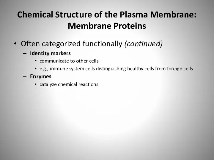
- 42. Chemical Structure of the Plasma Membrane: Membrane Proteins Often categorized functionally (continued) Identity markers communicate to
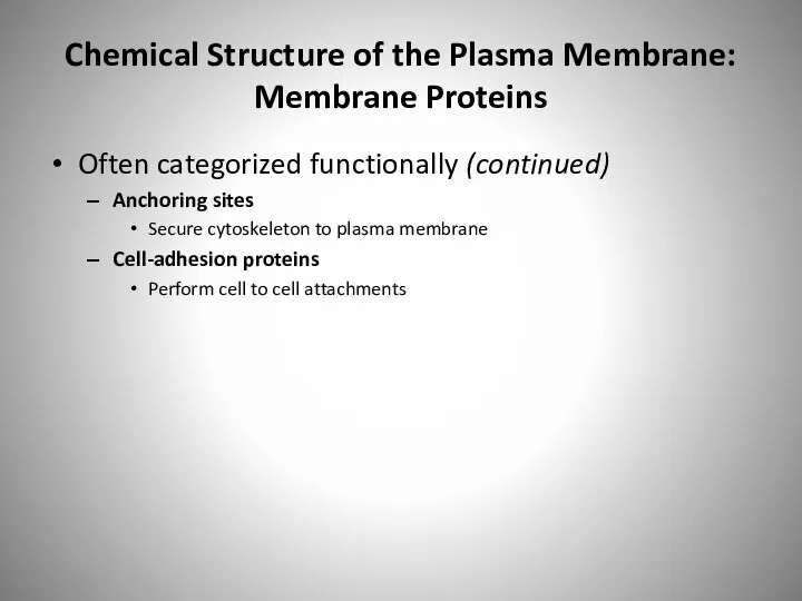
- 43. Chemical Structure of the Plasma Membrane: Membrane Proteins Often categorized functionally (continued) Anchoring sites Secure cytoskeleton
- 44. Figure 4.6 Copyright © The McGraw-Hill Companies, Inc. Permission required for reproduction or display. Interstitial fluid
- 45. Membrane Transport One important function of plasma membrane Regulating movement of materials into and out of
- 46. Membrane Transport Passive processes of membrane transport Do not require energy Depend on substances moving down
- 47. Membrane Transport Active processes of membrane transport Require energy E.g., movement of a substance up its
- 48. Membrane Transport— Passive Processes: Diffusion Environmental conditions affecting rate of diffusion “Steepness” of concentration gradient measure
- 49. Figure 4.7 Copyright © The McGraw-Hill Companies, Inc. Permission required for reproduction or display.
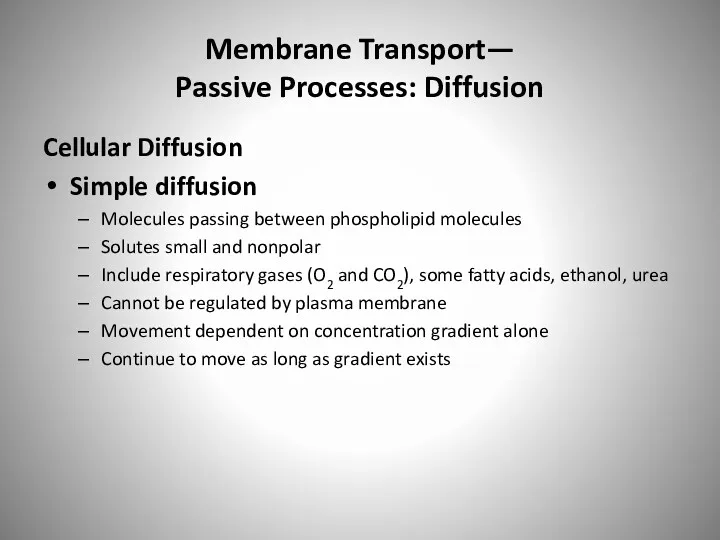
- 50. Membrane Transport— Passive Processes: Diffusion Cellular Diffusion Simple diffusion Molecules passing between phospholipid molecules Solutes small
- 51. Figure 4.8 Copyright © The McGraw-Hill Companies, Inc. Permission required for reproduction or display. Small nonpolar
- 52. Membrane Transport— Passive Processes: Diffusion Cellular Diffusion (continued) Facilitated diffusion Transport process for small charged or
- 53. Membrane Transport— Passive Processes: Diffusion Cellular Diffusion (continued) Facilitated diffusion Transport process for small charged or
- 54. Membrane Transport— Passive Processes: Diffusion Cellular Diffusion (continued) Channel-mediated diffusion Movement of small ions through water-filled
- 55. Figure 4.9a Copyright © The McGraw-Hill Companies, Inc. Permission required for reproduction or display. Ions move
- 56. Membrane Transport— Passive Processes: Diffusion Cellular Diffusion (continued) Na+ channels Na+ leak channels allow Na+ to
- 57. Membrane Transport— Passive Processes: Diffusion Cellular Diffusion (continued) Carrier-mediated diffusion Small, polar molecules assisted across membrane
- 58. Figure 4.9b Interstitial fluid Cytosol (b) Carrier-mediated diffusion Glucose carrier protein Carrier proteins change shape to
- 59. Membrane Transport— Passive Processes: Osmosis Osmosis Passive movement of water through selectively permeable membrane membrane allowing
- 60. Membrane Transport— Passive Processes: Osmosis Plasma Membrane: A Selectively Permeable Membrane Two ways water crosses membrane
- 61. Membrane Transport— Passive Processes: Osmosis Plasma Membrane: A Selectively Permeable Membrane (continued) Two types of solutes
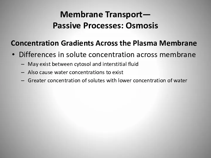
- 62. Membrane Transport— Passive Processes: Osmosis Concentration Gradients Across the Plasma Membrane Differences in solute concentration across
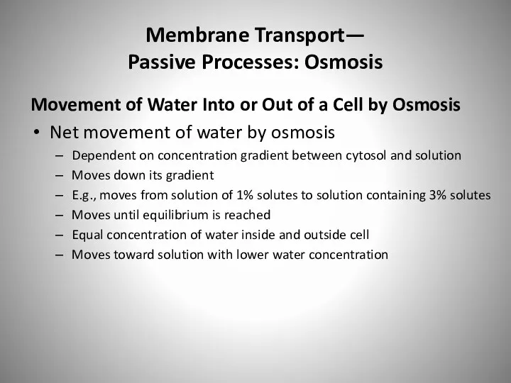
- 63. Membrane Transport— Passive Processes: Osmosis Movement of Water Into or Out of a Cell by Osmosis
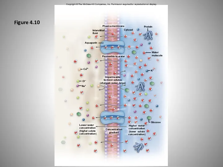
- 64. Plasma membrane Cytosol Protein Water molecule Interstitial fluid Aquaporin Ca2+ Cl-– Impermeable to most solutes (charged,
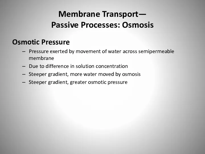
- 65. Membrane Transport— Passive Processes: Osmosis Osmotic Pressure Pressure exerted by movement of water across semipermeable membrane
- 66. Membrane Transport— Passive Processes: Osmosis Osmotic Pressure (continued) Figure 4.11 Semipermeable membrane allowing for passage of
- 67. Figure 4.11 Side A Side B Side A Side B Semipermeable membrane Final setup: Water moved
- 68. Membrane Transport— Passive Processes: Osmosis Osmotic Pressure (continued) Can be measured indirectly Could put stopper on
- 69. Membrane Transport— Passive Processes: Osmosis Osmosis and Tonicity Cell gains or loses water with osmosis Accompanying
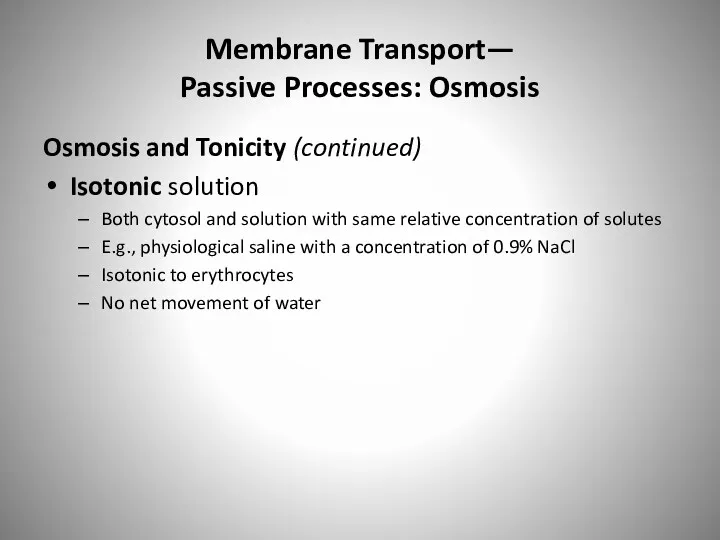
- 70. Membrane Transport— Passive Processes: Osmosis Osmosis and Tonicity (continued) Isotonic solution Both cytosol and solution with
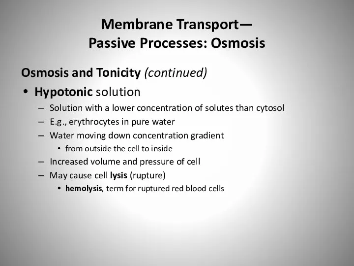
- 71. Membrane Transport— Passive Processes: Osmosis Osmosis and Tonicity (continued) Hypotonic solution Solution with a lower concentration
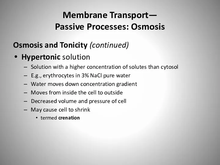
- 72. Membrane Transport— Passive Processes: Osmosis Osmosis and Tonicity (continued) Hypertonic solution Solution with a higher concentration
- 73. Figure 4.12 Copyright © The McGraw-Hill Companies, Inc. Permission required for reproduction or display. SEM 11,550x
- 74. Membrane Transport: Active Processes Active Transport Opposes the movement of solutes by diffusion Solutes moved against
- 75. Membrane Transport: Active Processes Active Transport (continued) Primary active transport Uses energy directly from breakdown of
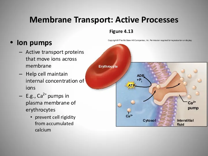
- 76. Membrane Transport: Active Processes Ion pumps Active transport proteins that move ions across membrane Help cell
- 77. Membrane Transport: Active Processes Active Transport (continued) Sodium-potassium pump Special kind of ion pump, an exchange
- 78. Membrane Transport: Active Processes Active Transport Sodium-potassium pump (continued) Maintains an electrochemical gradient electrical charge difference
- 79. Figure 4.14 Copyright © The McGraw-Hill Companies, Inc. Permission required for reproduction or display. Three sodium
- 80. Membrane Transport: Active Processes Active Transport (continued) Secondary active transport Moves substance against concentration gradient Uses
- 81. Membrane Transport: Active Processes Active Transport Secondary active transport (continued) Two substances moved in same direction
- 82. Membrane Transport: Active Processes Active Transport Secondary active transport (continued) Two substances moved in opposite directions
- 83. Figure 4.15 Copyright © The McGraw-Hill Companies, Inc. Permission required for reproduction or display. Na+ diffuses
- 84. Membrane Transport: Active Processes Vesicular Transport Requires vesicles membrane-bounded sac filled with materials Requires energy to
- 85. Membrane Transport: Active Processes Vesicular Transport (continued) Exocytosis How large substances are secreted from cell Macromolecules
- 86. Figure 4.16 Copyright © The McGraw-Hill Companies, Inc. Permission required for reproduction or display. 1 2
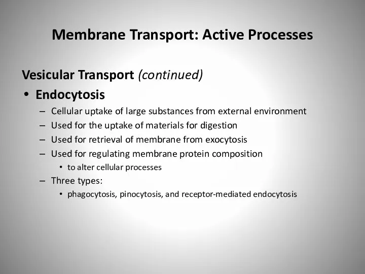
- 87. Membrane Transport: Active Processes Vesicular Transport (continued) Endocytosis Cellular uptake of large substances from external environment
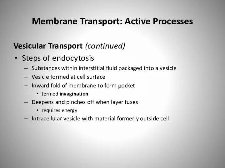
- 88. Membrane Transport: Active Processes Vesicular Transport (continued) Steps of endocytosis Substances within interstitial fluid packaged into
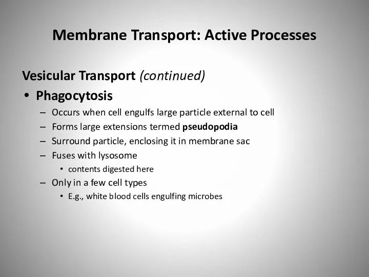
- 89. Membrane Transport: Active Processes Vesicular Transport (continued) Phagocytosis Occurs when cell engulfs large particle external to
- 90. Figure 4.17a Copyright © The McGraw-Hill Companies, Inc. Permission required for reproduction or display. (a) Phagocytosis
- 91. Membrane Transport: Active Processes Vesicular Transport (continued) Pinocytosis Internalization of droplets of interstitial fluid Multiple, small
- 92. Figure 4.17b Copyright © The McGraw-Hill Companies, Inc. Permission required for reproduction or display. (b) Pinocytosis
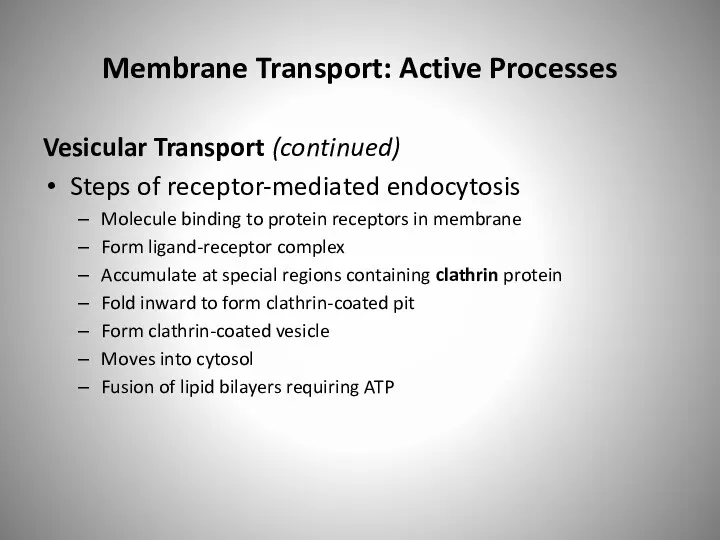
- 93. Membrane Transport: Active Processes Vesicular Transport (continued) Receptor-mediated endocytosis Movement of specific molecules from interstitial environment
- 94. Membrane Transport: Active Processes Vesicular Transport (continued) Steps of receptor-mediated endocytosis Molecule binding to protein receptors
- 95. Membrane Transport: Active Processes Vesicular Transport (continued) Steps of receptor-mediated endocytosis Molecule binding to protein receptors
- 96. Figure 4.17c Copyright © The McGraw-Hill Companies, Inc. Permission required for reproduction or display. (c) Receptor-mediated
- 97. Figure 4.18a DIFFUSION: Movement of a solute from an area of higher concentration to an area
- 98. Figure 4.18b Copyright © The McGraw-Hill Companies, Inc. Permission required for reproduction or display. Require expenditure
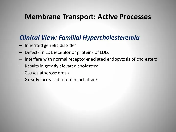
- 99. Membrane Transport: Active Processes Clinical View: Familial Hypercholesteremia Inherited genetic disorder Defects in LDL receptor or
- 101. Скачать презентацию
Understanding the Cell
All body processes dependent upon cells for their activities
Cells
Understanding the Cell
All body processes dependent upon cells for their activities
Cells

Introduction to Cells: How Cells Are Studied
Cells
Studied through the discipline of
Introduction to Cells: How Cells Are Studied
Cells
Studied through the discipline of
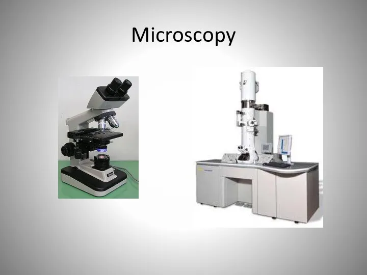
Microscopy
Microscopy
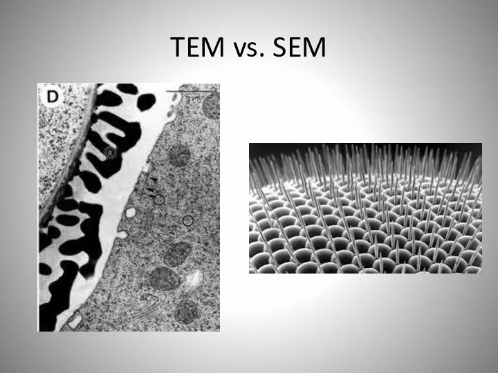
TEM vs. SEM
TEM vs. SEM
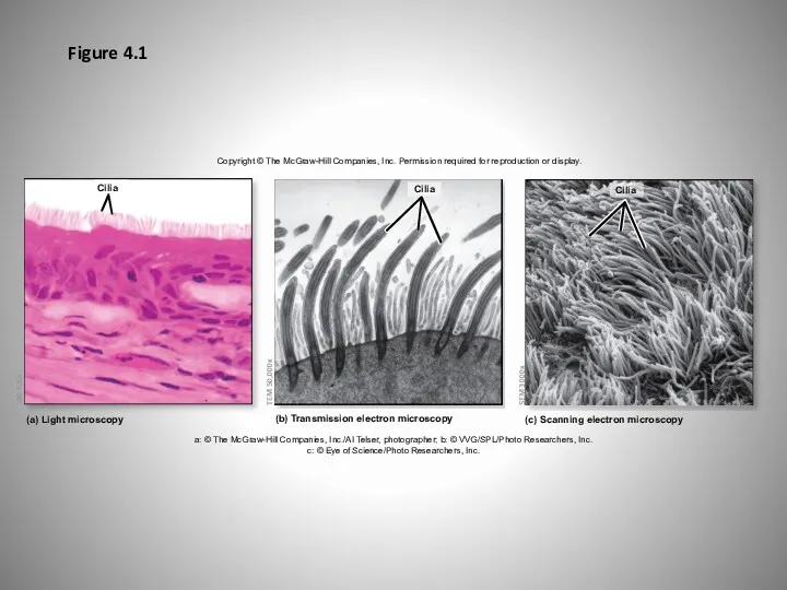
Figure 4.1
Copyright © The McGraw-Hill Companies, Inc. Permission required for reproduction
Figure 4.1
Copyright © The McGraw-Hill Companies, Inc. Permission required for reproduction
Introduction to Cells: Cell Size and Shape
Cells vary greatly in size
Introduction to Cells: Cell Size and Shape
Cells vary greatly in size
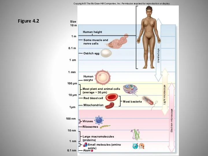
Figure 4.2
Copyright © The McGraw-Hill Companies, Inc. Permission required for reproduction
Figure 4.2
Copyright © The McGraw-Hill Companies, Inc. Permission required for reproduction
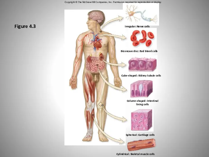
Figure 4.3
Copyright © The McGraw-Hill Companies, Inc. Permission required for reproduction
Figure 4.3
Copyright © The McGraw-Hill Companies, Inc. Permission required for reproduction

Introduction to Cells: Common Features
and General Functions
Overview of Cellular Components
Plasma
Introduction to Cells: Common Features
and General Functions
Overview of Cellular Components
Plasma

Plasma Membrane
Plasma Membrane
Introduction to Cells: Common Features
and General Functions
Overview of Cellular Components
Introduction to Cells: Common Features
and General Functions
Overview of Cellular Components
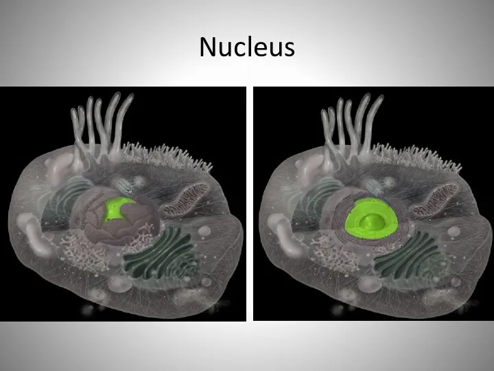
Nucleus
Nucleus
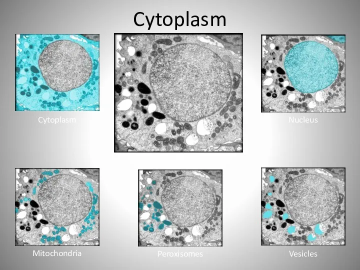
Cytoplasm
Cytoplasm
Nucleus
Mitochondria
Peroxisomes
Vesicles
Cytoplasm
Cytoplasm
Nucleus
Mitochondria
Peroxisomes
Vesicles
Introduction to Cells: Common Features
and General Functions
Cytoplasmic Components
Cytosol (intracellular fluid)
Viscous
Introduction to Cells: Common Features
and General Functions
Cytoplasmic Components
Cytosol (intracellular fluid)
Viscous
Introduction to Cells: Common Features
and General Functions
Cytoplasmic Components (continued)
Organelles
Organized structures
Introduction to Cells: Common Features
and General Functions
Cytoplasmic Components (continued)
Organelles
Organized structures
Introduction to Cells: Common Features
and General Functions
Cytoplasmic Components
Organelles (continued)
Non-membrane-bound organelles
not
Introduction to Cells: Common Features
and General Functions
Cytoplasmic Components
Organelles (continued)
Non-membrane-bound organelles
not
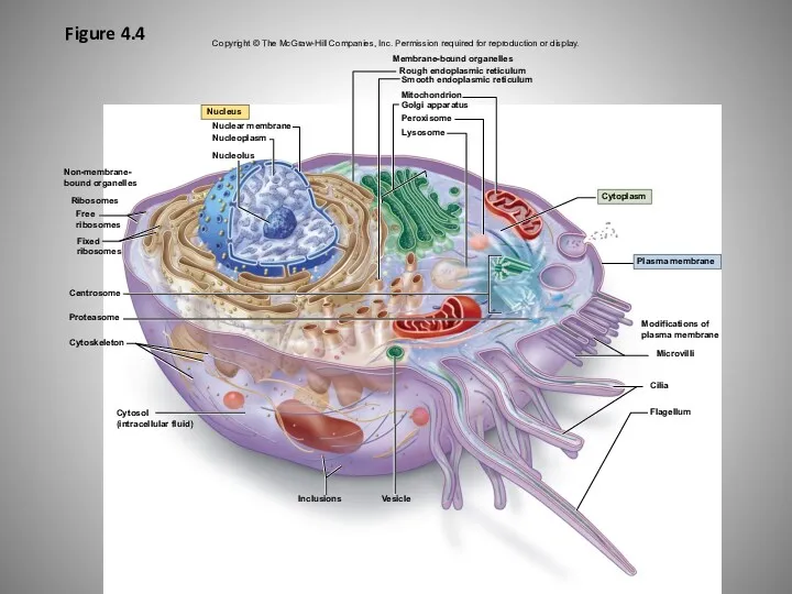
Figure 4.4
Copyright © The McGraw-Hill Companies, Inc. Permission required for reproduction
Figure 4.4
Copyright © The McGraw-Hill Companies, Inc. Permission required for reproduction
The Structure of a Cell
The Structure of a Cell
Introduction to Cells: Common Features
and General Functions
General Cell Functions
Performed by
Introduction to Cells: Common Features
and General Functions
General Cell Functions
Performed by
Introduction to Cells: Common Features
and General Functions
General Cell Functions (continued)
Performed
Introduction to Cells: Common Features
and General Functions
General Cell Functions (continued)
Performed
Plasma Membrane
Inner leaflet
Outer leaflet
Plasma membranes
Cytoplasm
Extracellular matrix
Plasma Membrane
Inner leaflet
Outer leaflet
Plasma membranes
Cytoplasm
Extracellular matrix
Membrane Lipids
Membrane Proteins
Membrane Carbohydrates
Components of Plasma Membrane
Membrane Lipids
Membrane Proteins
Membrane Carbohydrates
Components of Plasma Membrane
Plasma Membrane
Plasma Membrane
Chemical Structure of the Plasma Membrane: Lipid Components
Phospholipids
Most membrane lipids of
Chemical Structure of the Plasma Membrane: Lipid Components
Phospholipids
Most membrane lipids of
Phospholipid Bilayer
Phospholipid Bilayer
Phospholipid Molecules
Fatty Acid Tails
Polar Heads
Phospholipid Molecules
Fatty Acid Tails
Polar Heads
Outer Leaflet
Inner Leaflet
Outer Leaflet
Inner Leaflet
Chemical Structure of the Plasma Membrane: Lipid Components
Cholesterol
Scattered within phospholipid bilayer
Strengthens
Chemical Structure of the Plasma Membrane: Lipid Components
Cholesterol
Scattered within phospholipid bilayer
Strengthens
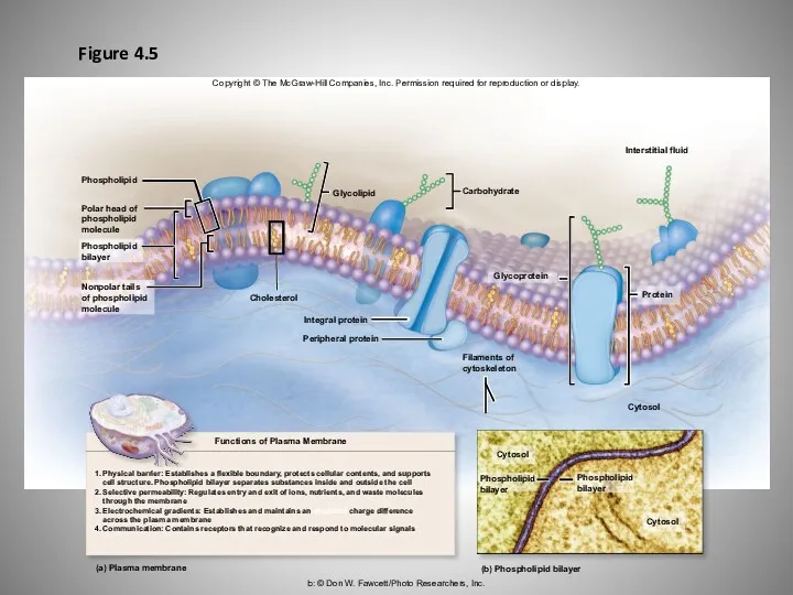
Figure 4.5
b: © Don W. Fawcett/Photo Researchers, Inc.
Copyright © The McGraw-Hill
Figure 4.5
b: © Don W. Fawcett/Photo Researchers, Inc.
Copyright © The McGraw-Hill
Membrane Lipid
Cholesterol
Membrane Lipid
Cholesterol
Membrane Lipid
Glycolipid
Membrane Lipid
Glycolipid
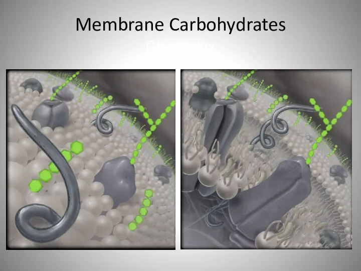
Membrane Carbohydrates
Glycocalyx
Membrane Carbohydrates
Glycocalyx
Chemical Structure of the Plasma Membrane: Membrane Proteins
Membrane proteins
Compose half of
Chemical Structure of the Plasma Membrane: Membrane Proteins
Membrane proteins
Compose half of
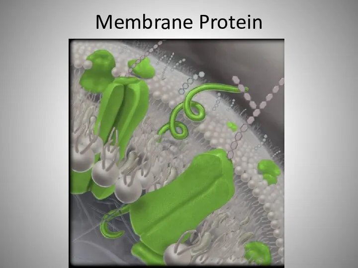
Membrane Protein
Membrane Protein
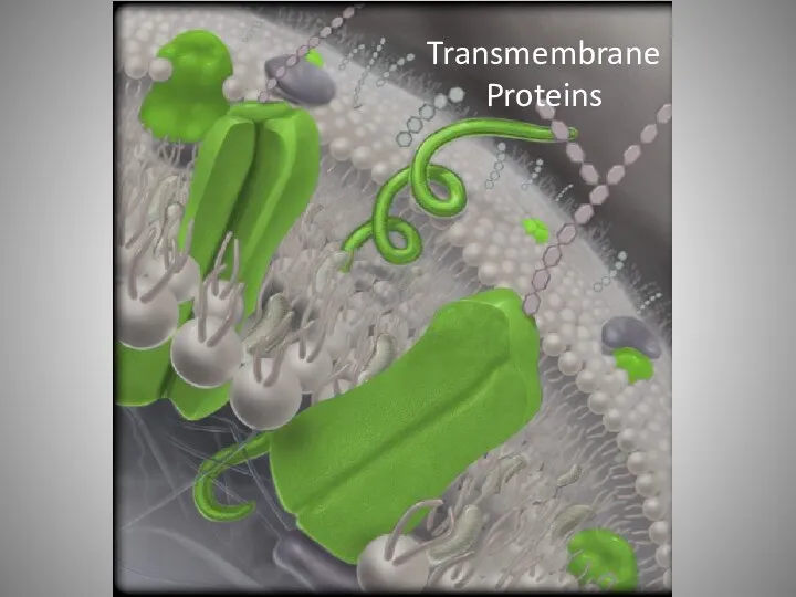
Transmembrane Proteins
Transmembrane Proteins
Chemical Structure of the Plasma Membrane: Membrane Proteins
Integral proteins
Embedded within and
Chemical Structure of the Plasma Membrane: Membrane Proteins
Integral proteins
Embedded within and
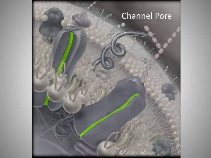
Channel Pore
Channel Pore
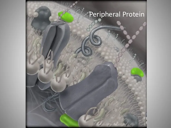
Peripheral Protein
Peripheral Protein
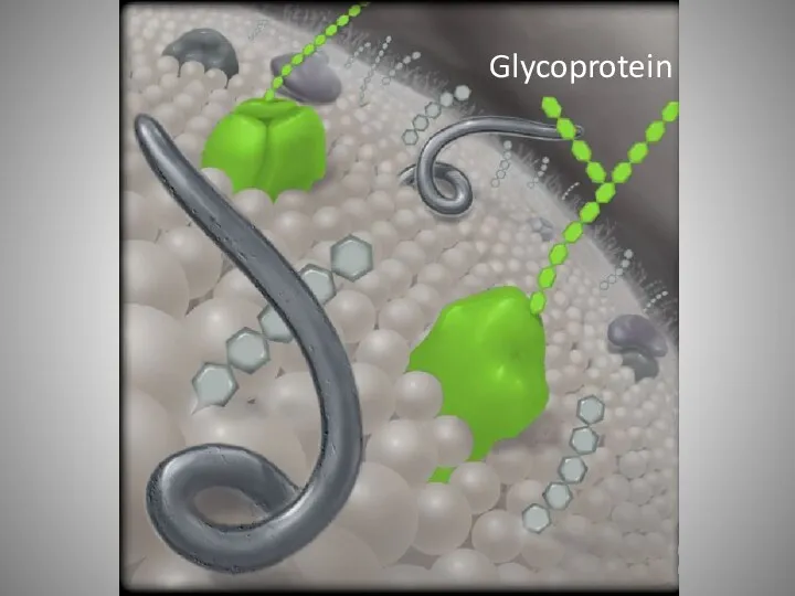
Glycoprotein
Glycoprotein
Chemical Structure of the Plasma Membrane: Membrane Proteins
Often categorized functionally
Transport proteins
regulate
Chemical Structure of the Plasma Membrane: Membrane Proteins
Often categorized functionally
Transport proteins
regulate
Chemical Structure of the Plasma Membrane: Membrane Proteins
Often categorized functionally (continued)
Identity
Chemical Structure of the Plasma Membrane: Membrane Proteins
Often categorized functionally (continued)
Identity
Chemical Structure of the Plasma Membrane: Membrane Proteins
Often categorized functionally (continued)
Anchoring
Chemical Structure of the Plasma Membrane: Membrane Proteins
Often categorized functionally (continued)
Anchoring
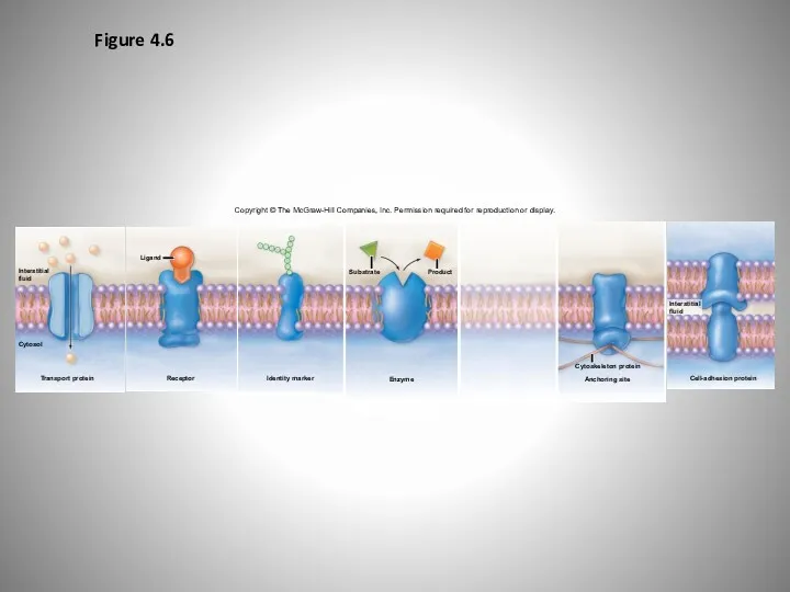
Figure 4.6
Copyright © The McGraw-Hill Companies, Inc. Permission required for reproduction
Figure 4.6
Copyright © The McGraw-Hill Companies, Inc. Permission required for reproduction
Membrane Transport
One important function of plasma membrane
Regulating movement of materials into
Membrane Transport
One important function of plasma membrane
Regulating movement of materials into
Membrane Transport
Passive processes of membrane transport
Do not require energy
Depend on substances
Membrane Transport
Passive processes of membrane transport
Do not require energy
Depend on substances
Membrane Transport
Active processes of membrane transport
Require energy
E.g., movement of a substance
Membrane Transport
Active processes of membrane transport
Require energy
E.g., movement of a substance
Membrane Transport—
Passive Processes: Diffusion
Environmental conditions affecting rate of diffusion
“Steepness” of concentration
Membrane Transport—
Passive Processes: Diffusion
Environmental conditions affecting rate of diffusion
“Steepness” of concentration
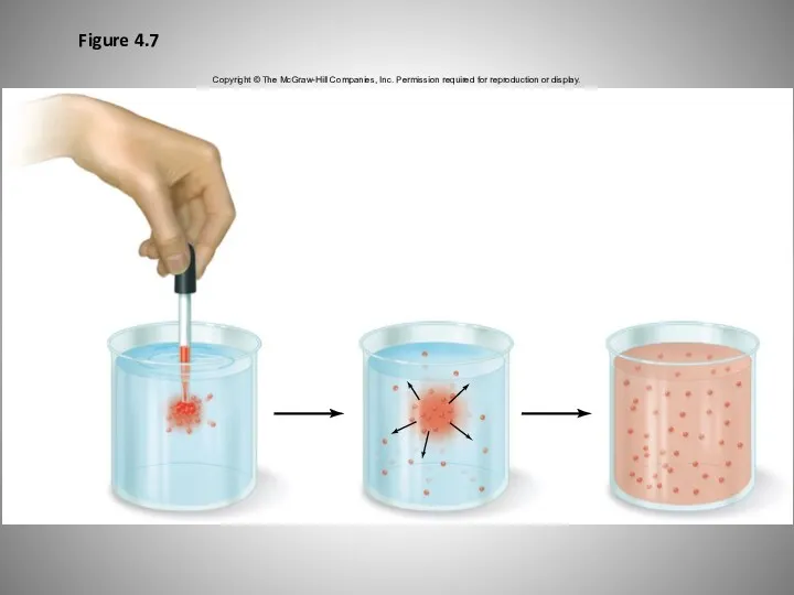
Figure 4.7
Copyright © The McGraw-Hill Companies, Inc. Permission required for reproduction
Figure 4.7
Copyright © The McGraw-Hill Companies, Inc. Permission required for reproduction
Membrane Transport—
Passive Processes: Diffusion
Cellular Diffusion
Simple diffusion
Molecules passing between phospholipid molecules
Solutes small
Membrane Transport—
Passive Processes: Diffusion
Cellular Diffusion
Simple diffusion
Molecules passing between phospholipid molecules
Solutes small
Figure 4.8
Copyright © The McGraw-Hill Companies, Inc. Permission required for reproduction
Figure 4.8
Copyright © The McGraw-Hill Companies, Inc. Permission required for reproduction
Membrane Transport—
Passive Processes: Diffusion
Cellular Diffusion (continued)
Facilitated diffusion
Transport process for small charged
Membrane Transport—
Passive Processes: Diffusion
Cellular Diffusion (continued)
Facilitated diffusion
Transport process for small charged

Membrane Transport—
Passive Processes: Diffusion
Cellular Diffusion (continued)
Facilitated diffusion
Transport process for small charged
Membrane Transport—
Passive Processes: Diffusion
Cellular Diffusion (continued)
Facilitated diffusion
Transport process for small charged
Membrane Transport—
Passive Processes: Diffusion
Cellular Diffusion (continued)
Channel-mediated diffusion
Movement of small ions through
Membrane Transport—
Passive Processes: Diffusion
Cellular Diffusion (continued)
Channel-mediated diffusion
Movement of small ions through
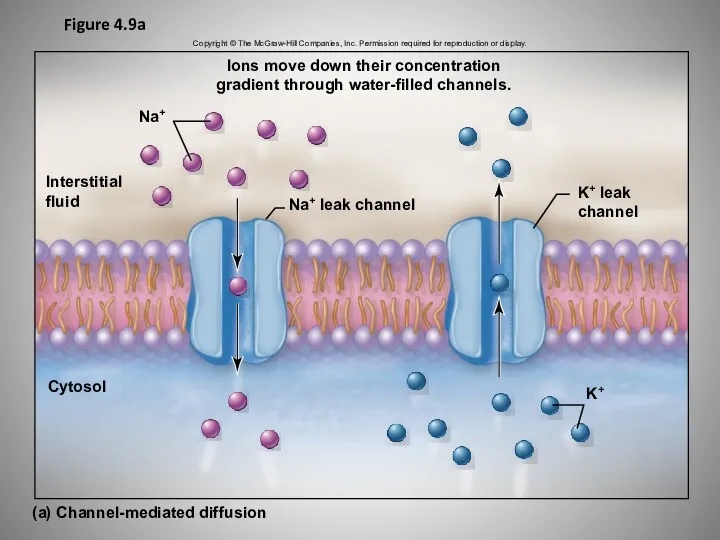
Figure 4.9a
Copyright © The McGraw-Hill Companies, Inc. Permission required for reproduction
Figure 4.9a
Copyright © The McGraw-Hill Companies, Inc. Permission required for reproduction
Membrane Transport—
Passive Processes: Diffusion
Cellular Diffusion (continued)
Na+ channels
Na+ leak channels
allow Na+
Membrane Transport—
Passive Processes: Diffusion
Cellular Diffusion (continued)
Na+ channels
Na+ leak channels
allow Na+
Membrane Transport—
Passive Processes: Diffusion
Cellular Diffusion (continued)
Carrier-mediated diffusion
Small, polar molecules assisted across
Membrane Transport—
Passive Processes: Diffusion
Cellular Diffusion (continued)
Carrier-mediated diffusion
Small, polar molecules assisted across
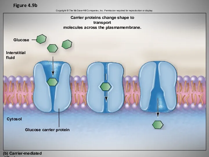
Figure 4.9b
Interstitial
fluid
Cytosol
(b) Carrier-mediated diffusion
Glucose carrier protein
Carrier proteins change shape to transport
molecules
Figure 4.9b
Interstitial
fluid
Cytosol
(b) Carrier-mediated diffusion
Glucose carrier protein
Carrier proteins change shape to transport
molecules
Membrane Transport—
Passive Processes: Osmosis
Osmosis
Passive movement of water through selectively permeable membrane
membrane
Membrane Transport—
Passive Processes: Osmosis
Osmosis
Passive movement of water through selectively permeable membrane
membrane
Membrane Transport—
Passive Processes: Osmosis
Plasma Membrane: A Selectively Permeable Membrane
Two ways water
Membrane Transport—
Passive Processes: Osmosis
Plasma Membrane: A Selectively Permeable Membrane
Two ways water
Membrane Transport—
Passive Processes: Osmosis
Plasma Membrane: A Selectively Permeable Membrane (continued)
Two types
Membrane Transport—
Passive Processes: Osmosis
Plasma Membrane: A Selectively Permeable Membrane (continued)
Two types
Membrane Transport—
Passive Processes: Osmosis
Concentration Gradients Across the Plasma Membrane
Differences in solute
Membrane Transport—
Passive Processes: Osmosis
Concentration Gradients Across the Plasma Membrane
Differences in solute
Membrane Transport—
Passive Processes: Osmosis
Movement of Water Into or Out of a
Membrane Transport—
Passive Processes: Osmosis
Movement of Water Into or Out of a
Plasma membrane
Cytosol
Protein
Water
molecule
Interstitial
fluid
Aquaporin
Ca2+
Cl-–
Impermeable
to most solutes
(charged, polar, large)
Lower water
concentration
(higher solute
concentration)
Concentration
gradient
Glucose
Higher water
concentration
(lower solute
concentration)
Permeable to
Plasma membrane
Cytosol
Protein
Water
molecule
Interstitial
fluid
Aquaporin
Ca2+
Cl-–
Impermeable
to most solutes
(charged, polar, large)
Lower water
concentration
(higher solute
concentration)
Concentration
gradient
Glucose
Higher water
concentration
(lower solute
concentration)
Permeable to
Membrane Transport—
Passive Processes: Osmosis
Osmotic Pressure
Pressure exerted by movement of water across
Membrane Transport—
Passive Processes: Osmosis
Osmotic Pressure
Pressure exerted by movement of water across
Membrane Transport—
Passive Processes: Osmosis
Osmotic Pressure (continued)
Figure 4.11
Semipermeable membrane allowing for passage
Membrane Transport—
Passive Processes: Osmosis
Osmotic Pressure (continued)
Figure 4.11
Semipermeable membrane allowing for passage
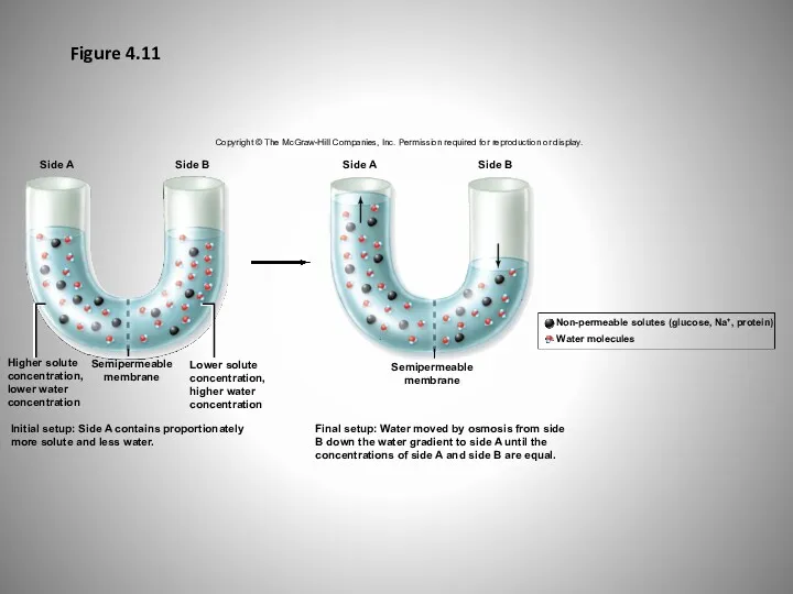
Figure 4.11
Side A
Side B
Side A
Side B
Semipermeable
membrane
Final setup: Water moved by osmosis
Figure 4.11
Side A
Side B
Side A
Side B
Semipermeable
membrane
Final setup: Water moved by osmosis
Membrane Transport—
Passive Processes: Osmosis
Osmotic Pressure (continued)
Can be measured indirectly
Could put stopper
Membrane Transport—
Passive Processes: Osmosis
Osmotic Pressure (continued)
Can be measured indirectly
Could put stopper
Membrane Transport—
Passive Processes: Osmosis
Osmosis and Tonicity
Cell gains or loses water with
Membrane Transport—
Passive Processes: Osmosis
Osmosis and Tonicity
Cell gains or loses water with
Membrane Transport—
Passive Processes: Osmosis
Osmosis and Tonicity (continued)
Isotonic solution
Both cytosol and solution
Membrane Transport—
Passive Processes: Osmosis
Osmosis and Tonicity (continued)
Isotonic solution
Both cytosol and solution
Membrane Transport—
Passive Processes: Osmosis
Osmosis and Tonicity (continued)
Hypotonic solution
Solution with a lower
Membrane Transport—
Passive Processes: Osmosis
Osmosis and Tonicity (continued)
Hypotonic solution
Solution with a lower
Membrane Transport—
Passive Processes: Osmosis
Osmosis and Tonicity (continued)
Hypertonic solution
Solution with a higher
Membrane Transport—
Passive Processes: Osmosis
Osmosis and Tonicity (continued)
Hypertonic solution
Solution with a higher
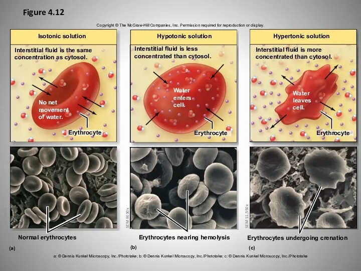
Figure 4.12
Copyright © The McGraw-Hill Companies, Inc. Permission required for reproduction
Figure 4.12
Copyright © The McGraw-Hill Companies, Inc. Permission required for reproduction
Membrane Transport: Active Processes
Active Transport
Opposes the movement of solutes by diffusion
Solutes
Membrane Transport: Active Processes
Active Transport
Opposes the movement of solutes by diffusion
Solutes
Membrane Transport: Active Processes
Active Transport (continued)
Primary active transport
Uses energy directly from
Membrane Transport: Active Processes
Active Transport (continued)
Primary active transport
Uses energy directly from
Membrane Transport: Active Processes
Ion pumps
Active transport proteins that move ions across
Membrane Transport: Active Processes
Ion pumps
Active transport proteins that move ions across
Membrane Transport: Active Processes
Active Transport (continued)
Sodium-potassium pump
Special kind of ion pump,
Membrane Transport: Active Processes
Active Transport (continued)
Sodium-potassium pump
Special kind of ion pump,
Membrane Transport: Active Processes
Active Transport
Sodium-potassium pump (continued)
Maintains an electrochemical gradient
electrical charge
Membrane Transport: Active Processes
Active Transport
Sodium-potassium pump (continued)
Maintains an electrochemical gradient
electrical charge
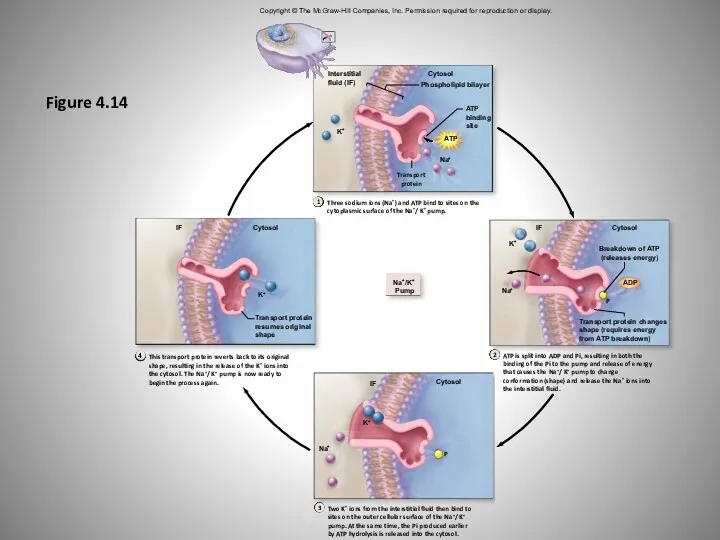
Figure 4.14
Copyright © The McGraw-Hill Companies, Inc. Permission required for reproduction
Figure 4.14
Copyright © The McGraw-Hill Companies, Inc. Permission required for reproduction
Membrane Transport: Active Processes
Active Transport (continued)
Secondary active transport
Moves substance against concentration
Membrane Transport: Active Processes
Active Transport (continued)
Secondary active transport
Moves substance against concentration
Membrane Transport: Active Processes
Active Transport
Secondary active transport (continued)
Two substances moved in
Membrane Transport: Active Processes
Active Transport
Secondary active transport (continued)
Two substances moved in
Membrane Transport: Active Processes
Active Transport
Secondary active transport (continued)
Two substances moved in
Membrane Transport: Active Processes
Active Transport
Secondary active transport (continued)
Two substances moved in
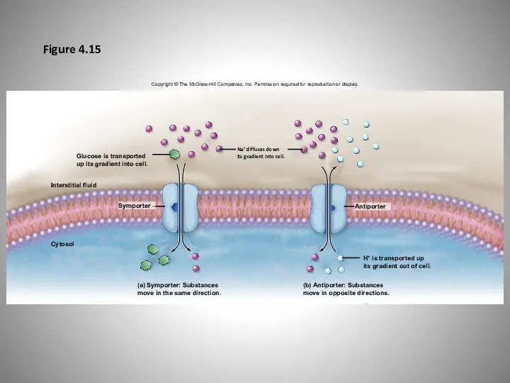
Figure 4.15
Copyright © The McGraw-Hill Companies, Inc. Permission required for reproduction
Figure 4.15
Copyright © The McGraw-Hill Companies, Inc. Permission required for reproduction
Membrane Transport: Active Processes
Vesicular Transport
Requires vesicles
membrane-bounded sac filled with materials
Requires energy
Membrane Transport: Active Processes
Vesicular Transport
Requires vesicles
membrane-bounded sac filled with materials
Requires energy
Membrane Transport: Active Processes
Vesicular Transport (continued)
Exocytosis
How large substances are secreted from
Membrane Transport: Active Processes
Vesicular Transport (continued)
Exocytosis
How large substances are secreted from
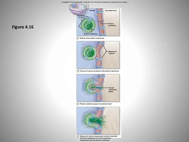
Figure 4.16
Copyright © The McGraw-Hill Companies, Inc. Permission required for reproduction
Figure 4.16
Copyright © The McGraw-Hill Companies, Inc. Permission required for reproduction
Membrane Transport: Active Processes
Vesicular Transport (continued)
Endocytosis
Cellular uptake of large substances from
Membrane Transport: Active Processes
Vesicular Transport (continued)
Endocytosis
Cellular uptake of large substances from
Membrane Transport: Active Processes
Vesicular Transport (continued)
Steps of endocytosis
Substances within interstitial fluid
Membrane Transport: Active Processes
Vesicular Transport (continued)
Steps of endocytosis
Substances within interstitial fluid
Membrane Transport: Active Processes
Vesicular Transport (continued)
Phagocytosis
Occurs when cell engulfs large particle
Membrane Transport: Active Processes
Vesicular Transport (continued)
Phagocytosis
Occurs when cell engulfs large particle
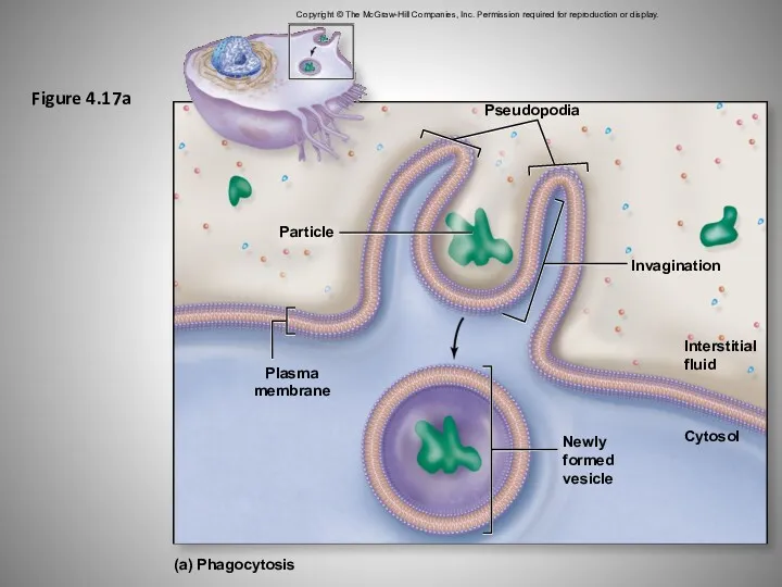
Figure 4.17a
Copyright © The McGraw-Hill Companies, Inc. Permission required for reproduction
Figure 4.17a
Copyright © The McGraw-Hill Companies, Inc. Permission required for reproduction
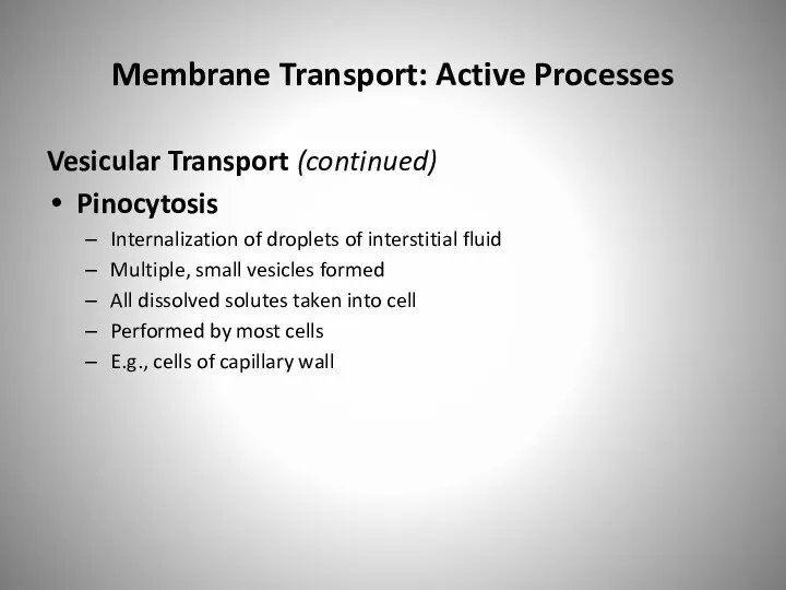
Membrane Transport: Active Processes
Vesicular Transport (continued)
Pinocytosis
Internalization of droplets of interstitial fluid
Multiple,
Membrane Transport: Active Processes
Vesicular Transport (continued)
Pinocytosis
Internalization of droplets of interstitial fluid
Multiple,
Figure 4.17b
Copyright © The McGraw-Hill Companies, Inc. Permission required for reproduction
Figure 4.17b
Copyright © The McGraw-Hill Companies, Inc. Permission required for reproduction
Membrane Transport: Active Processes
Vesicular Transport (continued)
Receptor-mediated endocytosis
Movement of specific molecules
Membrane Transport: Active Processes
Vesicular Transport (continued)
Receptor-mediated endocytosis
Movement of specific molecules

Membrane Transport: Active Processes
Vesicular Transport (continued)
Steps of receptor-mediated endocytosis
Molecule binding
Membrane Transport: Active Processes
Vesicular Transport (continued)
Steps of receptor-mediated endocytosis
Molecule binding
Membrane Transport: Active Processes
Vesicular Transport (continued)
Steps of receptor-mediated endocytosis
Molecule binding
Membrane Transport: Active Processes
Vesicular Transport (continued)
Steps of receptor-mediated endocytosis
Molecule binding
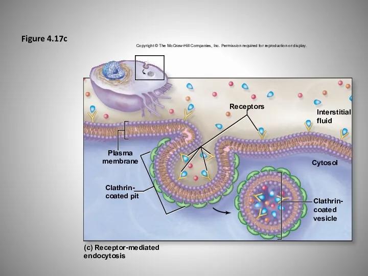
Figure 4.17c
Copyright © The McGraw-Hill Companies, Inc. Permission required for reproduction
Figure 4.17c
Copyright © The McGraw-Hill Companies, Inc. Permission required for reproduction
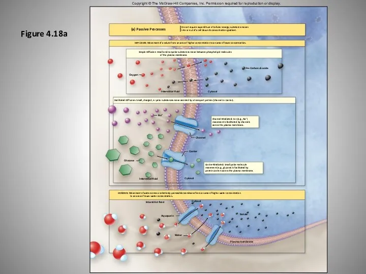
Figure 4.18a
DIFFUSION: Movement of a solute from an area of higher
Figure 4.18a
DIFFUSION: Movement of a solute from an area of higher
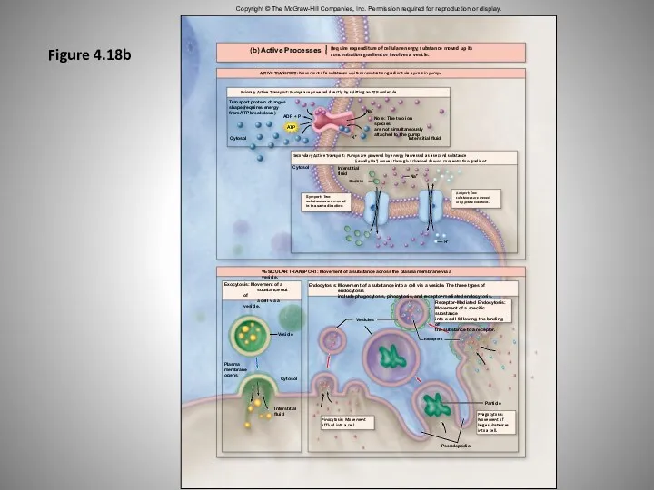
Figure 4.18b
Copyright © The McGraw-Hill Companies, Inc. Permission required for reproduction
Figure 4.18b
Copyright © The McGraw-Hill Companies, Inc. Permission required for reproduction
Membrane Transport: Active Processes
Clinical View: Familial Hypercholesteremia
Inherited genetic disorder
Defects in LDL
Membrane Transport: Active Processes
Clinical View: Familial Hypercholesteremia
Inherited genetic disorder
Defects in LDL
 Белки. Переваривание и всасывание
 Бактерии и их виды (часть 1)
 Презентация к уроку биологии в 8 классе транспортные системы организма
 Антропогенез. Человеческие расы
 Утворення перлин
 Характеристика семейства Розоцветные
 Презентация к уроку 5 класса ФГОС
 Соцветия. Виды соцветий
 Мир как система. Системный подход в современной науке
 Which statement about the properties of life is false?
 Цитоплазма. Химический состав
 Выборка вещи. Контрастный метод
 Изучение строения скелета человека
 Презентация к уроку биологии 8 класс Кожа. Строение и функции
 Язык животных
 Вода в жизни человека
 Тип Плоские черви
 Видоизменения побегов
 Клетка. Клеточная теория. Строение и функции клетки, органоиды клетки
 Энергетический и пластический обмен
 Класс насекомые
 Самые красивые цветы. Творческое название: красивейшие цветы. 6 класс
 Закономерности наследования, установленные Г. Менделем
 Терминология и символика, используемая в генетике.
 Вирусы
 Закономерности формирования и наследования признаков. Изменчивость
 Соцветия. Виды соцветий
 Биофизика цветного зрения