- Dissection Guide to the Rat

Содержание
- 2. Anatomical Terms Cranial-toward the head Caudal-toward the tail Dorsal-toward the backbone Ventral-toward the belly side Lateral-toward
- 3. The Skeleton of the Rat
- 4. Skinning the Rat Lay rat on its back Using a blunt probe, separate the skin from
- 5. Skinning the Rat
- 6. Muscles to Locate Locate the starred muscles. Draw and label them on your lab report.
- 7. Beginning the Muscle Identification The Triceps Brachii
- 8. The Internal Anatomy The first incisions were made down the center of the body cavity and
- 9. After the pinning is complete the interior muscle tissues are exposed, then by making careful incisions,
- 11. Скачать презентацию
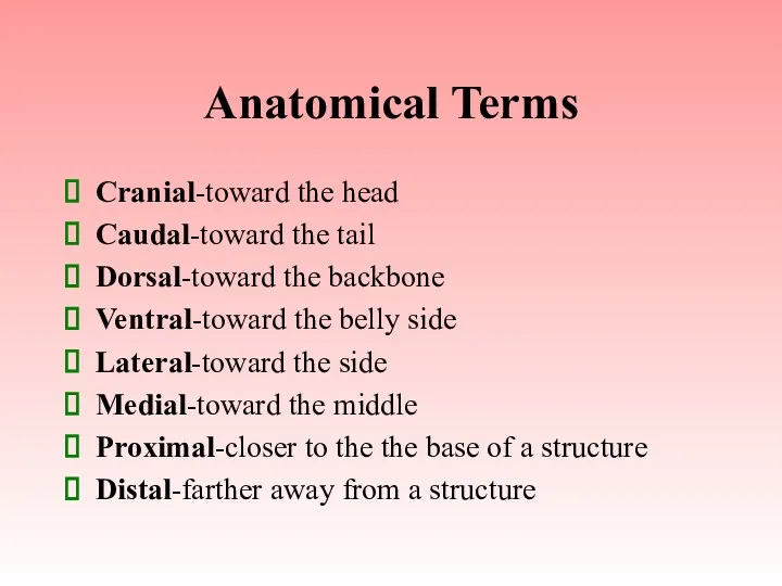
Anatomical Terms
Cranial-toward the head
Caudal-toward the tail
Dorsal-toward the backbone
Ventral-toward the belly side
Lateral-toward
Anatomical Terms
Cranial-toward the head
Caudal-toward the tail
Dorsal-toward the backbone
Ventral-toward the belly side
Lateral-toward
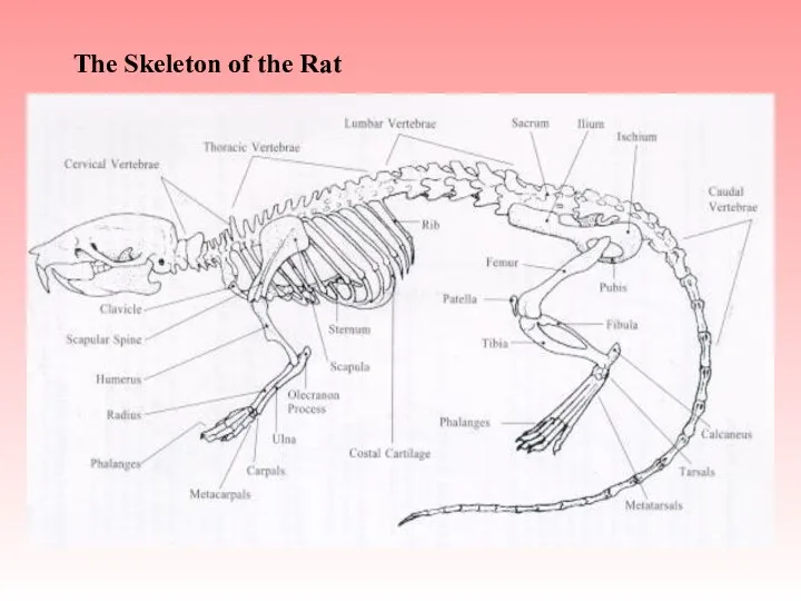
The Skeleton of the Rat
The Skeleton of the Rat
Skinning the Rat
Lay rat on its back
Using a blunt probe, separate
Skinning the Rat
Lay rat on its back
Using a blunt probe, separate
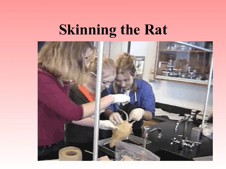
Skinning the Rat
Skinning the Rat
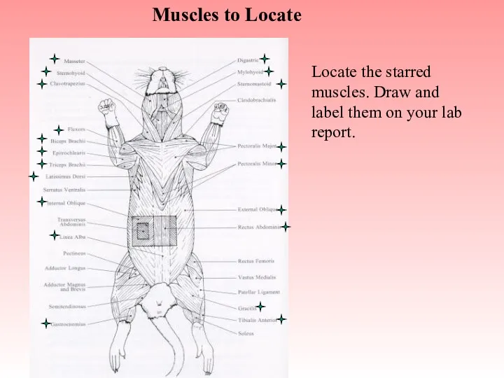
Muscles to Locate
Locate the starred muscles. Draw and label them on
Muscles to Locate
Locate the starred muscles. Draw and label them on
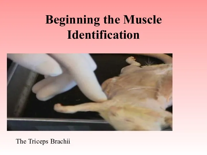
Beginning the Muscle Identification
The Triceps Brachii
Beginning the Muscle Identification
The Triceps Brachii
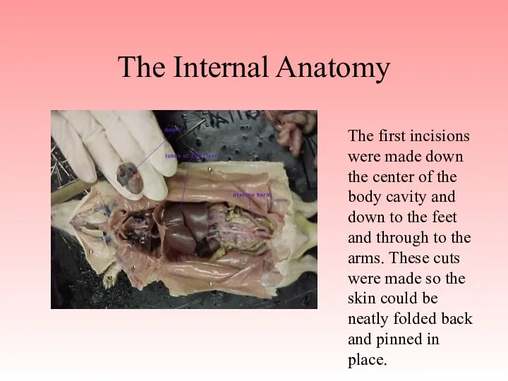
The Internal Anatomy
The first incisions were made down the center
The Internal Anatomy
The first incisions were made down the center
After the pinning is complete the interior muscle tissues are exposed,
After the pinning is complete the interior muscle tissues are exposed,
 Насекомые. Подготовительная группа
 Введение в зоологию
 Декоративные устройства для оформления объектов. Устройство и содержание цветников, вертикальное озеленение, каменистые участки
 Тип: хордовые. Подтипы: бесчерепные и черепные, позвоночные
 The Human-Animal Bond
 Топ 10 новых пород собак
 В гостях у природы
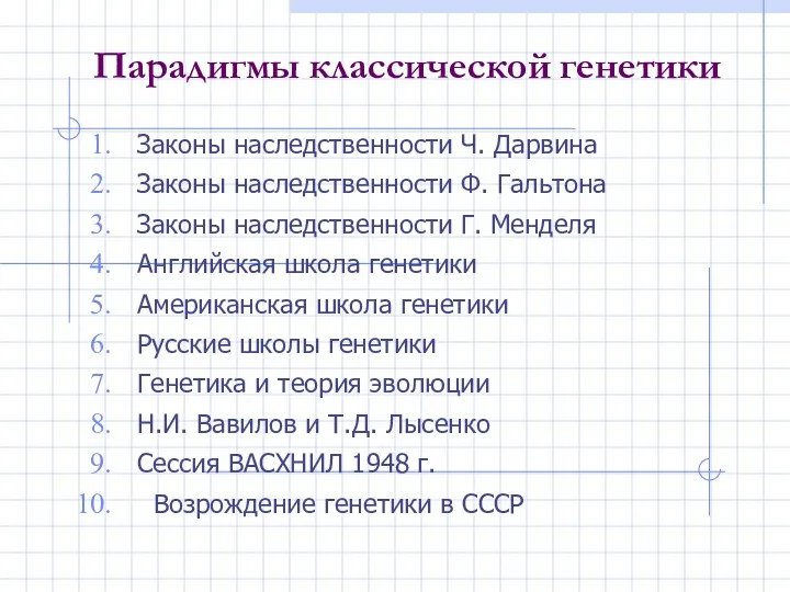 Парадигмы классической генетики
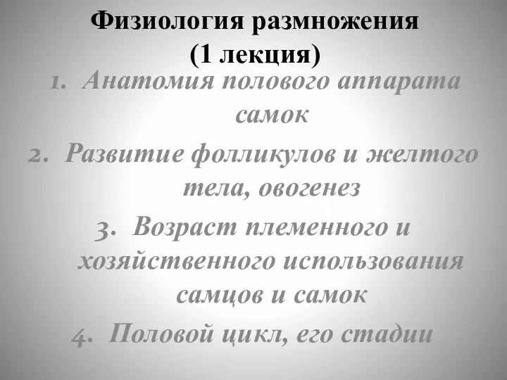 Физиология размножения животных
 Биосинтез белка
 Функции белков
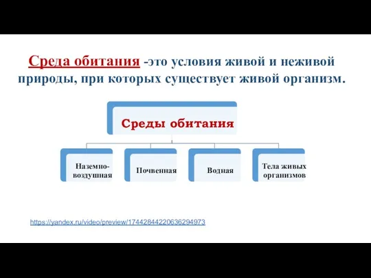 Наземно-воздушная среда обитания
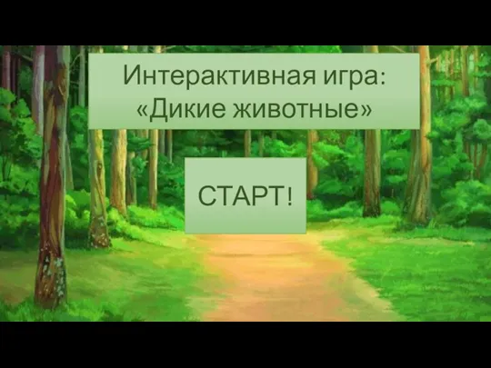 дикий интерактив конец
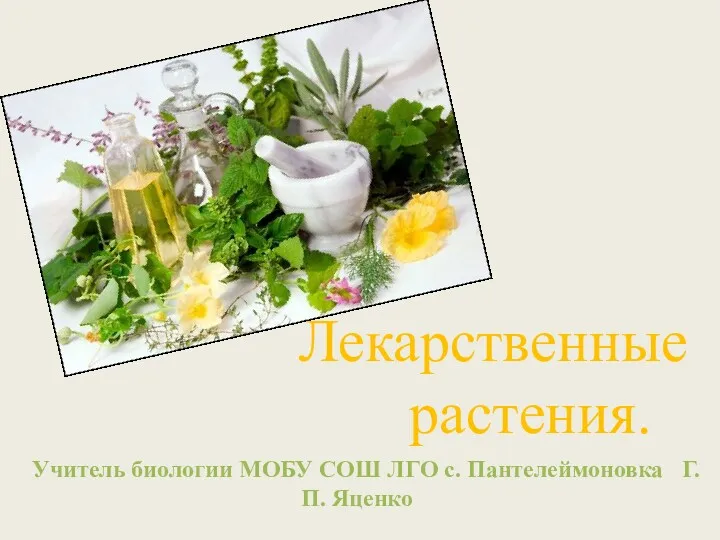 Лекарственные растения
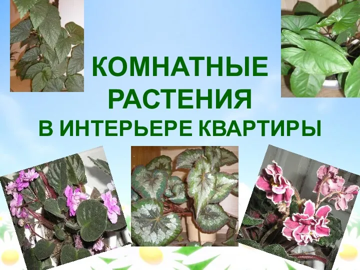 Комнатные растения
 Строение и работа сердца. Круги кровообращения. Движение крови и лимфы
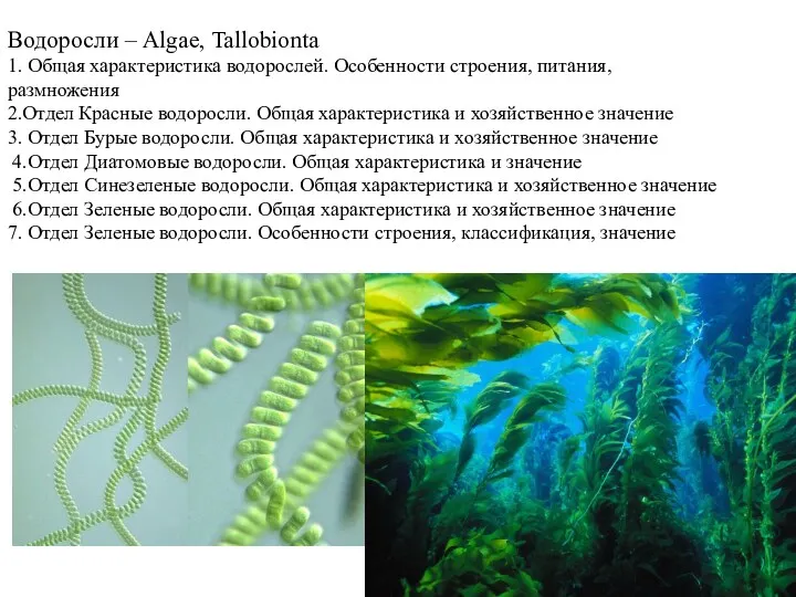 Водоросли. Особенности строения, питания, размножения
 Регуляція експресії генів. (Лекція 2)
 Как появился человек на Земле
 Презентация, конспект урока, карточки - задания к уроку биологии для 6 класса на тему Фотосинтез.
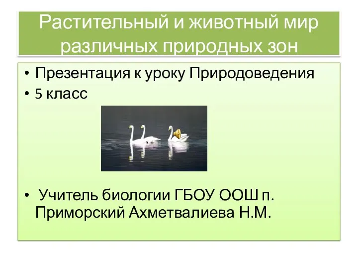 Растительный и животный мир различных природных зон
 Плесневые грибы и дрожжи
 Презентация для урока биологии. Мендель. Жизненный путь
 Еңбектенудің физиологиялық негізі
 Азотсодержащие гетероциклические соединения. Нуклеиновые кислоты
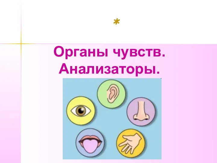 Органы чувств. Анализаторы
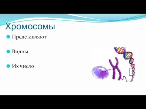 Хромосомы. Набор хромосом
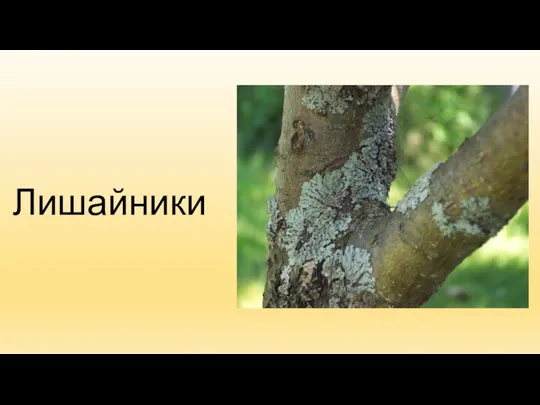 Лишайники 6 класс