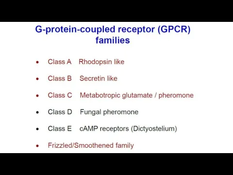
- G-protein-coupled receptors

Содержание
- 4. protein–coupled receptors are found only in eukaryotes, including yeast, and animals. The ligands that bind and
- 5. There are two principal signal transduction pathways involving the G protein–coupled receptors: the cAMP signal pathway
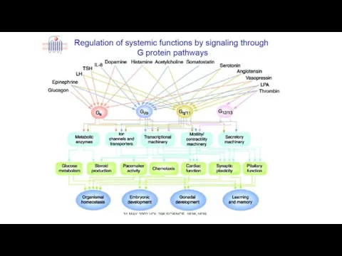
- 6. Physiological roles GPCRs are involved in a wide variety of physiological processes. Some examples of their
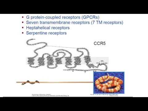
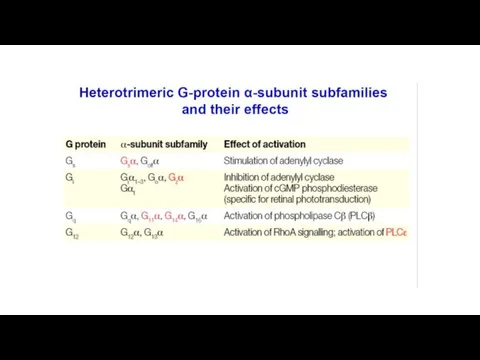
- 9. Structure - Single protein with 7 transmembrane regions
- 10. Ligands Monoamines e.g. dopamine, histamine, noradrenaline, acetylcholine (muscarinic) Nucleotides Lipids Hormones Glutamate Ca++ G-protein-coupled receptors (7-TM
- 11. Ligand binding site - varies depending on receptor type A) Monoamines - pocket in TM helices
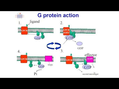
- 12. G-Protein alters shape GDP binding site distorted GDP binding weakened GDP departs a) Interaction of receptor
- 13. Induced fit G-protein alters shape Complex destabilised Process repeated for as long as ligand bound to
- 14. b) Interaction of αs with adenylate cyclase 3. G-protein-coupled receptors (7-TM receptors) 3.6 Signal transduction pathway
- 17. Adrenoreceptor
- 18. Localization and the main effects α1- и β1- receptors localized mainly in the postsynaptic membrane and
- 19. Механизм действия адренергических рецепторов. Эпинефрин и норадреналин являются лигандами для адренергических рецепторов α1, α2 или β.
- 22. Скачать презентацию
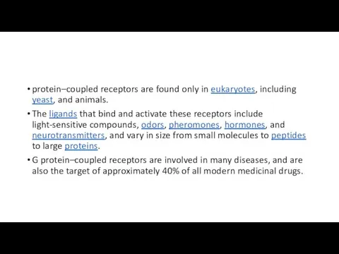
protein–coupled receptors are found only in eukaryotes, including yeast, and animals.
protein–coupled receptors are found only in eukaryotes, including yeast, and animals.
There are two principal signal transduction pathways involving the G protein–coupled
There are two principal signal transduction pathways involving the G protein–coupled
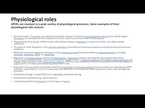
Physiological roles
GPCRs are involved in a wide variety of physiological processes.
Physiological roles GPCRs are involved in a wide variety of physiological processes.
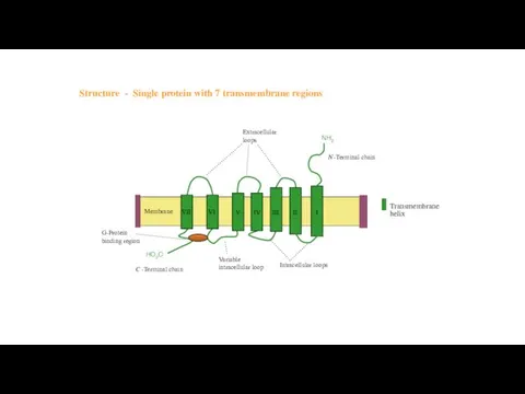
Structure - Single protein with 7 transmembrane regions
Structure - Single protein with 7 transmembrane regions
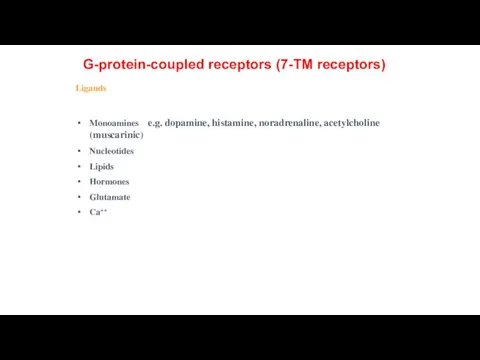
Ligands
Monoamines e.g. dopamine, histamine, noradrenaline, acetylcholine (muscarinic)
Nucleotides
Lipids
Hormones
Glutamate
Ca++
G-protein-coupled receptors
Ligands
Monoamines e.g. dopamine, histamine, noradrenaline, acetylcholine (muscarinic)
Nucleotides
Lipids
Hormones
Glutamate
Ca++
G-protein-coupled receptors
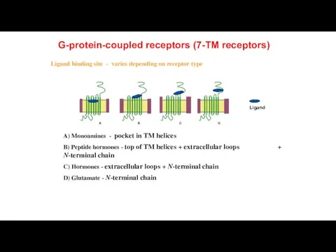
Ligand binding site - varies depending on receptor type
A) Monoamines
Ligand binding site - varies depending on receptor type
A) Monoamines
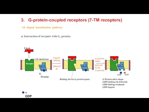
G-Protein alters shape
GDP binding site distorted
GDP binding weakened
GDP departs
a) Interaction of
G-Protein alters shape
GDP binding site distorted
GDP binding weakened
GDP departs
a) Interaction of
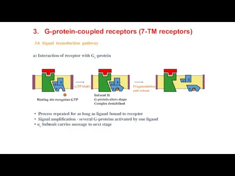
Induced fit
G-protein alters shape
Complex destabilised
Process repeated for as long
Induced fit
G-protein alters shape
Complex destabilised
Process repeated for as long
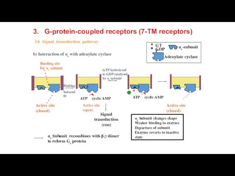
b) Interaction of αs with adenylate cyclase
3. G-protein-coupled receptors (7-TM receptors)
3.6
b) Interaction of αs with adenylate cyclase
3. G-protein-coupled receptors (7-TM receptors)
3.6
Adrenoreceptor
Adrenoreceptor
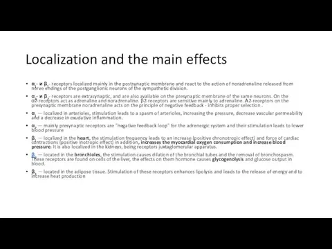
Localization and the main effects
α1- и β1- receptors localized mainly in
Localization and the main effects
α1- и β1- receptors localized mainly in
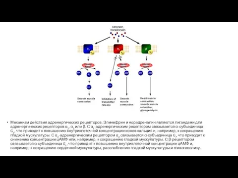
Механизм действия адренергических рецепторов. Эпинефрин и норадреналин являются лигандами для адренергических
Механизм действия адренергических рецепторов. Эпинефрин и норадреналин являются лигандами для адренергических
 Редкие виды растений в искусственных лесных насаждениях рядом с садоводческим товариществом Виктория
 Методы изучения биологического круговорота веществ в фитоценозах
 Сахарозаменители. Натуральные сахарозаменители
 Қызынаққа зиян келтіретін бунақденелердің таралуы, биологиясы, зияндылығы және қолданатын күресу шаралар жүйесі
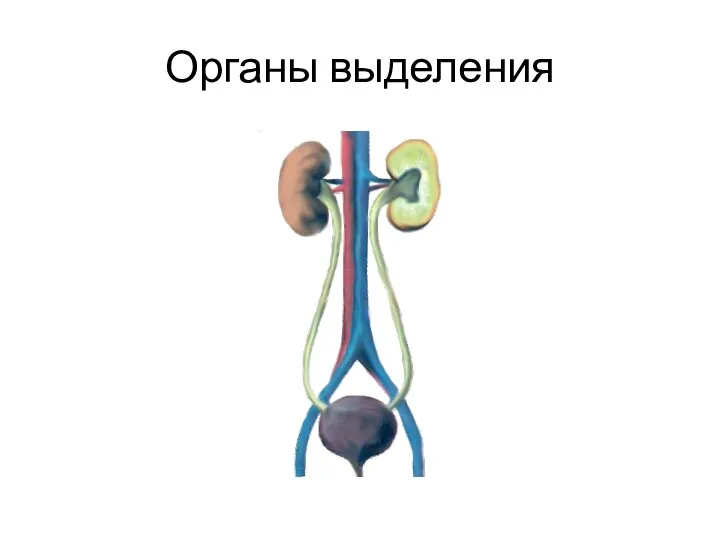 Органы выделения у человека
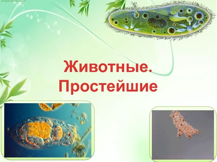 Животные. Простейшие
 Состав популяций
 Итоговая контрольная работа. 6 класс
 Биотехнологическое производство сыра
 Слуховая сенсорная система
 Органы растений. Рост, развитие и размножение растений. Цветок
 Адамның рефлекторлық реакциясын зерттеу
 Анатомия стебля растения
 Животные холодных и жарких стран
 Земледелие. Введение
 Хижі птахи
 Органеллы. Строение клеток эукариот
 Презентация: Роль бактерий в природе и жизни человека.
 Тест Полезные ископаемые (для 6 класса коррекционной школы 8 вида)
 Движение крови по сосудам
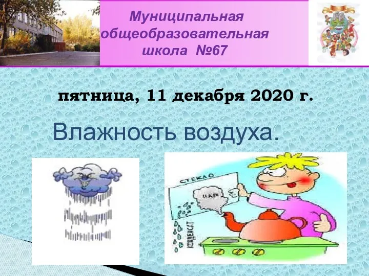 Влажность воздуха
 Бауырдың биохимиясы
 Репликация ДНК
 Митоздың биологиялық маңызы
 Парнокопытные и непарнокопытные животные
 Гербициттер микология
 Предмет и задачи цитологии
 Тест. Поджелудочна железа