History of Microbiology. Classification of Microorganisms. Morphology and Structure of Bacteria, Fungi, Spirochetes, Chlamydia презентация
- History of Microbiology. Classification of Microorganisms. Morphology and Structure of Bacteria, Fungi, Spirochetes, Chlamydia

Содержание
- 2. Historical Introduction Antony van First to observe live microorganisms, using a simple Leeuwenhoek microscope (1685) John
- 3. Definition of Microbiology Medical microbiology is the study of microbes that infect humans, the diseases they
- 4. Classification of Microorganisms Scientific nomenclature includes a hierarchial scheme. The lower down in the system the
- 5. Family Tree of Microorganisms Protozoa Bacteria Fungi Algae Cyanobacteria Archaebacteria Eukaryotes Prokaryotes Primitive Cells
- 6. Differences between Prokaryotic and Eukaryotic Cells Structure Prokaryotes Eukaryotes Nucleus Nuclear membrane Absent Present Nucleus Absent
- 7. Prokaryotic Cell Structure Prokaryotes are unicellular organisms of relatively simple construction. A prokaryotic cell has five
- 8. Cell structure
- 9. Characteristic of typical bacterial cell structures Sturcture Function(s) Predominant chemical composition Flagella Swimming movement Protein Pili
- 10. Plasma membrane Permeability barrier; transport of solutes; energy Phospholipid and protein generation; location of numerous enzyme
- 11. Appendages Flagella-are filamentous protein structures attached to the cell surface that provide the swimming movement for
- 12. Flagella may be variously distributed over the surface of bacterial cells. Arraingment of flagella: monotrichous, amphitrichous,
- 13. Detecting Bacterial Motility Flagellar stains (show their pattern of distribution) Motility test medium demonstrates if cells
- 14. Fimbriae Fimbriae and pili are short, hair-like structures they are composed of protein shorter, stiffer, smaller
- 15. The Cell Envelope The cell envelope consists: plasma membrane a cell wall a capsule
- 16. Capsules Polysaccharide layer outside of the cell wall polymer
- 17. The function of capsules: Mediate adherence of cells to surface Protect bacterial cells from engulfment by
- 18. Cell Wall is essential rigid structure for viability (protection cell protoplasm from mechanical damage and osmotic
- 19. Cell wall structure contains a unique type of peptidoglycan called murein- (N-acetylmuramic acid) By cell wall
- 20. Bacteria with Defective Cell Wall The synthesis of cell wall may be inhibited or interfered by
- 21. The Plasma Membrane Functions of the prokaryotic plasma membrane. 1. Osmotic or permeability barrier 2. Location
- 22. It is 5-10 nm thick elastic semipermiable layer which lies beneath the cell wall separating it
- 23. The Cytoplasm The bacterial cytoplasm is a colloidal system containing a variety of organic and inorganic
- 24. Nucleus Bacterial nucleus has no nuclear membrane or nucleolus The genomic DNA is double stranded in
- 25. Inclusions Often contained in the cytoplasm of prokaryotic cells is one or another of some type
- 26. Endospores A bacterial structure sometimes observed as an inclusion is actually a type of dormant cell
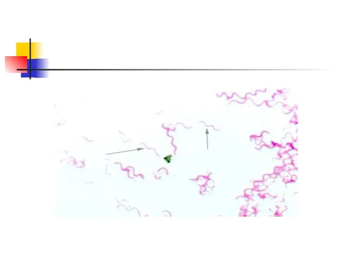
- 27. Morphology of the Spirochetes The spirochetes Long Thin Corkscrewlike Gram-negative Anaerobic bacteria There are three families
- 29. The spirochetes - very difficult to culture This is due to their extreme anaerobic requirements their
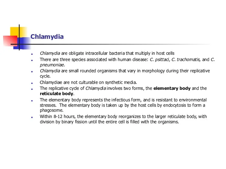
- 30. Chlamydia Chlamydia are obligate intracellular bacteria that multiply in host cells There are three species associated
- 31. A transmission electron microscope picture of a thin section through an elementary body of C. psittaci
- 32. Mycoplasma Mycoplasma are bacteria that lacks cell walls. Two human species are associated with disease: M.
- 33. Rickettsia The rickettsia are bacteria which are obligate intracellular parasites. They are considered a separate group
- 34. Gimenez stain of tick hemolymph cells infected with R. rickettsii
- 36. Скачать презентацию
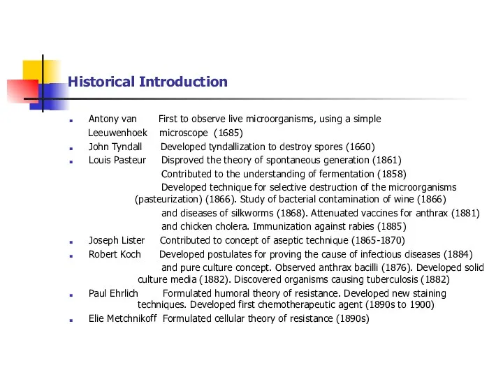
Historical Introduction
Antony van First to observe live microorganisms, using a simple
Historical Introduction
Antony van First to observe live microorganisms, using a simple
Definition of Microbiology
Medical microbiology is the study of microbes that infect
Definition of Microbiology
Medical microbiology is the study of microbes that infect
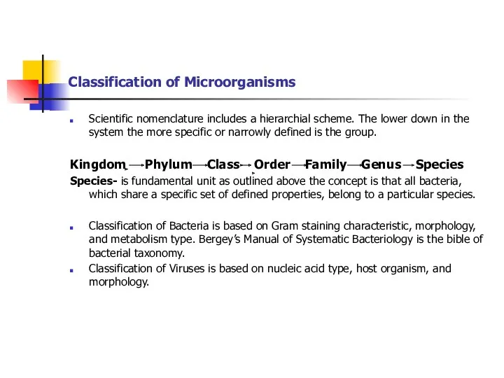
Classification of Microorganisms
Scientific nomenclature includes a hierarchial scheme. The lower down
Classification of Microorganisms
Scientific nomenclature includes a hierarchial scheme. The lower down
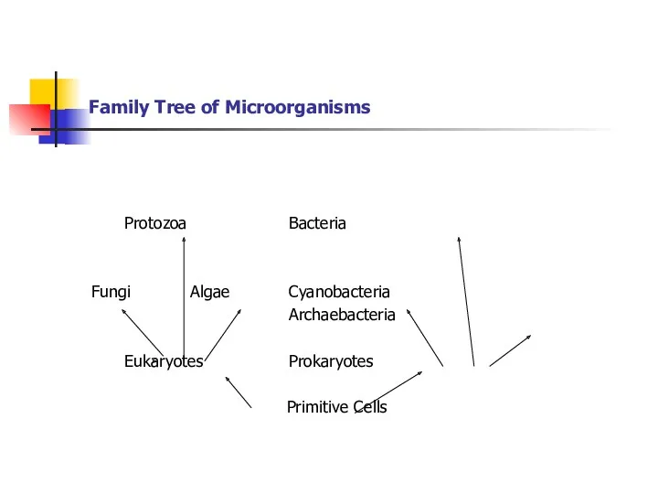
Family Tree of Microorganisms
Protozoa Bacteria
Fungi Algae Cyanobacteria
Archaebacteria
Eukaryotes Prokaryotes
Primitive Cells
Family Tree of Microorganisms
Protozoa Bacteria
Fungi Algae Cyanobacteria
Archaebacteria
Eukaryotes Prokaryotes
Primitive Cells
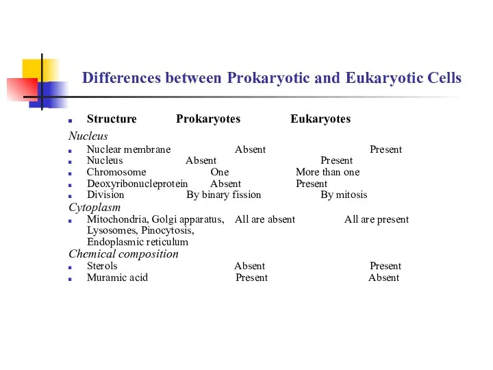
Differences between Prokaryotic and Eukaryotic Cells
Structure Prokaryotes Eukaryotes
Nucleus
Nuclear membrane
Differences between Prokaryotic and Eukaryotic Cells
Structure Prokaryotes Eukaryotes
Nucleus
Nuclear membrane
Prokaryotic Cell Structure
Prokaryotes are unicellular organisms of relatively simple construction.
A
Prokaryotic Cell Structure
Prokaryotes are unicellular organisms of relatively simple construction.
A
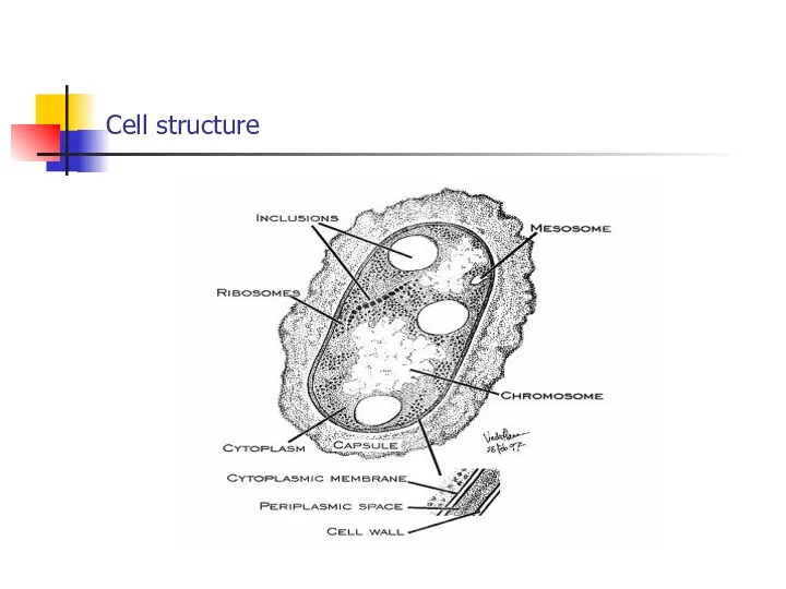
Cell structure
Cell structure
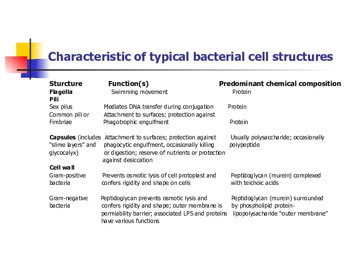
Characteristic of typical bacterial cell structures
Sturcture Function(s) Predominant chemical composition
Flagella Swimming
Characteristic of typical bacterial cell structures
Sturcture Function(s) Predominant chemical composition
Flagella Swimming
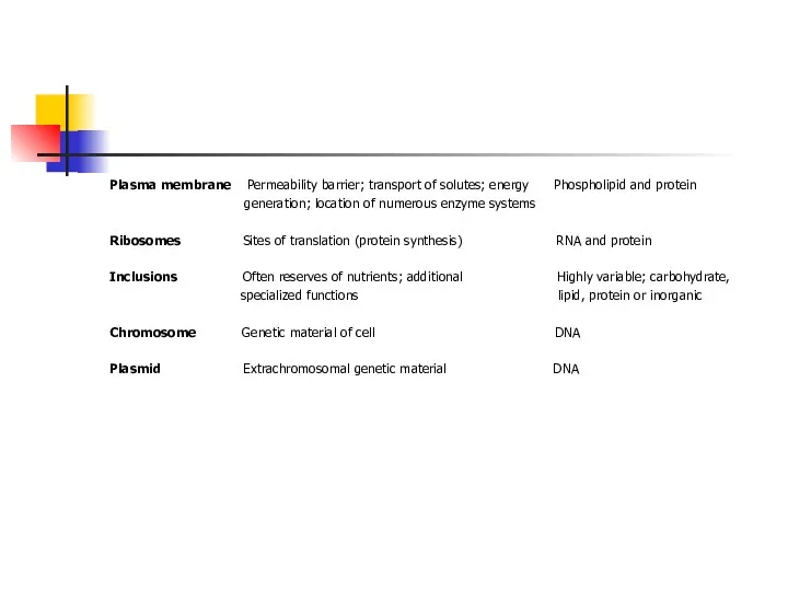
Plasma membrane Permeability barrier; transport of solutes; energy Phospholipid and protein
Plasma membrane Permeability barrier; transport of solutes; energy Phospholipid and protein
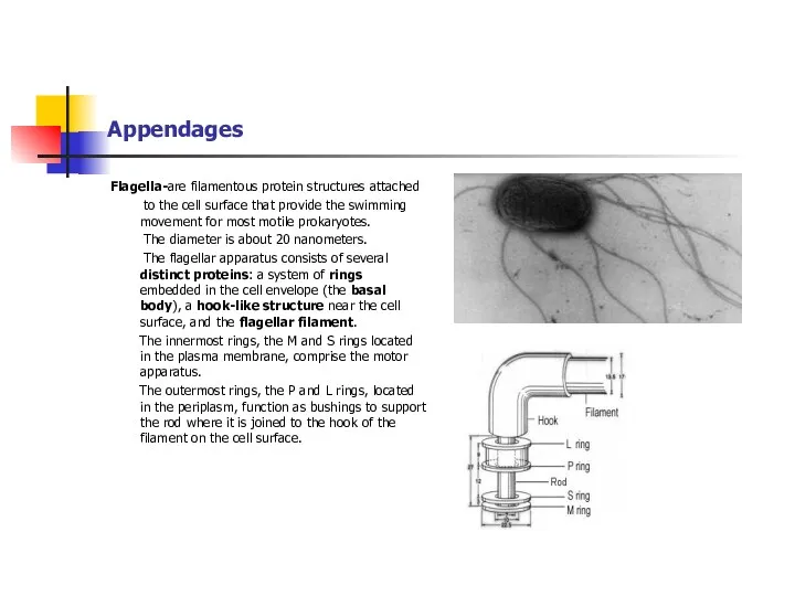
Appendages
Flagella-are filamentous protein structures attached
to the cell surface that provide
Appendages
Flagella-are filamentous protein structures attached
to the cell surface that provide
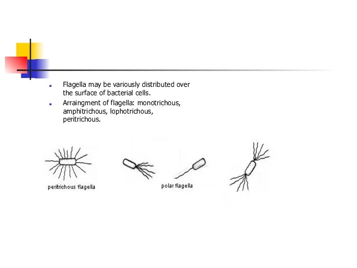
Flagella may be variously distributed over the surface of bacterial cells.
Arraingment
Flagella may be variously distributed over the surface of bacterial cells.
Arraingment
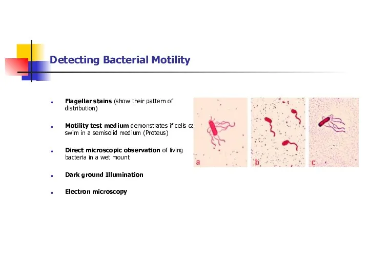
Detecting Bacterial Motility
Flagellar stains (show their pattern of distribution)
Motility test medium
Detecting Bacterial Motility
Flagellar stains (show their pattern of distribution)
Motility test medium
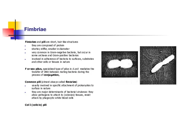
Fimbriae
Fimbriae and pili are short, hair-like structures
they are composed of
Fimbriae
Fimbriae and pili are short, hair-like structures
they are composed of
The Cell Envelope
The cell envelope consists:
plasma membrane
a
The Cell Envelope
The cell envelope consists:
plasma membrane
a
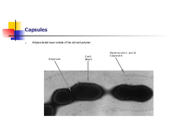
Capsules
Polysaccharide layer outside of the cell wall polymer
Capsules
Polysaccharide layer outside of the cell wall polymer
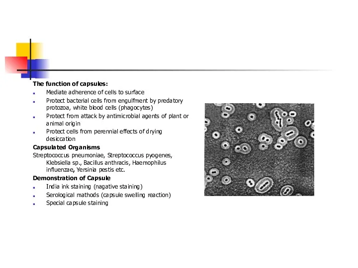
The function of capsules:
Mediate adherence of cells to surface
Protect bacterial cells
The function of capsules:
Mediate adherence of cells to surface
Protect bacterial cells
Cell Wall
is essential rigid structure for viability (protection cell protoplasm from
Cell Wall
is essential rigid structure for viability (protection cell protoplasm from
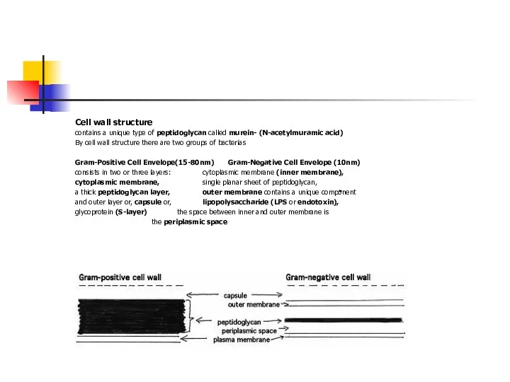
Cell wall structure
contains a unique type of peptidoglycan called murein- (N-acetylmuramic
Cell wall structure
contains a unique type of peptidoglycan called murein- (N-acetylmuramic
Bacteria with Defective Cell Wall
The synthesis of cell wall may be
Bacteria with Defective Cell Wall
The synthesis of cell wall may be
The Plasma Membrane
Functions of the prokaryotic plasma membrane.
1. Osmotic or
The Plasma Membrane
Functions of the prokaryotic plasma membrane.
1. Osmotic or
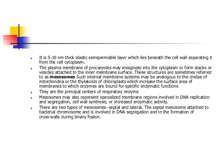
It is 5-10 nm thick elastic semipermiable layer which lies beneath
The Cytoplasm
The bacterial cytoplasm is a colloidal system containing a variety
The Cytoplasm
The bacterial cytoplasm is a colloidal system containing a variety
Nucleus
Bacterial nucleus has no nuclear membrane or nucleolus
The genomic DNA
Nucleus
Bacterial nucleus has no nuclear membrane or nucleolus
The genomic DNA
Inclusions
Often contained in the cytoplasm of prokaryotic cells is one or
Inclusions
Often contained in the cytoplasm of prokaryotic cells is one or
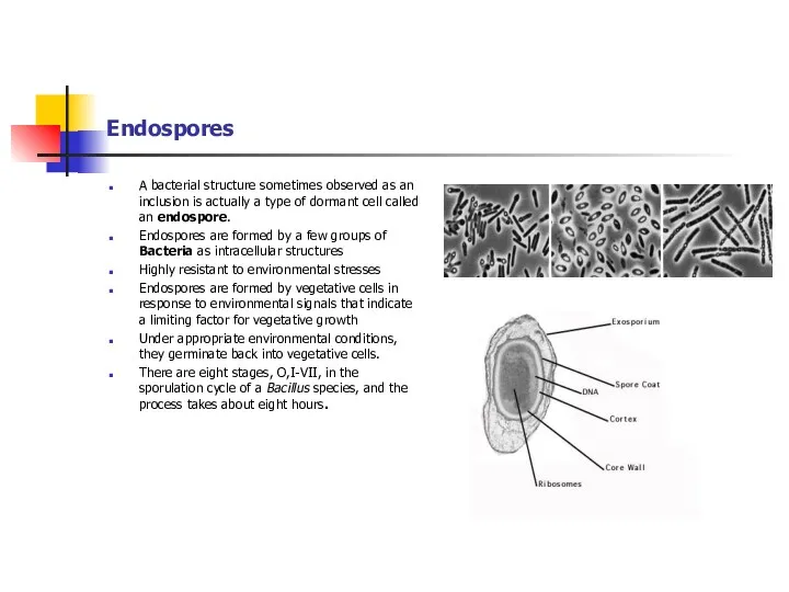
Endospores
A bacterial structure sometimes observed as an inclusion is actually
Endospores
A bacterial structure sometimes observed as an inclusion is actually
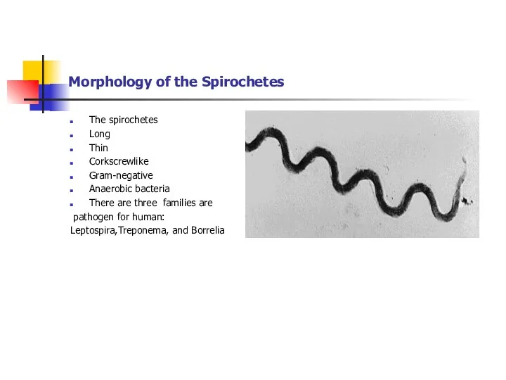
Morphology of the Spirochetes
The spirochetes
Long
Thin
Corkscrewlike
Gram-negative
Anaerobic
Morphology of the Spirochetes
The spirochetes
Long
Thin
Corkscrewlike
Gram-negative
Anaerobic
The spirochetes - very difficult to culture
This is due to
The spirochetes - very difficult to culture
This is due to
Chlamydia
Chlamydia are obligate intracellular bacteria that multiply in host cells
There
Chlamydia
Chlamydia are obligate intracellular bacteria that multiply in host cells
There
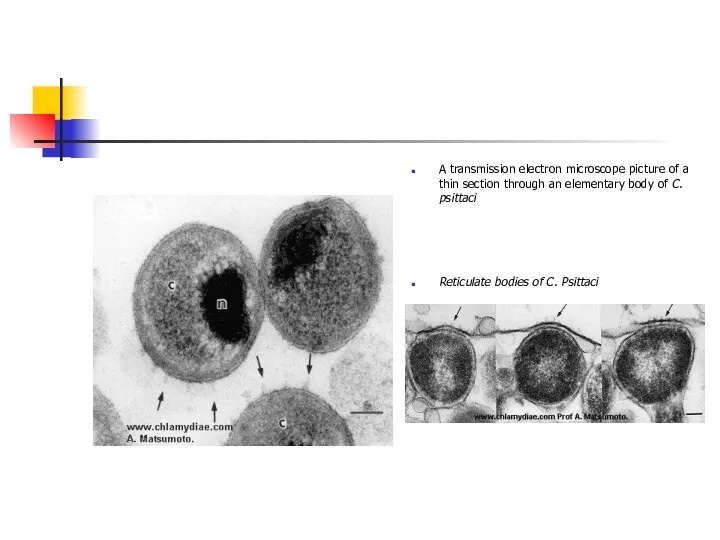
A transmission electron microscope picture of a thin section through an
A transmission electron microscope picture of a thin section through an
Mycoplasma
Mycoplasma are bacteria that lacks cell walls.
Two human species are associated
Mycoplasma
Mycoplasma are bacteria that lacks cell walls.
Two human species are associated
Rickettsia
The rickettsia are bacteria which are obligate intracellular parasites.
They
Rickettsia
The rickettsia are bacteria which are obligate intracellular parasites.
They
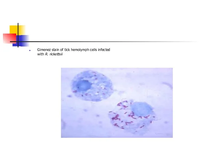
Gimenez stain of tick hemolymph cells infected with R. rickettsii
Gimenez stain of tick hemolymph cells infected with R. rickettsii
 Функциональная анатомия вегетативной нервной системы. Парасимпатическая часть ВНС
 Анатомо-физиологические особенности органа слуха и равновесия
 Вегетативные органы растений: корень
 Удивительные животные
 Нервная система человека
 Семейство Лилейные. Класс Однодольные
 Микроорганизмдердің қореқтенуі
 МАТЕМАТИКА и БИОЛОГИЯ ... НА КУХНЕ?
 Закономерности изменчивости
 Саморазвитие экосистем
 Строение и функции белков
 Типы питания растений
 Покровы тела. Сравнительная характеристика
 Митоз Способы деления клетки
 Клонирование и этическая проблема
 Презентация педагогического опыта по теме Методологические основы интегрированных уроков
 Фізіологія дихання
 Выращивание томатов
 Курс подготовки к ЕГЭ по биологии
 Эволюция опорно- двигательной системы у животных
 Роль витаминов в формировании потребительских свойств продовольственных товаров
 Відходи технічних виробництв та харчові відходи
 Ядовитые растения, произрастающие в Ленинградской области
 Круговорот углерода в природе
 Технологии высокопроизводительного паралельного секвенирования (NGS)
 История подсолнечника
 Генетика
 Серцевий цикл. Робота серця