- Interstitial Lung Disease

Содержание
- 2. Objectives Interstitium Pleural disease Chest wall disease
- 3. Interstitial disease What is the interstitium? What does the interstitium do? What are the pathophysiological effects
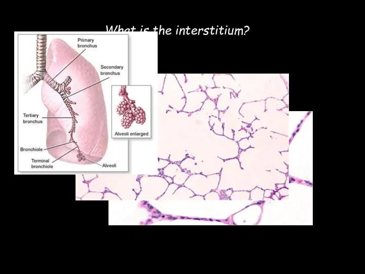
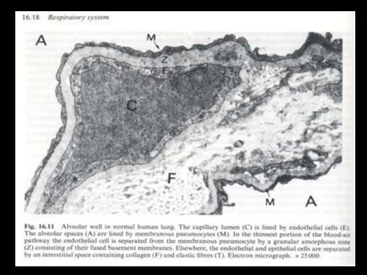
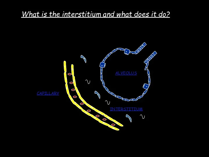
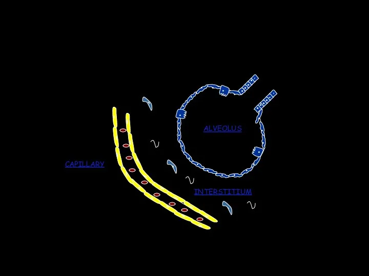
- 4. What is the interstitium?
- 6. . . . . . . . . . . . . . . . .
- 7. Does interstitial disease effect just the interstitium? NO ! Structures affected: Acini Alveoli lumen Bronchiolar lumen
- 8. Ventilation Diffusion Perfusion O2 CO2
- 9. Pulmonary function tests Volume (l) Time (s)
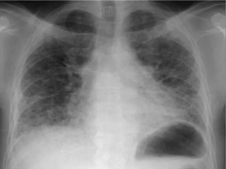
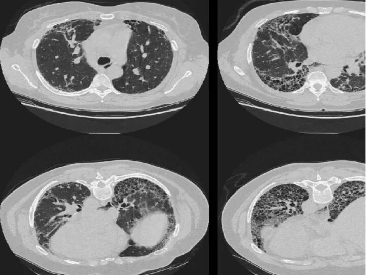
- 10. 59 year old male Shortness of breath & dry cough, increasing 1 year - breathless with
- 13. Symptoms & history taking Interstitial Lung Disease Respiratory symptoms Shortness of breath Dry Cough
- 14. . . . . . . . . . . . . . . . .
- 15. Common clinical features Symptoms 1-Chronic dry cough 2-Exertional dyspnea Signs 1-Clubbing 2-Basal inspiratory crepitations Laboratory 1-High
- 16. Pulmonary function tests Spirometry 1-Decreased FEV1,FEV (Normal FEV1/FVC) 2-Decreased TLC 3-Mildly Decreased PEF 4-Markedly Decreased DLCO
- 17. Clubbing Course crackles Tachypnoea Signs of right heart failure Signs of underlying disease Cyanosis ↓chest movement
- 18. Blood tests Interstitial Lung Disease Occupational Treatment related Connective tissue disease Idiopathic Asbestosis Silicosis Coal Workers
- 19. Idiopathic interstitial pneumonitis (IIP) A variety of histological descriptions (UIP,NSIP,DIP,RB-ILD, BOOP) Histological descriptions - high inter
- 20. Asbestos
- 21. Asbestos Asbestos plaques Diffuse pleural thickening Benign asbestos pleural effusions (BAPE) ASBESTOSIS Mesothelioma Bronchogenic lung cancer
- 22. Drug induced ILD Methotrexate Bleomycin Amiodarone Nitrofurantoin Methotrexate Treatment & cause of lung disease Dose &
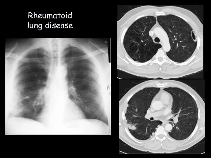
- 23. Rheumatoid lung disease
- 24. Connective tissue disease Dermatomyositis/ Polymyositis Sjogren’s Syndrome Systemic Lupus erythematosis Schleroderma Rheumatoid arthritis Rheumatoid lung disease
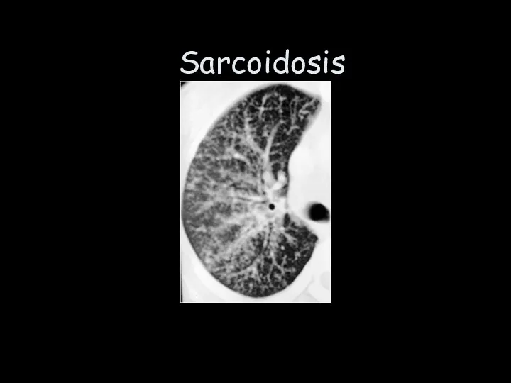
- 25. Sarcoidosis
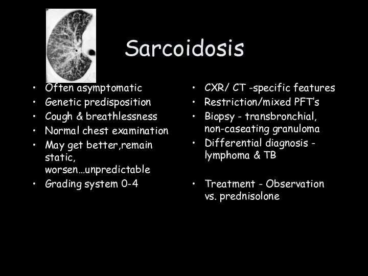
- 26. Sarcoidosis Often asymptomatic Genetic predisposition Cough & breathlessness Normal chest examination May get better,remain static, worsen…unpredictable
- 27. Interstitial disease What is the interstitium? What does the interstitium do? What are the pathophysiological effects
- 28. Objectives Interstitium Pleural disease Chest wall disease
- 29. Pleural Disease Anatomy Effusions Malignancy
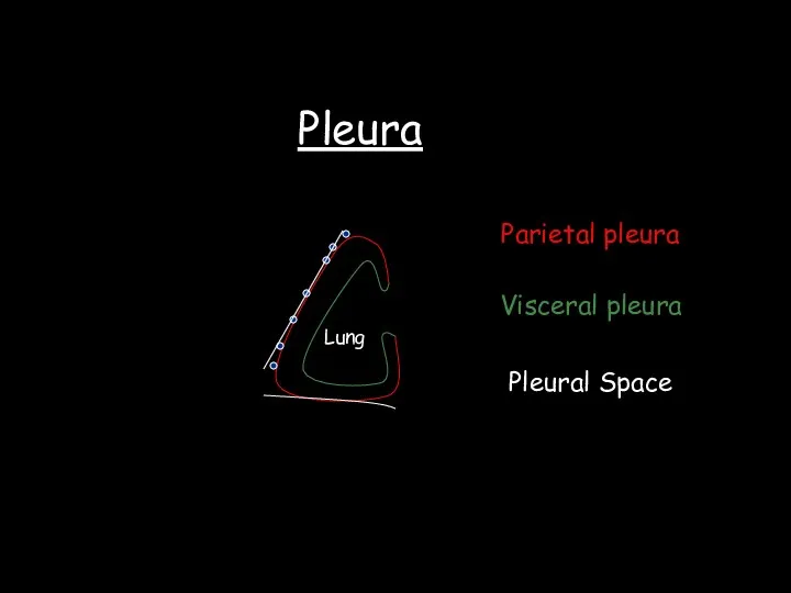
- 30. Pleura Lung Parietal pleura Visceral pleura Pleural Space
- 31. Visceral pleura Parietal pleura Fat pad Endothoracic fascia Innermost intercostal Intercostal fat & vessels Intercostal muscles
- 32. Functions of the pleural space Allow movement of lung and chest wall Coupling of chest wall
- 33. Blood supply Parietal pleura Intercostals & IMA - subclavian artery Venous drainage - peribronchial and venae
- 34. Lymphatic drainage Parietal Intercostal and internal mammary lymph vessels Visceral Pulmonary lymphatics Lung
- 35. Pleura - innervation Lung Parietal pleura - somatic, sympathetic & parasympathetic Phrenic & intercostal nerves Visceral
- 36. Pleural fluid turnover 15ml per day ( can increase to 300 ml/day) Production - Capillary filtration(Starling
- 37. Pathogenesis of pleural fluid accumulation Increased production Lung interstitial fluid increase Hydrostatic pressure increase Permeability increase
- 38. Pleural effusions Transudate Exudate Hydrothorax Haemothorax Chylothora Empyema Thoracocentesis
- 39. PLEURAL EMPYEMA
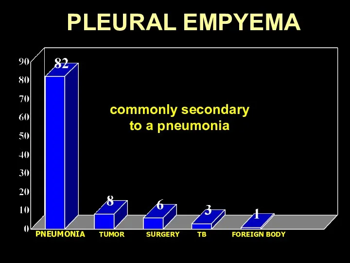
- 40. PLEURAL EMPYEMA PNEUMONIA TUMOR SURGERY TB FOREIGN BODY Collection of pus in the pleural cavity commonly
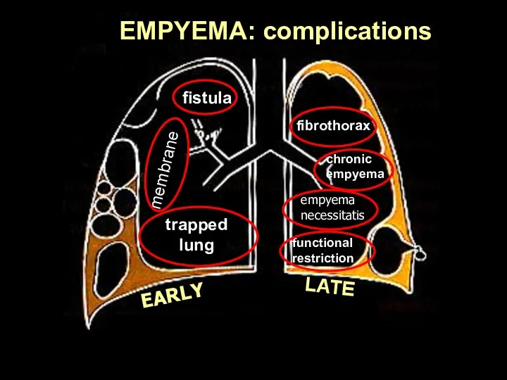
- 41. EARLY LATE fistula trapped lung membranes fibrothorax chronic empyema empyema necessitatis functional restriction EMPYEMA: complications
- 42. PLEURAL EMPYEMA
- 43. PLEURAL EMPYEMA
- 44. Pleural malignancy Metastatic Primary - mesothelioma Mesothelioma Asbestos exposure Pain, breathlessness Effusion, mediastinal pleural enhancement Chemotherapy,
- 45. Pleural Disease Anatomy Effusions Malignancy
- 46. Objectives Interstitium Pleural disease Chest wall disease
- 47. Chest wall disease Congenital Pectus deformities Scoliosis Kyphosis Muscular dystrophy Acquired Trauma Iatrogenic Ankylosing spondylitis Motor
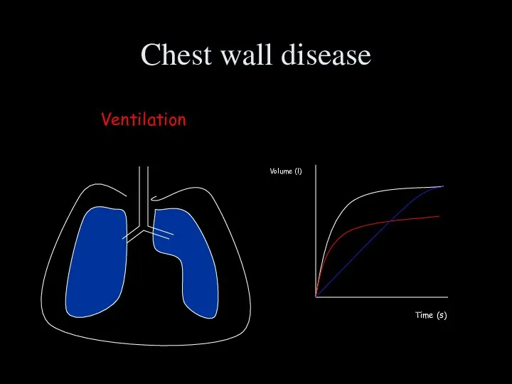
- 48. Chest wall disease Ventilation Volume (l) Time (s)
- 49. Chest wall disease Ventilation Sleep disordered breathing Poor clearance of secretions Atelectasis Pneumonia
- 51. Скачать презентацию
Objectives
Interstitium
Pleural disease
Chest wall disease
Objectives
Interstitium
Pleural disease
Chest wall disease

Interstitial disease
What is the interstitium?
What does the interstitium do?
What are the
Interstitial disease
What is the interstitium?
What does the interstitium do?
What are the
What is the interstitium?
What is the interstitium?
.
.
.
.
.
.
.
.
.
.
.
.
.
.
.
.
.
.
.
.
.
.
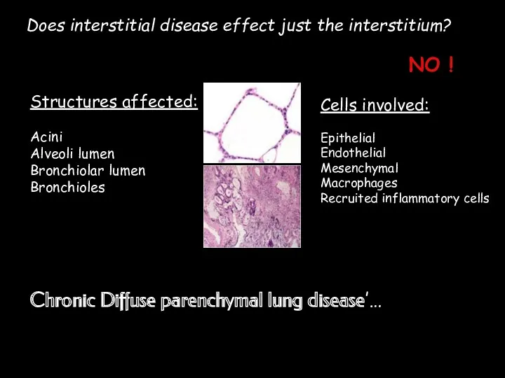
Does interstitial disease effect just the interstitium?
NO !
Structures affected:
Acini
Alveoli lumen
Bronchiolar lumen
Bronchioles
Cells
Does interstitial disease effect just the interstitium?
NO !
Structures affected:
Acini
Alveoli lumen
Bronchiolar lumen
Bronchioles
Cells
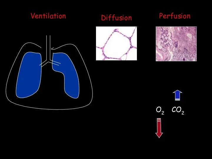
Ventilation
Diffusion
Perfusion
O2
CO2
Ventilation
Diffusion
Perfusion
O2
CO2
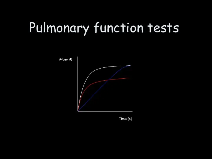
Pulmonary function tests
Volume (l)
Time (s)
Pulmonary function tests
Volume (l)
Time (s)
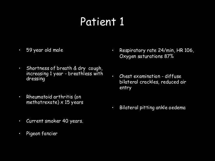
59 year old male
Shortness of breath & dry cough, increasing 1
59 year old male
Shortness of breath & dry cough, increasing 1
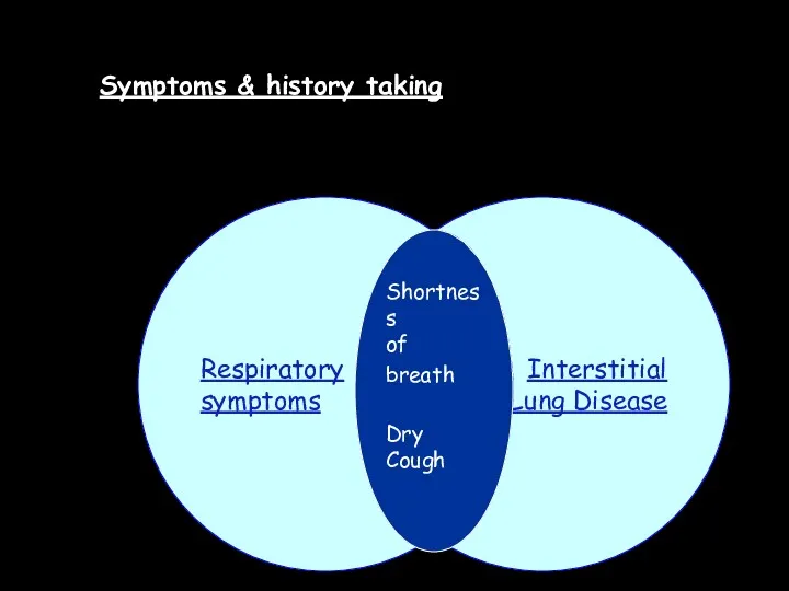
Symptoms & history taking
Interstitial
Lung Disease
Respiratory
symptoms
Shortness
of breath
Dry Cough
Symptoms & history taking
Interstitial
Lung Disease
Respiratory
symptoms
Shortness
of breath
Dry Cough
.
.
.
.
.
.
.
.
.
.
.
.
.
.
.
.
.
.
.
.
.
.
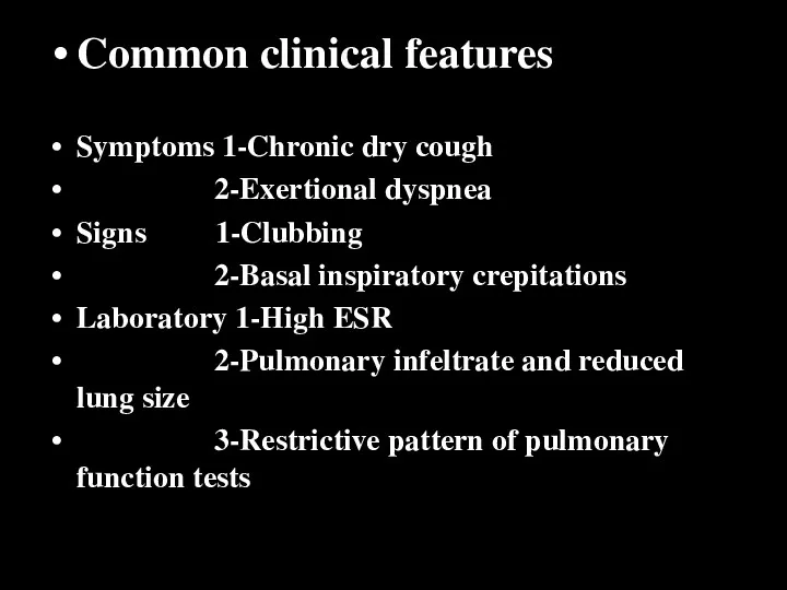
Common clinical features
Symptoms 1-Chronic dry cough
2-Exertional dyspnea
Signs 1-Clubbing
2-Basal
Common clinical features
Symptoms 1-Chronic dry cough
2-Exertional dyspnea
Signs 1-Clubbing
2-Basal
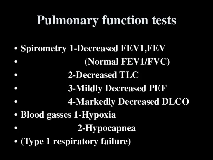
Pulmonary function tests
Spirometry 1-Decreased FEV1,FEV
(Normal FEV1/FVC)
2-Decreased TLC
3-Mildly
Pulmonary function tests
Spirometry 1-Decreased FEV1,FEV
(Normal FEV1/FVC)
2-Decreased TLC
3-Mildly
Clubbing
Course crackles
Tachypnoea
Signs of right heart failure
Signs of underlying disease
Cyanosis
↓chest movement
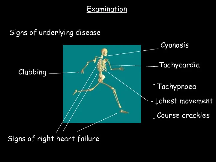
Tachycardia
Examination
Clubbing
Course crackles
Tachypnoea
Signs of right heart failure
Signs of underlying disease
Cyanosis
↓chest movement
Tachycardia
Examination
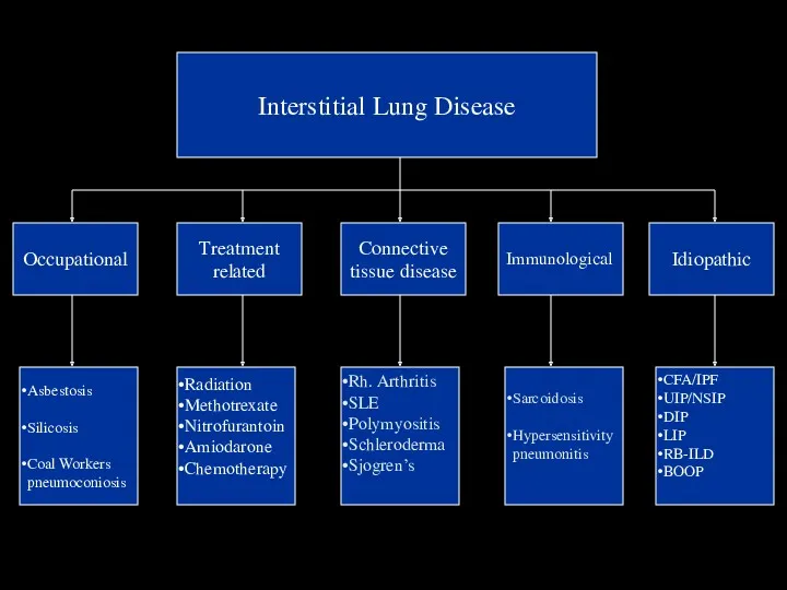
Blood tests
Interstitial Lung Disease
Occupational
Treatment
related
Connective
tissue disease
Idiopathic
Asbestosis
Silicosis
Coal Workers
pneumoconiosis
Radiation
Methotrexate
Nitrofurantoin
Amiodarone
Chemotherapy
Sarcoidosis
Hypersensitivity
pneumonitis
CFA/IPF
UIP/NSIP
DIP
LIP
RB-ILD
BOOP
Immunological
Rh. Arthritis
SLE
Polymyositis
Schleroderma
Sjogren’s
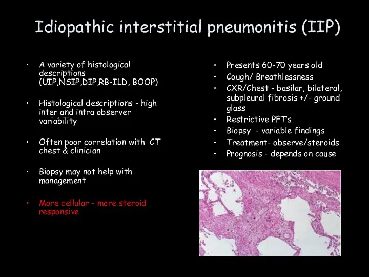
Idiopathic interstitial pneumonitis (IIP)
A variety of histological descriptions (UIP,NSIP,DIP,RB-ILD, BOOP)
Histological descriptions
Idiopathic interstitial pneumonitis (IIP)
A variety of histological descriptions (UIP,NSIP,DIP,RB-ILD, BOOP)
Histological descriptions
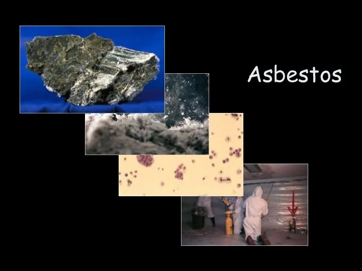
Asbestos
Asbestos
Asbestos
Asbestos plaques
Diffuse pleural thickening
Benign asbestos pleural effusions (BAPE)
ASBESTOSIS
Mesothelioma
Bronchogenic lung cancer
Rounded atelectasis
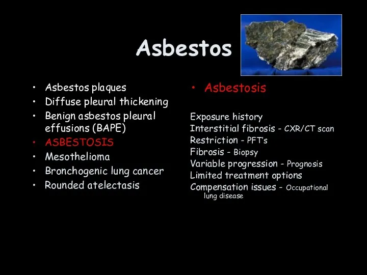
Asbestos
Asbestos plaques
Diffuse pleural thickening
Benign asbestos pleural effusions (BAPE)
ASBESTOSIS
Mesothelioma
Bronchogenic lung cancer
Rounded atelectasis
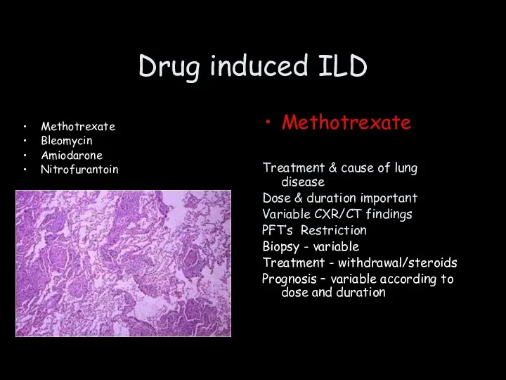
Drug induced ILD
Methotrexate
Bleomycin
Amiodarone
Nitrofurantoin
Methotrexate
Treatment & cause of lung disease
Dose & duration important
Drug induced ILD
Methotrexate
Bleomycin
Amiodarone
Nitrofurantoin
Methotrexate
Treatment & cause of lung disease
Dose & duration important
Rheumatoid
lung disease
Rheumatoid
lung disease
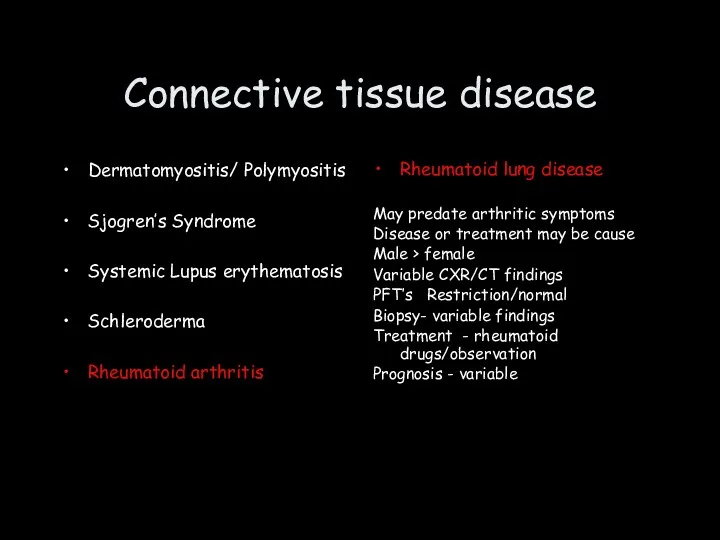
Connective tissue disease
Dermatomyositis/ Polymyositis
Sjogren’s Syndrome
Systemic Lupus erythematosis
Schleroderma
Rheumatoid arthritis
Rheumatoid lung disease
May predate
Connective tissue disease
Dermatomyositis/ Polymyositis
Sjogren’s Syndrome
Systemic Lupus erythematosis
Schleroderma
Rheumatoid arthritis
Rheumatoid lung disease
May predate
Sarcoidosis
Sarcoidosis
Sarcoidosis
Often asymptomatic
Genetic predisposition
Cough & breathlessness
Normal chest examination
May get better,remain static, worsen…unpredictable
Grading
Sarcoidosis
Often asymptomatic
Genetic predisposition
Cough & breathlessness
Normal chest examination
May get better,remain static, worsen…unpredictable
Grading
Interstitial disease
What is the interstitium?
What does the interstitium do?
What are the
Interstitial disease
What is the interstitium?
What does the interstitium do?
What are the
Objectives
Interstitium
Pleural disease
Chest wall disease
Objectives
Interstitium
Pleural disease
Chest wall disease
Pleural Disease
Anatomy
Effusions
Malignancy
Pleural Disease
Anatomy
Effusions
Malignancy
Pleura
Lung
Parietal pleura
Visceral pleura
Pleural Space
Pleura
Lung
Parietal pleura
Visceral pleura
Pleural Space
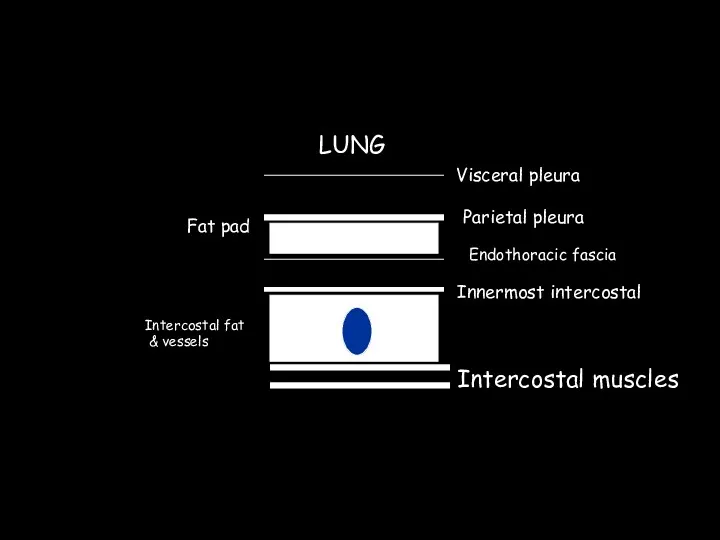
Visceral pleura
Parietal pleura
Fat pad
Endothoracic fascia
Innermost intercostal
Intercostal fat
& vessels
Intercostal muscles
LUNG
Visceral pleura
Parietal pleura
Fat pad
Endothoracic fascia
Innermost intercostal
Intercostal fat
& vessels
Intercostal muscles
LUNG
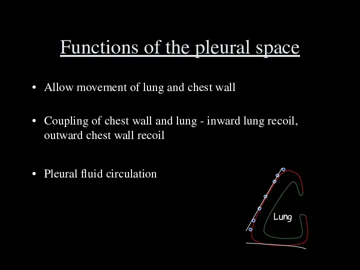
Functions of the pleural space
Allow movement of lung and chest wall
Coupling
Functions of the pleural space
Allow movement of lung and chest wall
Coupling
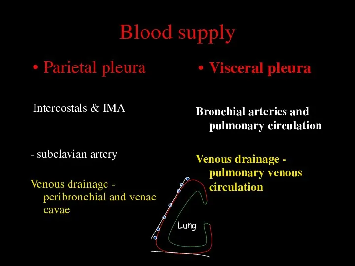
Blood supply
Parietal pleura
Intercostals & IMA
- subclavian artery
Venous drainage - peribronchial
Blood supply
Parietal pleura
Intercostals & IMA
- subclavian artery
Venous drainage - peribronchial
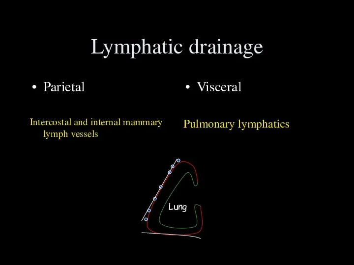
Lymphatic drainage
Parietal
Intercostal and internal mammary lymph vessels
Visceral
Pulmonary lymphatics
Lung
Lymphatic drainage
Parietal
Intercostal and internal mammary lymph vessels
Visceral
Pulmonary lymphatics
Lung
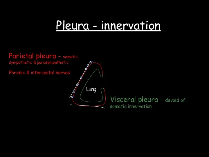
Pleura - innervation
Lung
Parietal pleura - somatic,
sympathetic & parasympathetic
Phrenic & intercostal
Pleura - innervation
Lung
Parietal pleura - somatic,
sympathetic & parasympathetic
Phrenic & intercostal
Pleural fluid turnover
15ml per day ( can increase to 300 ml/day)
Pleural fluid turnover
15ml per day ( can increase to 300 ml/day)
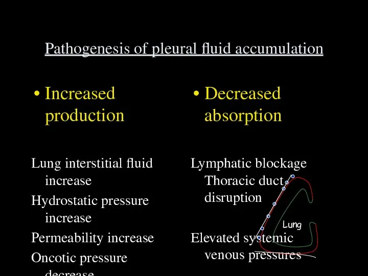
Pathogenesis of pleural fluid accumulation
Increased production
Lung interstitial fluid increase
Hydrostatic pressure increase
Permeability
Pathogenesis of pleural fluid accumulation
Increased production
Lung interstitial fluid increase
Hydrostatic pressure increase
Permeability
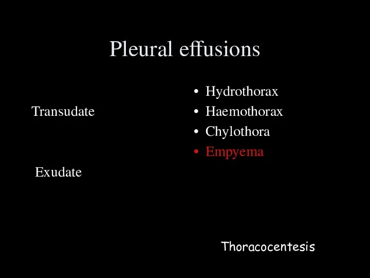
Pleural effusions
Transudate
Exudate
Hydrothorax
Haemothorax
Chylothora
Empyema
Thoracocentesis
Pleural effusions
Transudate
Exudate
Hydrothorax
Haemothorax
Chylothora
Empyema
Thoracocentesis
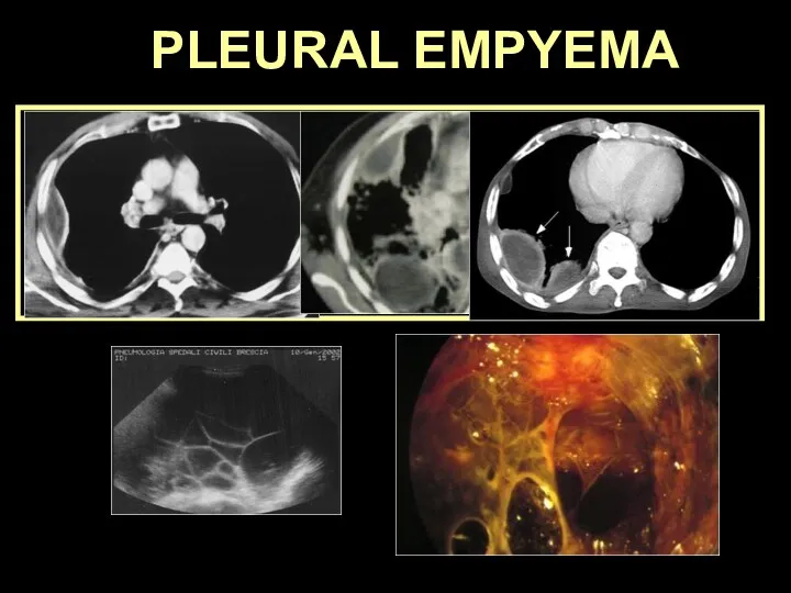
PLEURAL EMPYEMA
PLEURAL EMPYEMA
PLEURAL EMPYEMA
PNEUMONIA TUMOR SURGERY TB FOREIGN BODY
Collection of pus in
the
PLEURAL EMPYEMA
PNEUMONIA TUMOR SURGERY TB FOREIGN BODY
Collection of pus in
the
EARLY
LATE
fistula
trapped
lung
membranes
fibrothorax
chronic
empyema
empyema
necessitatis
functional
restriction
EMPYEMA: complications
EARLY
LATE
fistula
trapped
lung
membranes
fibrothorax
chronic
empyema
empyema
necessitatis
functional
restriction
EMPYEMA: complications
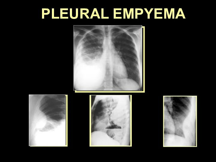
PLEURAL EMPYEMA
PLEURAL EMPYEMA
PLEURAL EMPYEMA
PLEURAL EMPYEMA
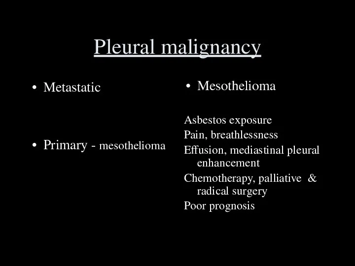
Pleural malignancy
Metastatic
Primary - mesothelioma
Mesothelioma
Asbestos exposure
Pain, breathlessness
Effusion, mediastinal pleural enhancement
Chemotherapy, palliative &
Pleural malignancy
Metastatic
Primary - mesothelioma
Mesothelioma
Asbestos exposure
Pain, breathlessness
Effusion, mediastinal pleural enhancement
Chemotherapy, palliative &

Pleural Disease
Anatomy
Effusions
Malignancy
Pleural Disease
Anatomy
Effusions
Malignancy
Objectives
Interstitium
Pleural disease
Chest wall disease
Objectives
Interstitium
Pleural disease
Chest wall disease
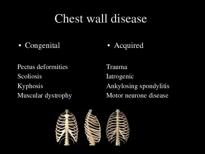
Chest wall disease
Congenital
Pectus deformities
Scoliosis
Kyphosis
Muscular dystrophy
Acquired
Trauma
Iatrogenic
Ankylosing spondylitis
Motor neurone disease
Chest wall disease
Congenital
Pectus deformities
Scoliosis
Kyphosis
Muscular dystrophy
Acquired
Trauma
Iatrogenic
Ankylosing spondylitis
Motor neurone disease
Chest wall disease
Ventilation
Volume (l)
Time (s)
Chest wall disease
Ventilation
Volume (l)
Time (s)
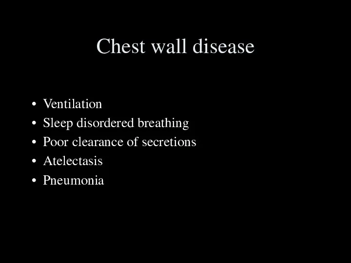
Chest wall disease
Ventilation
Sleep disordered breathing
Poor clearance of secretions
Atelectasis
Pneumonia
Chest wall disease
Ventilation
Sleep disordered breathing
Poor clearance of secretions
Atelectasis
Pneumonia
 Усачи, или дровосеки. Личинки. (Лекция 13)
 Достижения современной селекции
 Размножение организмов
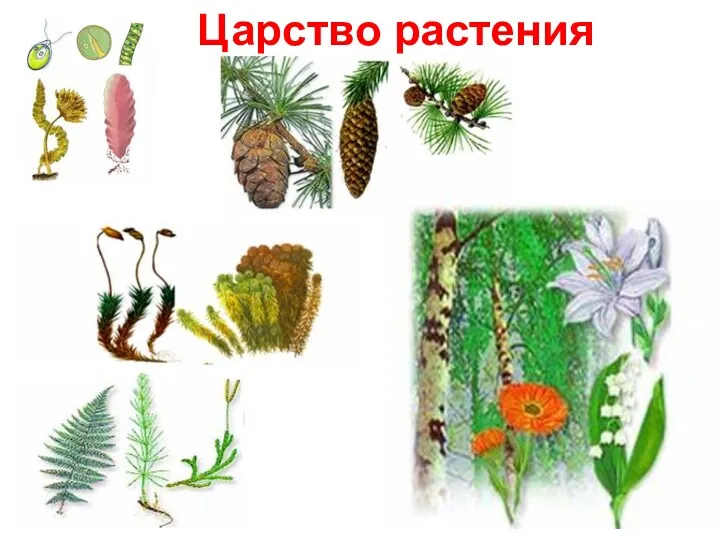 Царство растений. Многообразие растений
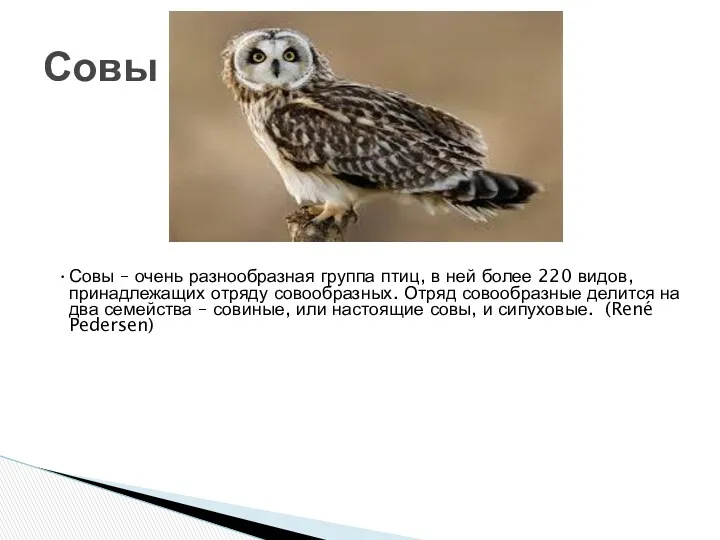 Совы. Виды
 Чудеса селекции. Самые любопытные фрукты и овощи, которые появились на свет благодаря селекции
 Забота животных о потомстве
 Красная книга Красноярского края
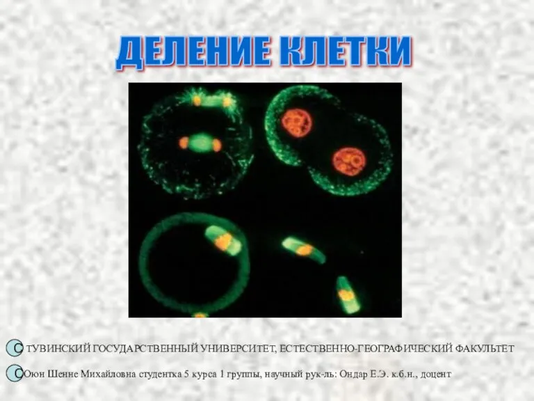 Клеточный цикл
 Геохронологическая история Земли
 Чарлз Роберт Дарвин
 Перелітні птахи
 Морские свинки
 Сердечно-сосудистая система
 Отдел Красные водоросли
 Ядро клетки
 Факторы, лимитирующие первичную продукцию в наземных и водных сообществах
 СОСТАВ КРОВИ.
 Многообразие и значение грибов
 Структурная организация микробной клетки
 Оживление грибов дрожжей
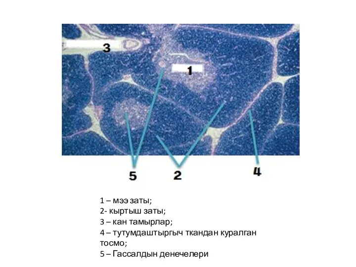 Ткань жана анын мааниси
 Открытый урок на тему: Многообразие земноводных.
 Тренажёр Вода
 Биологические задачи. Подготовка к ЕГЭ
 Дыхательная система
 Эволюция. Доказательства эволюции (часть 6)
 Жизнь организмов в морях и океанах