- Joint Programs – Health Sciences

Содержание
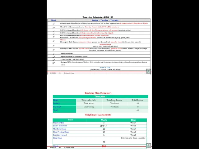
- 2. BIOLOGY 2019 Dr. Amin Al-Doaiss 3 Week Sunday – Tuesday – Thursday 1st Chemistry of life:
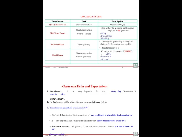
- 3. BIOLOGY 2019 Dr. Amin Al-Doaiss Examination Topic Description Quiz & homework Sheet examination Quizzes (MCQs) Mid-Term
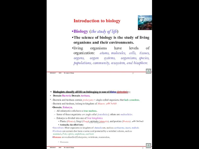
- 4. BIOLOGY 2019 Dr. Amin Al-Doaiss Introduction to biology Biology (the study of life) The science of
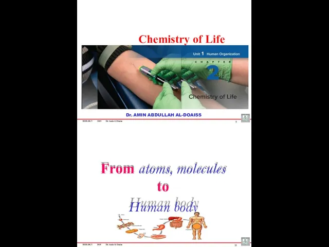
- 5. BIOLOGY 2019 Dr. Amin Al-Doaiss Chemistry of Life 9 Dr. AMIN ABDULLAH AL-DOAISS From atoms, molecules
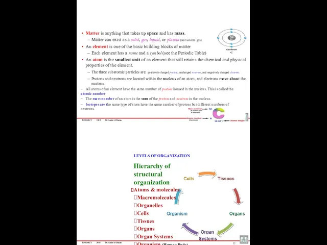
- 6. BIOLOGY 2019 Dr. Amin Al-Doaiss Matter is anything that takes up space and has mass. Matter
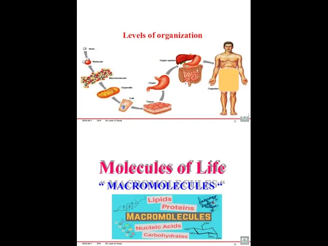
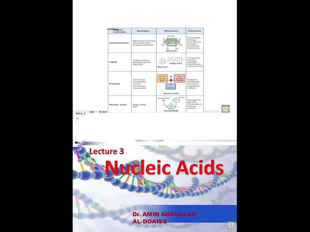
- 7. BIOLOGY 2019 Dr. Amin Al-Doaiss 13 Levels of organization Molecules of Life “ MACROMOLECULES “ BIOLOGY
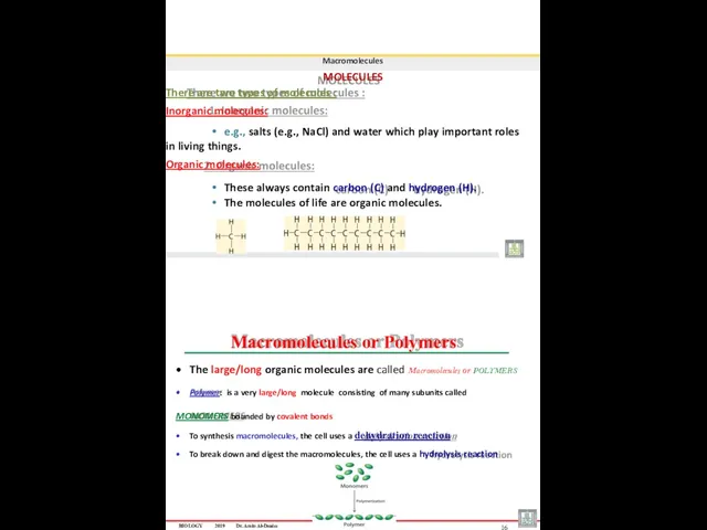
- 8. Macromolecules MOLECULES There are two types of molecules : Inorganic molecules: e.g., salts (e.g., NaCl) and
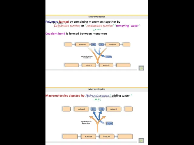
- 9. Macromolecules Polymers formed by combining monomers together by Dehydration reaction, or “condensation reaction” “removing water” ءاملا
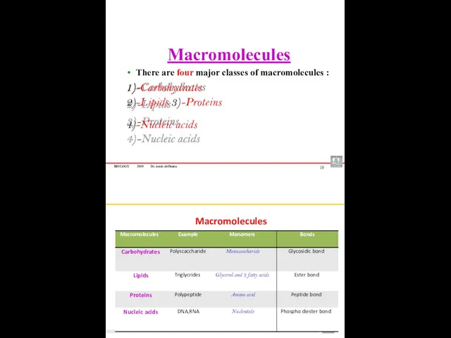
- 10. BIOLOGY 2019 Dr. Amin Al-Doaiss Macromolecules There are four major classes of macromolecules : 1)-Carbohydrates 2)-Lipids
- 11. Chemical Bond Macromolecules Building blocks of the cell Monosaccharaides FATTY ACIDS AMINO ACIDS NUCLEOTIDES Larger units
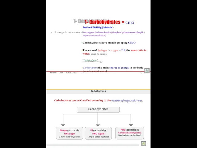
- 12. BIOLOGY 2019 Dr. Amin Al-Doaiss Fuel and Building Materials Are organic macromolecules composed of monomers (simple
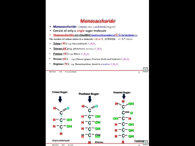
- 13. BIOLOGY 2019 Dr. Amin Al-Doaiss Monosaccharide : (mono, one; saccharide, sugar) Consist of only a single
- 14. •Examples of Pentoses Carbohydrates Ribose and deoxyribose: found in RNA and DNA respectively . DEOXYRIBOSE (in
- 15. Disaccharide Disaccharide : (di, “two”; saccharide, “sugar”) Composed of Two monosaccharides joining together by glycosidic bond.
- 16. Disaccharides Example: When glucose and fructose join, the disaccharide sucrose forms Sucrose, derived from sugarcane and
- 17. Carbohydrates STARCH : It is a Long chain of glucose (up to 4,000 units) linked together
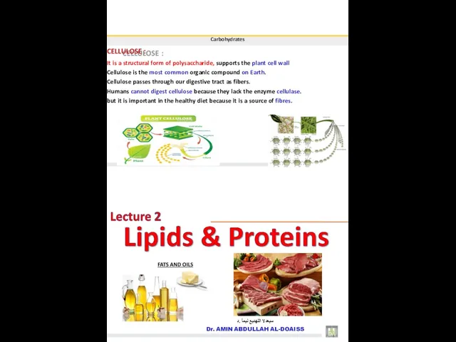
- 18. Carbohydrates CELLULOSE : It is a structural form of polysaccharide, supports the plant cell wall Cellulose
- 19. BIOLOGY 2019 Dr. Amin Al-Doaiss 2 Lipids Phospholipids are type of lipids that form the major
- 20. BIOLOGY 2019 Dr. Amin mponent of cell memb Al-Doaiss 4 Lipids Functions of lipids : Long
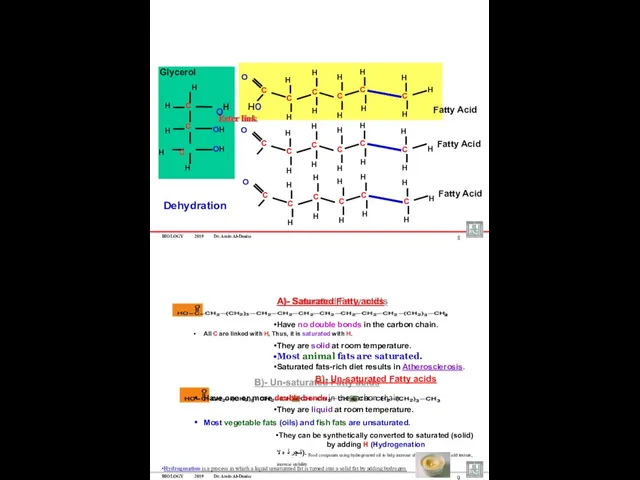
- 21. BIOLOGY 2019 Dr. Amin Al-Doaiss 6 Lipids Triglycerides are formed when glycerol combine with three fatty
- 22. BIOLOGY 2019 Dr. Amin Al-Doaiss OH H H C C C C C C H H
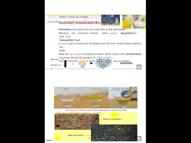
- 23. بلحتسملا و تابلِ حتسملا و بلاحتسلاا Emulsifiers, Emulsification & Emulsion Emulsifiers are substances can cause fats
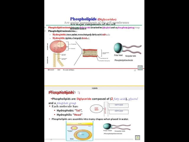
- 24. BIOLOGY 2019 Dr. Amin Al-Doaiss Phospholipid molecule has two fatty acids attached to glycerol and a
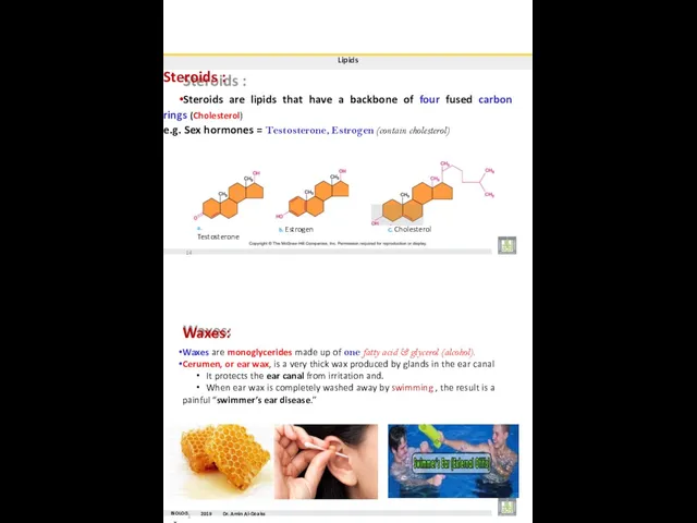
- 25. Lipids Steroids : Steroids are lipids that have a backbone of four fused carbon rings (Cholesterol)
- 26. BIOLOGY 2019 Dr. Amin Al-Doaiss The Omega-3 Fatty Acids A special class of unsaturated fatty acids,
- 27. - The side chain R links with different compounds - Differences in R groups produce the
- 28. Proteins Amino acids : Monomers of protein There are 20 different kinds of Amino Acids peptide
- 29. Proteins Peptides : When two amino acids join together a dipeptide is formed. • • Three
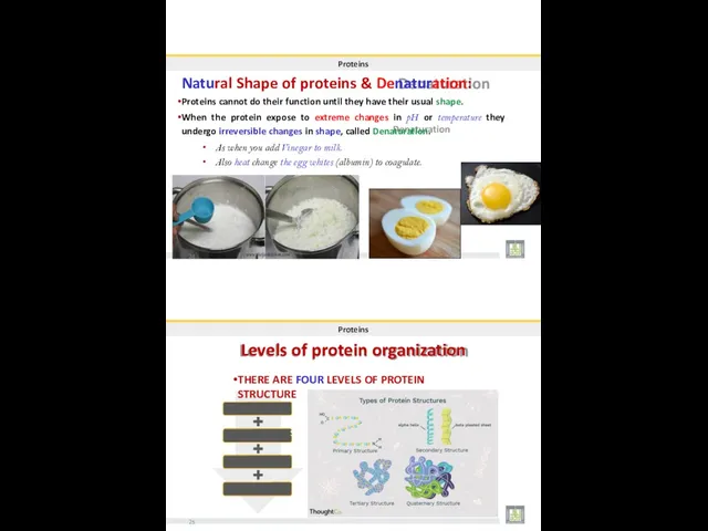
- 30. Proteins Natural Shape of proteins & Denaturation: Proteins cannot do their function until they have their
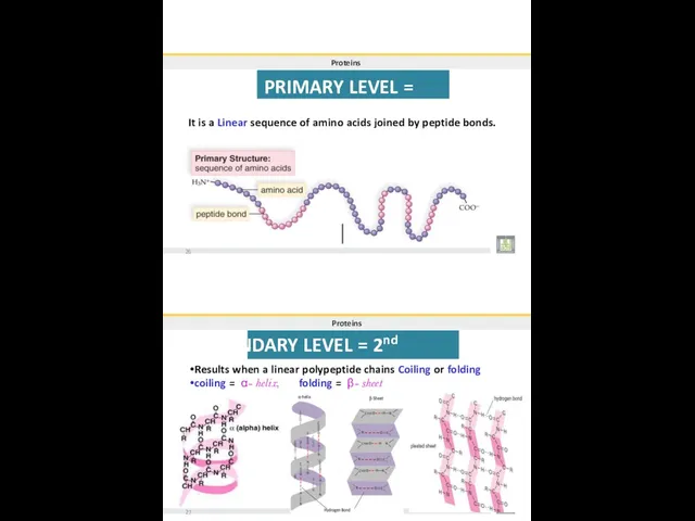
- 31. Proteins PRIMARY LEVEL = 1st It is a Linear sequence of amino acids joined by peptide
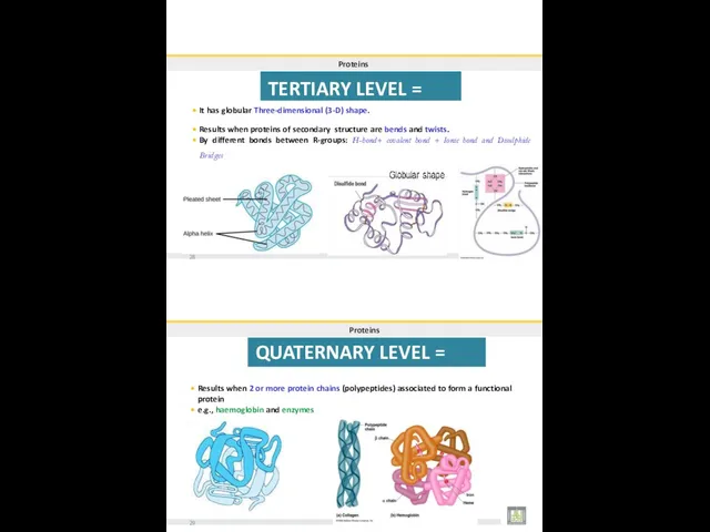
- 32. Proteins TERTIARY LEVEL = 3rd It has globular Three-dimensional (3-D) shape. Results when proteins of secondary
- 33. FOUR LEVELS OF PROTEIN STRUCTURE 30 BIOLOG31Y 2019 Dr. Amin Al-Doaiss
- 34. BIOLO3G2Y 2019 Dr. Ami n Al-Doaiss Dr. AMIN ABDULLAH AL-DOAISS
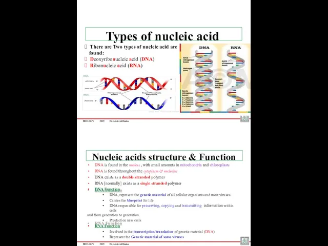
- 35. BIOLOGY 2019 Dr. Amin Al-Doaiss ? There are Two types of nucleic acid are found: ?
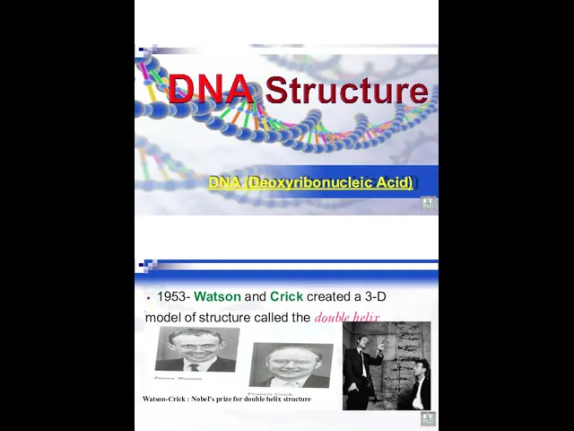
- 36. DNA (Deoxyribonucleic Acid) 1953- Watson and Crick created a 3-D model of structure called the double
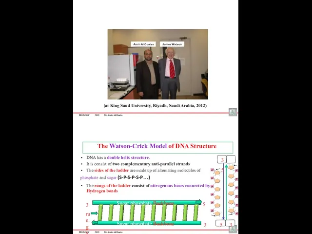
- 37. BIOLOGY 2019 Dr. Amin Al-Doaiss (at King Saud University, Riyadh, Saudi Arabia, 2012) James Watson Amin
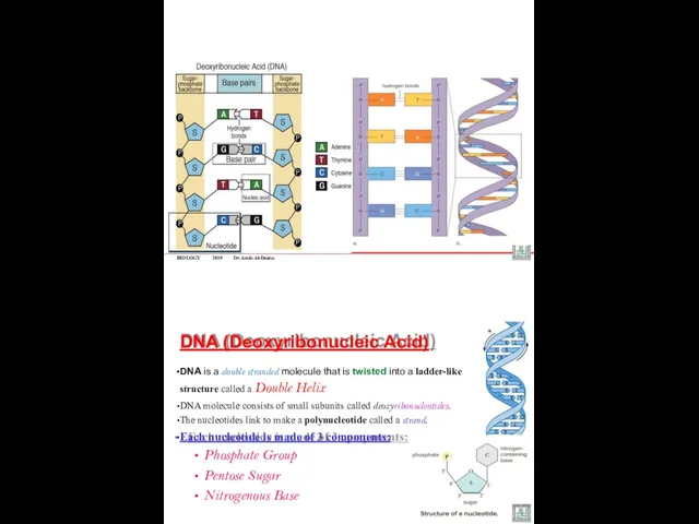
- 38. BIOLOGY 2019 Dr. Amin Al-Doaiss DNA (Deoxyribonucleic Acid) DNA is a double stranded molecule that is
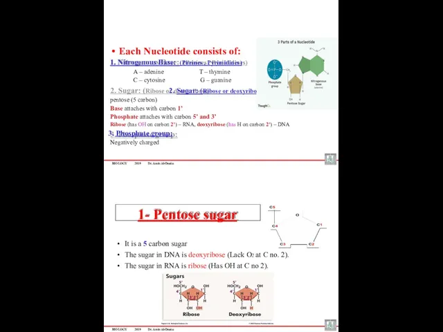
- 39. BIOLOGY 2019 Dr. Amin Al-Doaiss Each Nucleotide consists of: 1. Nitrogenous Base: (Purines , Pyrimidines) A
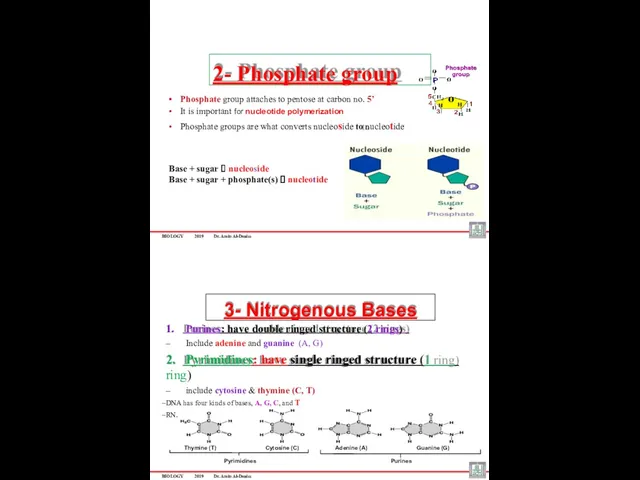
- 40. BIOLOGY 2019 Dr. Amin Al-Doaiss Phosphate group attaches to pentose at carbon no. 5’ It is
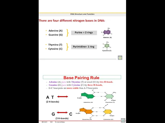
- 41. DNA Structure and Function There are four different nitrogen bases in DNA: Adenine (A) Guanine (G)
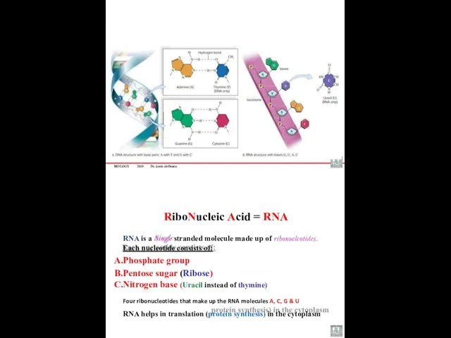
- 42. BIOLOGY 2019 Dr. Amin Al-Doaiss RiboNucleic Acid = RNA RNA is a Single stranded molecule made
- 43. There are three major types of RNA Ribosomal RNA (rRNA). Messenger RNA (mRNA). Transfer RNA (tRNA).
- 44. 2- Messenger RNA (mRNA) Messenger RNA (mRNA) is produced in the nucleus From the DNA by
- 45. Codon-Anticodon base pairing tRNA has Two ends: Anticodon loop interacts with codon on the mRNA. Amino
- 46. DNA-RNA Similarities and differences Dr. AMIN ABDULLAH AL-DOAISS BIOLOGY 2019 Dr. Amin Al-Doaiss 1
- 47. BIOLOGY 2019 Dr. Amin Al-Doaiss 2 THE CELL The term of “ cell” comes from the
- 48. BIOLOGY 2019 Dr. Amin Al-Doaiss CELL THEORY Cell theory refers to an idea that said: cells
- 49. BIOLOGY 2019 Dr. Amin Al-Doaiss 6 1 mm 2 mm Cell size, why are the cells
- 50. BIOLOGY 2019 Dr. Amin Al-Doaiss Cell Types Cells can be classify into two categories: Prokaryotes (prokaryotic
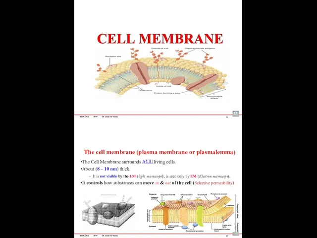
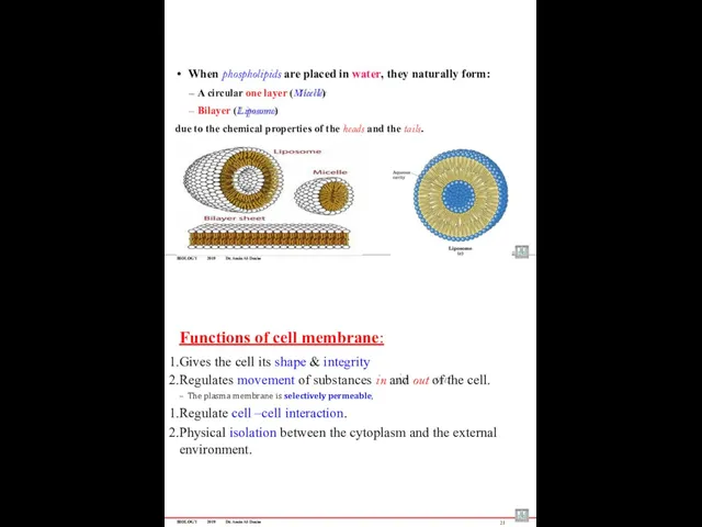
- 51. BIOLOGY 2019 Dr. Amin Al-Doaiss 10 Plasma membrane (cell membrane or plasmalemma) Gives the cell its
- 52. BIOLOGY 2019 Dr. Amin Al-Doaiss 12 Cytoplasmic Organelles (Organelle = “little organ”) An organelle is very
- 53. BIOLOGY 2019 Dr. Amin Al-Doaiss ËĪæĒäĚ ÊĤÖĊ ءÄäöÞ ÊàĪÎêijÈ ĦĤėÞĖÄ äÄàÖĖÄ ËĪĞĪÈ ÆĤďÒ 14 BIOLOGY 2019
- 54. BIOLOGY 2019 Dr. Amin Al-Doaiss 16 The cell membrane (plasma membrane or plasmalemma) The Cell Membrane
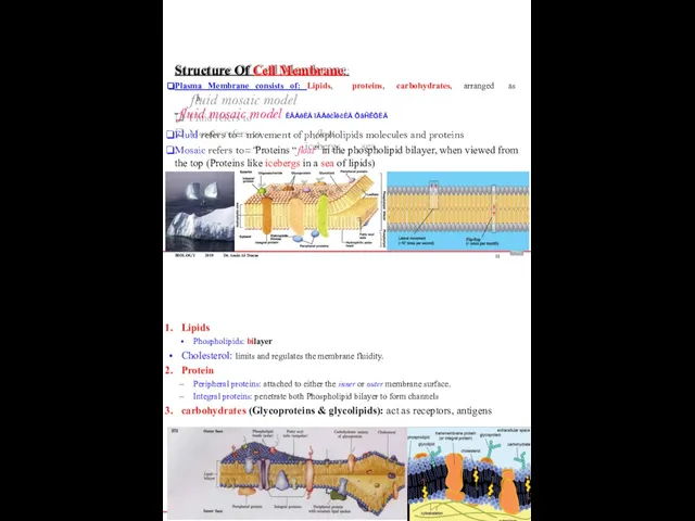
- 55. BIOLOGY 2019 Dr. Amin Al-Doaiss Structure Of Cell Membrane: Plasma Membrane consists of: Lipids, proteins, carbohydrates,
- 56. BIOLOGY 2019 Dr. Amin Al-Doaiss When phospholipids are placed in water, they naturally form: A circular
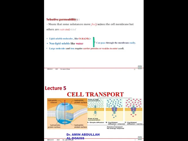
- 57. BIOLOGY 2019 Dr. Amin Al-Doaiss Selective permeability : – Means that some substances move freely across
- 58. BIOLOGY 2019 Dr. Amin Al-Doaiss Cell Transport is moving materials into, out of, or within the
- 59. BIOLOGY 2019 Dr. Amin Al-Doaiss 4 1 Passive transport (No Energy Required) )ًبلسلا( لماخلا لقنلا BIOLOGY
- 60. BIOLOGY 2019 Dr. Amin Al-Doaiss Passive transport includes: Simple diffusion طٌسبلا راشتنلاا Facilitated diffusion رسٌملا لقنلا
- 61. BIOLOGY 2019 Dr. Amin Al-Doaiss 2- Facilitated diffusion ) لهدلما( ردولما لقنلا Facilitated diffusion is a
- 62. BIOLOGY 2019 Dr. Amin Al-Doaiss Osmosis ةٌزومسلأا Osmosis is a transport of water from the hypotonic
- 63. BIOLOGY 2019 Dr. Amin Al-Doaiss 12 Tonicity Tonicity: comparing the concentration of solute in two solutions.
- 64. BIOLOGY 2019 Dr. Amin Al-Doaiss 14 2. Hypotonic Hypotonic solution has lower solute concentration and higher
- 65. BIOLOGY 2019 Dr. Amin Al-Doaiss If Animal cells & Plant Cells placed in: 16 If Animal
- 66. BIOLOGY 2019 Dr. Amin Al-Doaiss 18 Normal plant cells Plasmolytic plant cells BIOLOGY 2019 Dr. Amin
- 67. Phagocytosis Pinocytosis Receptor-mediated endocytosis ةمعلبلا Cellular eating يولخلا برشلا Cellular drinking تلابقتسملا قٌرط نع يولخلا لاخدلاا
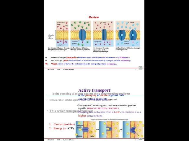
- 68. BIOLOGY 2019 Dr. Amin Al-Doaiss 22 Small uncharged (non-polar) molecules enter or leave the cell membrane
- 69. BIOLOGY 2019 Dr. Amin Al-Doaiss 24 Phagocytosis Pinocytosis Receptor-mediated endocytosis يولخلا لكلاا Cellular eating يولخلا برشلا
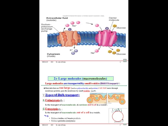
- 70. BIOLOGY 2019 Dr. Amin Al-Doaiss 26 Large molecules are transported by small vesicles (Bulk Transport) Materials
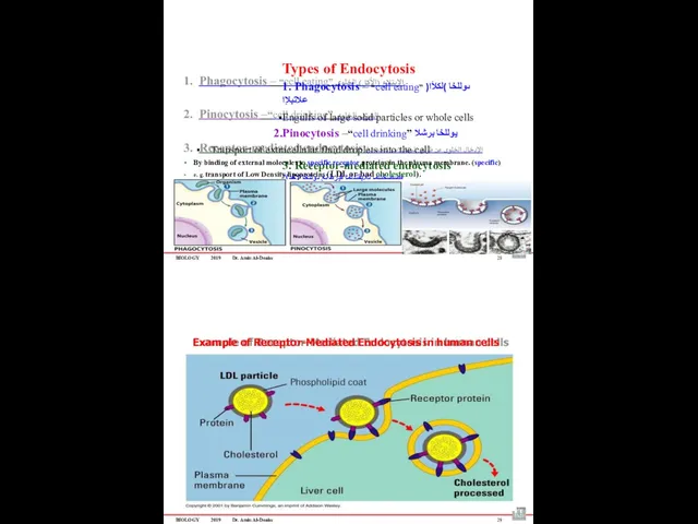
- 71. BIOLOGY 2019 Dr. Amin Al-Doaiss Types of Endocytosis 1. Phagocytosis – “cell eating” ىوللخا )لكلأا( علاتبلإا
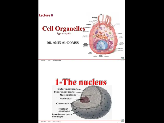
- 72. BIOLOGY 2019 Dr. Amin Al-Doaiss Dr. Amin Al-Doaiss 1 1-The nucleus BIOLOGY 2019 Dr. Amin Al-Doaiss
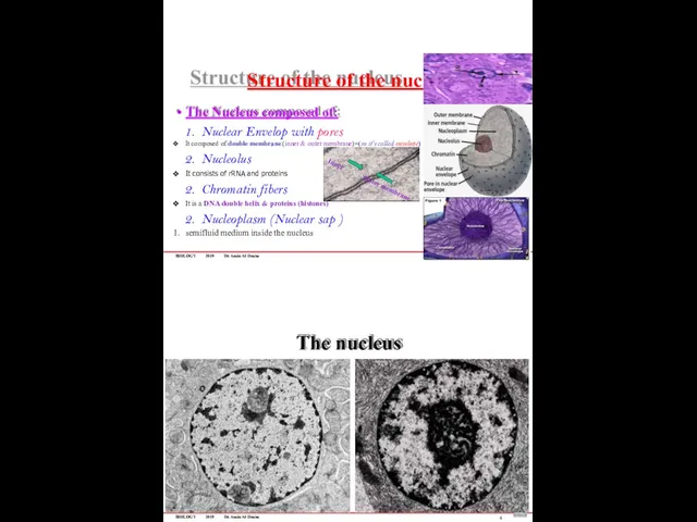
- 73. BIOLOGY 2019 Dr. Amin Al-Doaiss Structure of the nucleus 3 The Nucleus composed of: Nuclear Envelop
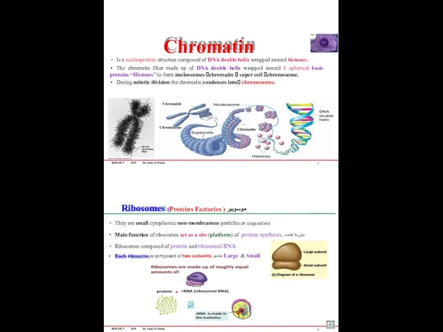
- 74. BIOLOGY 2019 Dr. Amin Al-Doaiss Chromatin Is a nucleoprotein structure composed of DNA double helix wrapped
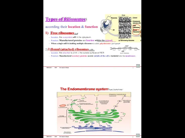
- 75. BIOLOGY 2019 Dr. Amin Al-Doaiss Types of Ribosomes according their location & function 1) Free ribosomes
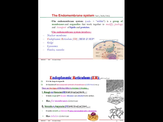
- 76. BIOLOGY 2019 Dr. Amin Al-Doaiss The Endomembrane system يلخادلا يئاشغلا زاهجلا The endomembrane system: (endo- =
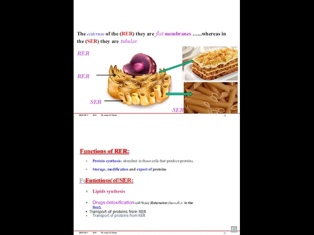
- 77. BIOLOGY 2019 Dr. Amin Al-Doaiss 11 The cisternae of the (RER) they are flat membranes …...whereas
- 78. BIOLOGY 2019 Dr. Amin Al-Doaiss GOLGI APPARATUS (COMPLEX) يجلوج-دقعم-زاهج Discovered by Camillo Golgi Golgi apparatus is
- 79. BIOLOGY 2019 Dr. Amin Al-Doaiss Function of Golgi apparatus Manufacturing, Packaging, Sorting, Modifications, Concentration shipping لقن
- 80. BIOLOGY 2019 Dr. Amin Al-Doaiss THE LYSOSOME Digestive bodies Trash collector of the cell 17 ة
- 81. BIOLOGY 2019 Dr. Amin Al-Doaiss Energy-Related Organelles The two energy-related organelles of eukaryotes are chloroplasts and
- 82. BIOLOGY 2019 Dr. Amin Al-Doaiss 21 ATP-ADP Cycle ATP is the energy of the cell, it
- 83. BIOLOGY 2019 Dr. Amin Al-Doaiss ATP molecule Adenosine triphosphate The three parts of an ATP molecule
- 84. BIOLOGY 2019 Dr. Amin Al-Doaiss The Cytoskeleton It is a network of protein fibers & filaments
- 85. BIOLOGY 2019 Dr. Amin Al-Doaiss 4 5 1- Microfilaments (Actin filaments) The smallest & thinnest fibers
- 86. 6 BIOLOGY 2019 Dr. Amin Al-Doaiss Medium sized filaments They are rope-like filaments Composed of fibrous
- 87. BIOLOGY 2019 Dr. Amin Al-Doaiss 8 Microtubule Motor Protein P Microtubules act as tracks that: Move
- 88. BIOLOGY 2019 Dr. Amin Al-Doaiss Cytoskeleton Tubular structure consist of α & β Tubulin protein Consist
- 89. BIOLOGY 2019 Dr. Amin Al-Doaiss Centrosome & Centrioles Centrosome: pair of centrioles ⮚ Centrioles: paired cylindrical
- 90. BIOLOGY 2019 Dr. Amin Al-Doaiss CILIA (cilium) AND FLAGELLA (flagellum) They are finger-like projections from the
- 91. BIOLOGY 2019 Dr. Amin Al-Doaiss Dr. Amin Al-Doaiss 1 Cellular metabolism The sum of all of
- 92. BIOLOGY 2019 Dr. Amin Al-Doaiss Types of metabolic reactions : 1. Anabolism: A metabolic reaction that
- 93. BIOLOGY 2019 Dr. Amin Al-Doaiss 5 Cellular Metabolism Metabolic pathway: Sequence steps of chemical reactions enzyme.
- 94. BIOLOGY 2019 Dr. Amin Al-Doaiss 7 Cellular Metabolism Enzymes : Majority of Enzymes are proteins Enzyme’s
- 95. BIOLOGY 2019 Dr. Amin Al-Doaiss 9 Cellular Metabolism Enzyme-Substrate Complex Enzymes form a complex with their
- 96. Cellular Respiration Aerobic respiration Cellular respiration converts the energy in the bonds of glucose into usable
- 97. Cellular Metabolism Glycolysis the process of splitting a sugar. glycolysis is the first step in both
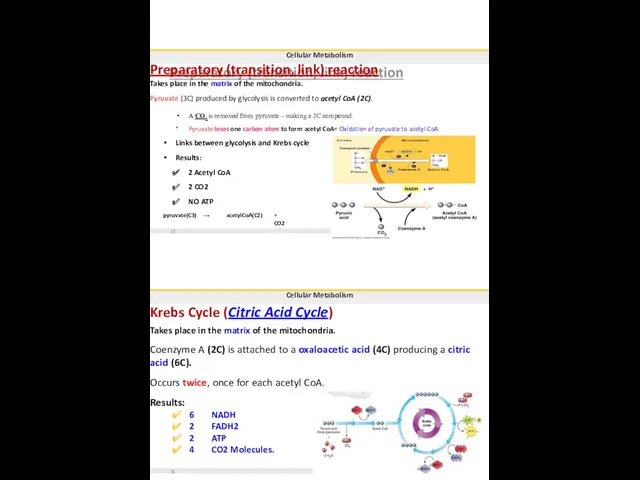
- 98. Cellular Metabolism Preparatory (transition, link) reaction Takes place in the matrix of the mitochondria. Pyruvate (3C)
- 99. Cellular Metabolism citric acid Input Acetic acid ADP Krebs Cycle 3 NAD+ FAD oxaloacetic acid 17
- 101. Скачать презентацию
BIOLOGY
2019
Dr. Amin Al-Doaiss
3
Week
Sunday – Tuesday – Thursday
1st
Chemistry of life: Introduction to
BIOLOGY
2019
Dr. Amin Al-Doaiss
3
Week
Sunday – Tuesday – Thursday
1st
Chemistry of life: Introduction to
BIOLOGY
2019
Dr. Amin Al-Doaiss
Examination
Topic
Description
Quiz & homework
Sheet examination
Quizzes (MCQs)
Mid-Term Exam
Sheet examination Written (1
BIOLOGY
2019
Dr. Amin Al-Doaiss
Examination
Topic
Description
Quiz & homework
Sheet examination
Quizzes (MCQs)
Mid-Term Exam
Sheet examination Written (1
BIOLOGY
2019
Dr. Amin Al-Doaiss
Introduction to biology
Biology (the study of life)
The science of
BIOLOGY
2019
Dr. Amin Al-Doaiss
Introduction to biology
Biology (the study of life)
The science of
BIOLOGY
2019
Dr. Amin Al-Doaiss
Chemistry of Life
9
Dr. AMIN ABDULLAH AL-DOAISS
From atoms, molecules
to
Human body
BIOLOGY
2019
Dr.
BIOLOGY
2019
Dr. Amin Al-Doaiss
Chemistry of Life
9
Dr. AMIN ABDULLAH AL-DOAISS
From atoms, molecules
to
Human body
BIOLOGY
2019
Dr.
BIOLOGY
2019
Dr. Amin Al-Doaiss
Matter is anything that takes up space and has
BIOLOGY
2019
Dr. Amin Al-Doaiss
Matter is anything that takes up space and has
BIOLOGY
2019
Dr. Amin Al-Doaiss
13
Levels of organization
Molecules of Life
“ MACROMOLECULES “
BIOLOGY
2019
Dr. Amin Al-Doaiss
14
BIOLOGY
2019
Dr. Amin Al-Doaiss
13
Levels of organization
Molecules of Life
“ MACROMOLECULES “
BIOLOGY
2019
Dr. Amin Al-Doaiss
14
Macromolecules
MOLECULES
There are two types of molecules :
Inorganic molecules:
e.g., salts (e.g., NaCl)
Macromolecules
MOLECULES
There are two types of molecules :
Inorganic molecules:
e.g., salts (e.g., NaCl)
Macromolecules
Polymers formed by combining monomers together by
Dehydration reaction, or “condensation reaction”
Macromolecules
Polymers formed by combining monomers together by
Dehydration reaction, or “condensation reaction”
BIOLOGY
2019
Dr. Amin Al-Doaiss
Macromolecules
There are four major classes of macromolecules
BIOLOGY
2019
Dr. Amin Al-Doaiss
Macromolecules
There are four major classes of macromolecules
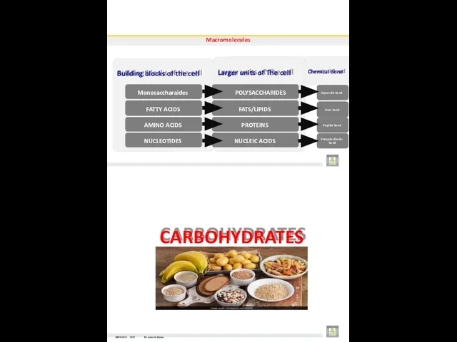
Chemical Bond
Macromolecules
Building blocks of the cell
Monosaccharaides
FATTY ACIDS
AMINO ACIDS
NUCLEOTIDES
Larger units of The
Chemical Bond
Macromolecules
Building blocks of the cell
Monosaccharaides
FATTY ACIDS
AMINO ACIDS
NUCLEOTIDES
Larger units of The
BIOLOGY
2019
Dr. Amin Al-Doaiss
Fuel and Building Materials
Are organic macromolecules composed of monomers
BIOLOGY
2019
Dr. Amin Al-Doaiss
Fuel and Building Materials
Are organic macromolecules composed of monomers
BIOLOGY
2019
Dr. Amin Al-Doaiss
Monosaccharide : (mono, one; saccharide, sugar)
Consist of only a
BIOLOGY
2019
Dr. Amin Al-Doaiss
Monosaccharide : (mono, one; saccharide, sugar)
Consist of only a
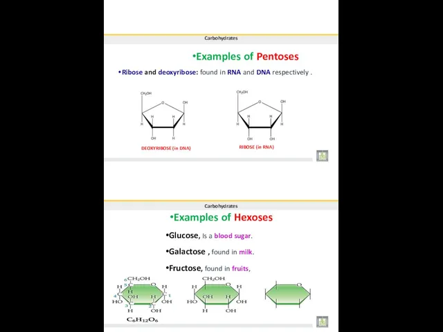
•Examples of Pentoses
Carbohydrates
Ribose and deoxyribose: found in RNA and DNA respectively
•Examples of Pentoses
Carbohydrates
Ribose and deoxyribose: found in RNA and DNA respectively
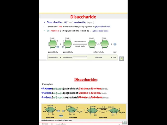
Disaccharide
Disaccharide : (di, “two”; saccharide, “sugar”)
Composed of Two monosaccharides joining together
Disaccharide
Disaccharide : (di, “two”; saccharide, “sugar”)
Composed of Two monosaccharides joining together
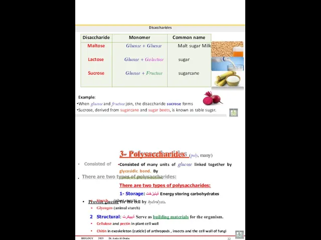
Disaccharides
Example:
When glucose and fructose join, the disaccharide sucrose forms
Sucrose, derived from
Disaccharides
Example:
When glucose and fructose join, the disaccharide sucrose forms
Sucrose, derived from
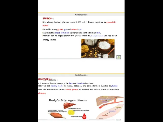
Carbohydrates
STARCH :
It is a Long chain of glucose (up to 4,000
Carbohydrates
STARCH :
It is a Long chain of glucose (up to 4,000
Carbohydrates
CELLULOSE :
It is a structural form of polysaccharide, supports the plant
Carbohydrates
CELLULOSE :
It is a structural form of polysaccharide, supports the plant
BIOLOGY
2019
Dr. Amin Al-Doaiss
2
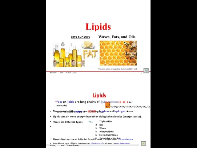
Lipids
Phospholipids are type of lipids that form the major
BIOLOGY
2019
Dr. Amin Al-Doaiss
2
Lipids
Phospholipids are type of lipids that form the major
BIOLOGY
2019
Dr. Amin
mponent of cell memb
Al-Doaiss
4
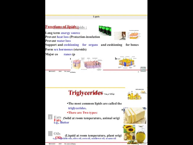
Lipids
Functions of lipids :
Long term energy source
Prevent
BIOLOGY
2019
Dr. Amin
mponent of cell memb
Al-Doaiss
4
Lipids
Functions of lipids :
Long term energy source
Prevent
BIOLOGY
2019
Dr. Amin Al-Doaiss
6
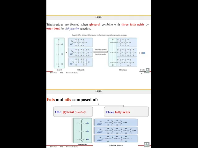
Lipids
Triglycerides are formed when glycerol combine with three fatty
BIOLOGY
2019
Dr. Amin Al-Doaiss
6
Lipids
Triglycerides are formed when glycerol combine with three fatty
BIOLOGY
2019
Dr. Amin Al-Doaiss
OH
H
H
C
C
C
C
C
C
H
H
H
H
H
H
H
H
H
O
H
OH
Glycerol
H
H C
H C
H C
OH
OH
Dehydration
Fatty Acid
Ester link
8
H
H
C
C
C
C
C
C
H
H
H H
H
H
H
H
H
H
C
C
C
C
C
C
H Fatty Acid
H
H
H
O
H Fatty Acid
H
H
H
H
H
O
A)- Saturated
BIOLOGY
2019
Dr. Amin Al-Doaiss
OH
H
H
C
C
C
C
C
C
H
H
H
H
H
H
H
H
H
O
H
OH
Glycerol
H
H C
H C
H C
OH
OH
Dehydration
Fatty Acid
Ester link
8
H
H
C
C
C
C
C
C
H
H
H H
H
H
H
H
H
H
C
C
C
C
C
C
H Fatty Acid
H
H
H
O
H Fatty Acid
H
H
H
H
H
O
A)- Saturated
بلحتسملا و تابلِ حتسملا و بلاحتسلاا
Emulsifiers, Emulsification & Emulsion
Emulsifiers are substances
بلحتسملا و تابلِ حتسملا و بلاحتسلاا
Emulsifiers, Emulsification & Emulsion
Emulsifiers are substances
BIOLOGY
2019
Dr. Amin Al-Doaiss
Phospholipid molecule has two fatty acids attached to glycerol
BIOLOGY
2019
Dr. Amin Al-Doaiss
Phospholipid molecule has two fatty acids attached to glycerol
Lipids
Steroids :
Steroids are lipids that have a backbone of four fused
Lipids
Steroids :
Steroids are lipids that have a backbone of four fused
BIOLOGY 2019
Dr. Amin Al-Doaiss
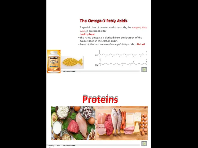
The Omega-3 Fatty Acids
A special class of unsaturated fatty
BIOLOGY 2019
Dr. Amin Al-Doaiss
The Omega-3 Fatty Acids
A special class of unsaturated fatty
- The side chain R links with different compounds
- Differences in
- The side chain R links with different compounds
- Differences in
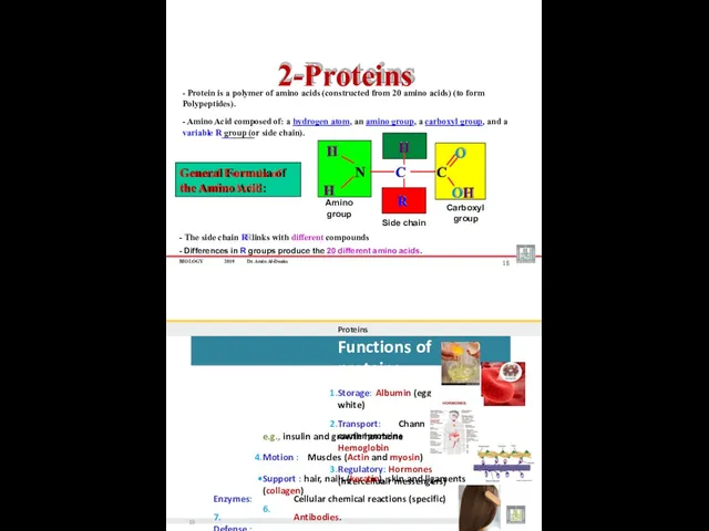
Proteins
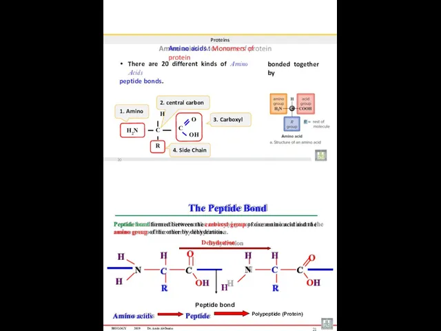
Amino acids : Monomers of protein
There are 20 different kinds of
Proteins
Amino acids : Monomers of protein
There are 20 different kinds of
Proteins
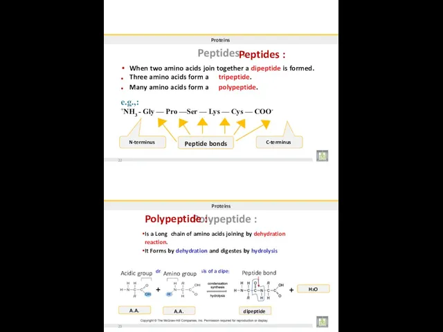
Peptides :
When two amino acids join together a dipeptide is formed.
•
•
Three
Proteins
Peptides :
When two amino acids join together a dipeptide is formed.
•
•
Three
Proteins
Natural Shape of proteins & Denaturation:
Proteins cannot do their function until they
Proteins
Natural Shape of proteins & Denaturation:
Proteins cannot do their function until they
Proteins
PRIMARY LEVEL = 1st
It is a Linear sequence of amino acids
Proteins
PRIMARY LEVEL = 1st
It is a Linear sequence of amino acids
Proteins
TERTIARY LEVEL = 3rd
It has globular Three-dimensional (3-D) shape.
Results when proteins
Proteins
TERTIARY LEVEL = 3rd
It has globular Three-dimensional (3-D) shape.
Results when proteins
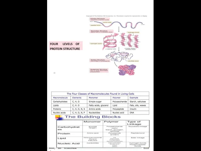
FOUR LEVELS OF
PROTEIN STRUCTURE
30
BIOLOG31Y
2019
Dr. Amin Al-Doaiss
FOUR LEVELS OF
PROTEIN STRUCTURE
30
BIOLOG31Y
2019
Dr. Amin Al-Doaiss
BIOLO3G2Y
2019
Dr. Ami
n Al-Doaiss
Dr. AMIN ABDULLAH AL-DOAISS
BIOLO3G2Y
2019
Dr. Ami
n Al-Doaiss
Dr. AMIN ABDULLAH AL-DOAISS
BIOLOGY
2019
Dr. Amin Al-Doaiss
? There are Two types of nucleic acid are
BIOLOGY
2019
Dr. Amin Al-Doaiss
? There are Two types of nucleic acid are
DNA (Deoxyribonucleic Acid)
1953- Watson and Crick created a 3-D
model of structure
DNA (Deoxyribonucleic Acid)
1953- Watson and Crick created a 3-D
model of structure
BIOLOGY
2019
Dr. Amin Al-Doaiss
(at King Saud University, Riyadh, Saudi Arabia, 2012)
James Watson
Amin
BIOLOGY
2019
Dr. Amin Al-Doaiss
(at King Saud University, Riyadh, Saudi Arabia, 2012)
James Watson
Amin
BIOLOGY
2019
Dr. Amin Al-Doaiss
DNA (Deoxyribonucleic Acid)
DNA is a double stranded molecule that
BIOLOGY
2019
Dr. Amin Al-Doaiss
DNA (Deoxyribonucleic Acid)
DNA is a double stranded molecule that
BIOLOGY
2019
Dr. Amin Al-Doaiss
Each Nucleotide consists of:
1. Nitrogenous Base: (Purines , Pyrimidines)
A
BIOLOGY
2019
Dr. Amin Al-Doaiss
Each Nucleotide consists of:
1. Nitrogenous Base: (Purines , Pyrimidines)
A
BIOLOGY
2019
Dr. Amin Al-Doaiss
Phosphate group attaches to pentose at carbon no. 5’
It
BIOLOGY
2019
Dr. Amin Al-Doaiss
Phosphate group attaches to pentose at carbon no. 5’
It
DNA Structure and Function
There are four different nitrogen bases in DNA:
Adenine
DNA Structure and Function
There are four different nitrogen bases in DNA:
Adenine
BIOLOGY
2019
Dr. Amin Al-Doaiss
RiboNucleic Acid = RNA
RNA is a Single stranded molecule
BIOLOGY
2019
Dr. Amin Al-Doaiss
RiboNucleic Acid = RNA
RNA is a Single stranded molecule
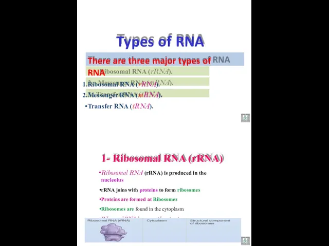
There are three major types of RNA
Ribosomal RNA (rRNA).
Messenger RNA (mRNA).
Transfer
There are three major types of RNA
Ribosomal RNA (rRNA).
Messenger RNA (mRNA).
Transfer
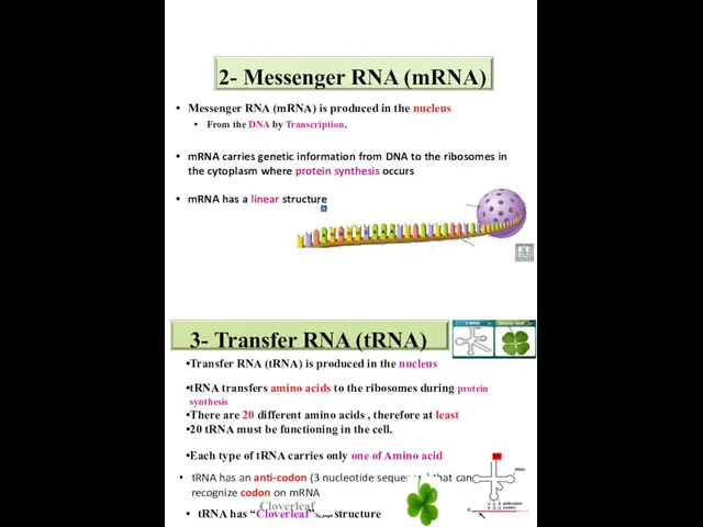
2- Messenger RNA (mRNA)
Messenger RNA (mRNA) is produced in the nucleus
From
2- Messenger RNA (mRNA)
Messenger RNA (mRNA) is produced in the nucleus
From
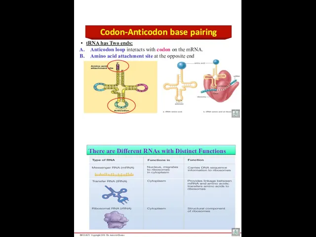
Codon-Anticodon base pairing
tRNA has Two ends:
Anticodon loop interacts with codon on
Codon-Anticodon base pairing
tRNA has Two ends:
Anticodon loop interacts with codon on
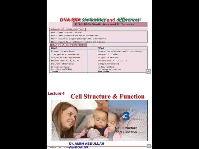
DNA-RNA Similarities and differences
Dr. AMIN ABDULLAH AL-DOAISS
BIOLOGY
2019
Dr. Amin Al-Doaiss
1
DNA-RNA Similarities and differences
Dr. AMIN ABDULLAH AL-DOAISS
BIOLOGY
2019
Dr. Amin Al-Doaiss
1
BIOLOGY
2019
Dr. Amin Al-Doaiss
2
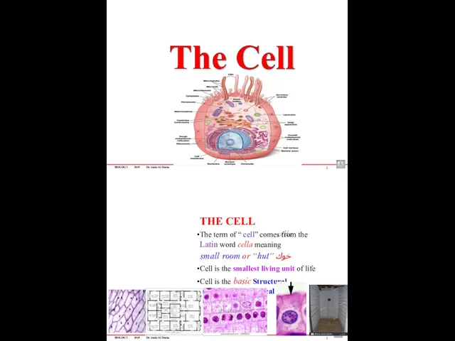
THE CELL
The term of “ cell” comes from the
BIOLOGY
2019
Dr. Amin Al-Doaiss
2
THE CELL
The term of “ cell” comes from the
BIOLOGY
2019
Dr. Amin Al-Doaiss
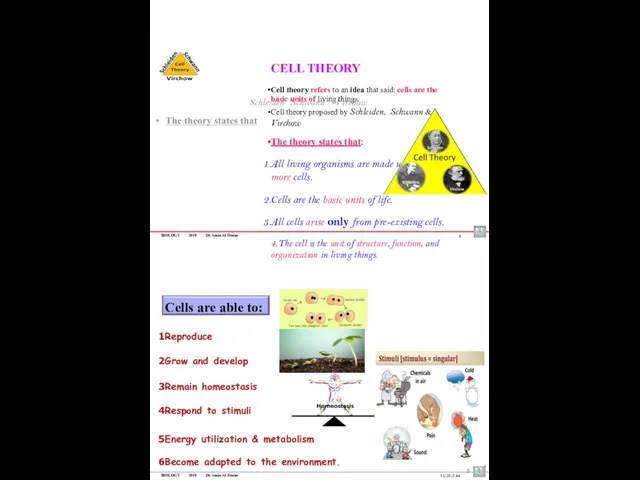
CELL THEORY
Cell theory refers to an idea that said:
BIOLOGY
2019
Dr. Amin Al-Doaiss
CELL THEORY
Cell theory refers to an idea that said:
BIOLOGY
2019
Dr. Amin Al-Doaiss
6
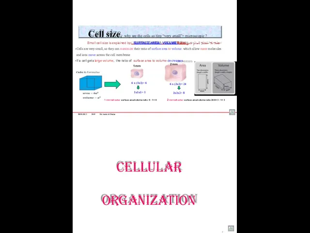
1 mm
2 mm
Cell size, why are the cells so
BIOLOGY
2019
Dr. Amin Al-Doaiss
6
1 mm
2 mm
Cell size, why are the cells so
BIOLOGY 2019
Dr. Amin Al-Doaiss
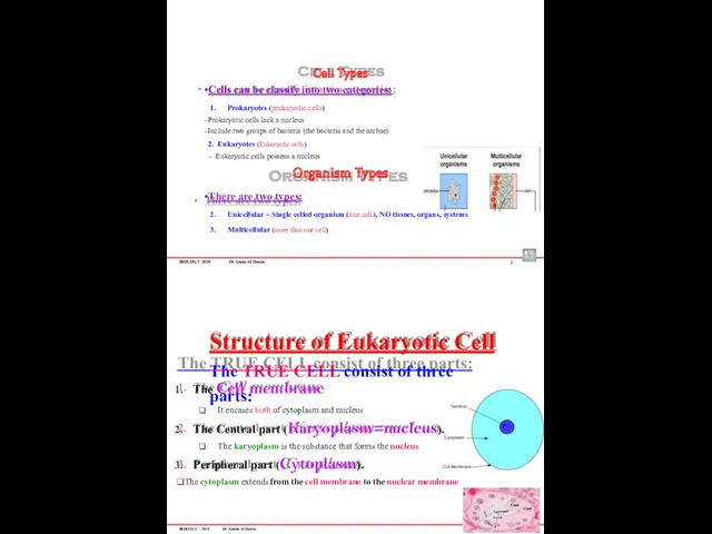
Cell Types
Cells can be classify into two categories:
Prokaryotes (prokaryotic
BIOLOGY 2019
Dr. Amin Al-Doaiss
Cell Types
Cells can be classify into two categories:
Prokaryotes (prokaryotic
BIOLOGY 2019
Dr. Amin Al-Doaiss
10
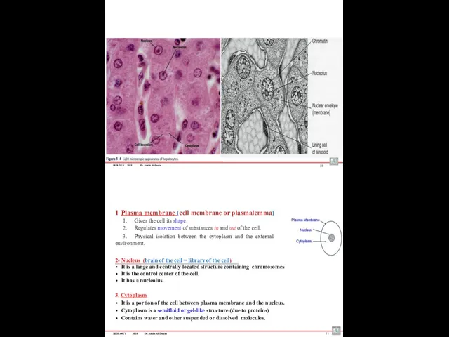
Plasma membrane (cell membrane or plasmalemma)
Gives the cell its
BIOLOGY 2019
Dr. Amin Al-Doaiss
10
Plasma membrane (cell membrane or plasmalemma)
Gives the cell its
BIOLOGY
2019
Dr. Amin Al-Doaiss
12
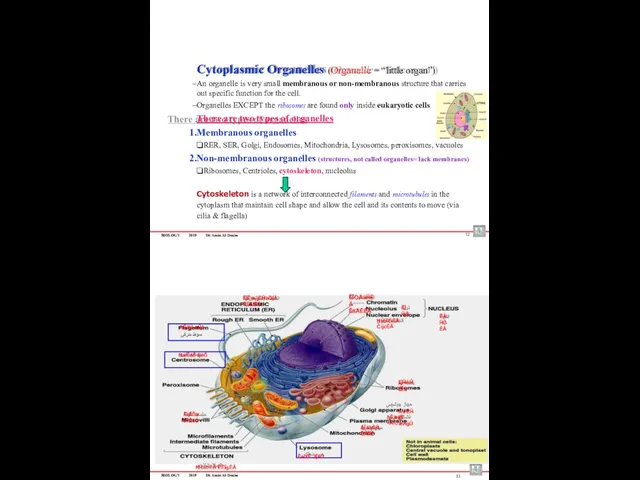
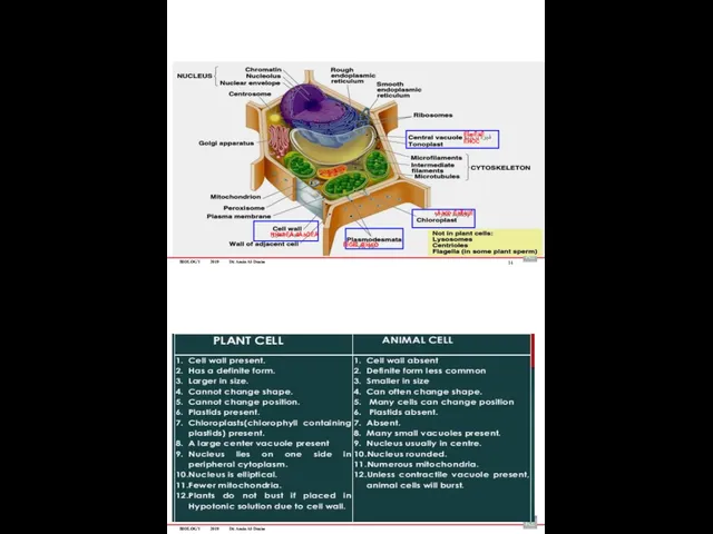
Cytoplasmic Organelles (Organelle = “little organ”)
An organelle is very
BIOLOGY
2019
Dr. Amin Al-Doaiss
12
Cytoplasmic Organelles (Organelle = “little organ”)
An organelle is very
BIOLOGY
2019
Dr. Amin Al-Doaiss
ËĪæĒäĚ ÊĤÖĊ
ءÄäöÞ ÊàĪÎêijÈ
ĦĤėÞĖÄ äÄàÖĖÄ
ËĪĞĪÈ ÆĤďÒ
14
BIOLOGY
2019
Dr. Amin Al-Doaiss
BIOLOGY
2019
Dr. Amin Al-Doaiss
ËĪæĒäĚ ÊĤÖĊ
ءÄäöÞ ÊàĪÎêijÈ
ĦĤėÞĖÄ äÄàÖĖÄ
ËĪĞĪÈ ÆĤďÒ
14
BIOLOGY
2019
Dr. Amin Al-Doaiss
BIOLOGY
2019
Dr. Amin Al-Doaiss
16
The cell membrane (plasma membrane or plasmalemma)
The Cell Membrane
BIOLOGY
2019
Dr. Amin Al-Doaiss
16
The cell membrane (plasma membrane or plasmalemma)
The Cell Membrane
BIOLOGY
2019
Dr. Amin Al-Doaiss
Structure Of Cell Membrane:
Plasma Membrane consists of: Lipids, proteins, carbohydrates, arranged as a
“fluid mosaic
BIOLOGY
2019
Dr. Amin Al-Doaiss
Structure Of Cell Membrane:
Plasma Membrane consists of: Lipids, proteins, carbohydrates, arranged as a
“fluid mosaic
BIOLOGY
2019
Dr. Amin Al-Doaiss
When phospholipids are placed in water, they naturally form:
A
BIOLOGY
2019
Dr. Amin Al-Doaiss
When phospholipids are placed in water, they naturally form:
A
BIOLOGY
2019
Dr. Amin Al-Doaiss
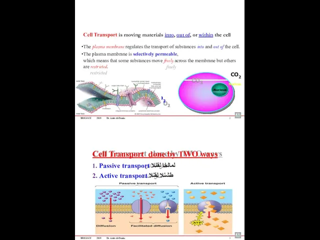
Selective permeability :
– Means that some substances move freely
BIOLOGY
2019
Dr. Amin Al-Doaiss
Selective permeability :
– Means that some substances move freely
BIOLOGY
2019
Dr. Amin Al-Doaiss
Cell Transport is moving materials into, out of, or
BIOLOGY
2019
Dr. Amin Al-Doaiss
Cell Transport is moving materials into, out of, or
BIOLOGY 2019
Dr. Amin Al-Doaiss
4
1
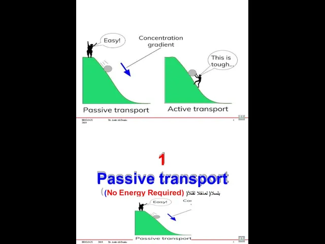
Passive transport
(No Energy Required) )ًبلسلا( لماخلا لقنلا
BIOLOGY
2019
Dr. Amin Al-Doaiss
5
BIOLOGY 2019
Dr. Amin Al-Doaiss
4
1
Passive transport
(No Energy Required) )ًبلسلا( لماخلا لقنلا
BIOLOGY
2019
Dr. Amin Al-Doaiss
5
BIOLOGY
2019
Dr. Amin Al-Doaiss
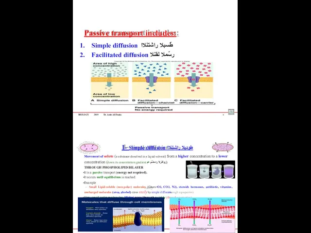
Passive transport includes:
Simple diffusion طٌسبلا راشتنلاا
Facilitated diffusion رسٌملا لقنلا
6
BIOLOGY 2019 Dr. Amin
BIOLOGY
2019
Dr. Amin Al-Doaiss
Passive transport includes:
Simple diffusion طٌسبلا راشتنلاا
Facilitated diffusion رسٌملا لقنلا
6
BIOLOGY 2019 Dr. Amin
BIOLOGY
2019
Dr. Amin Al-Doaiss
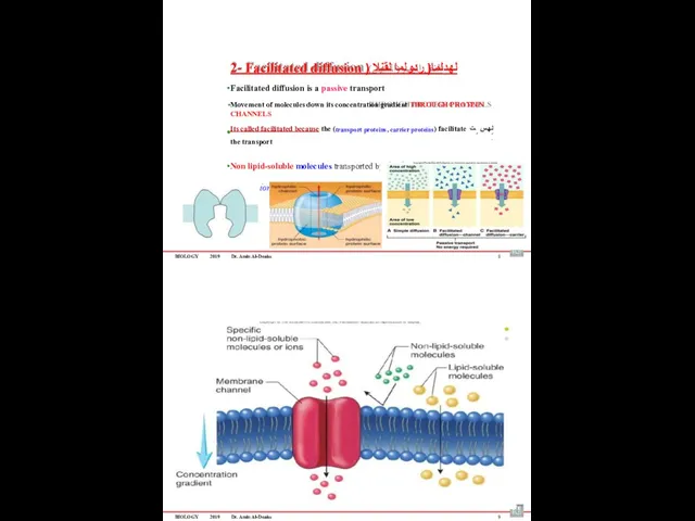
2- Facilitated diffusion ) لهدلما( ردولما لقنلا
Facilitated diffusion
BIOLOGY
2019
Dr. Amin Al-Doaiss
2- Facilitated diffusion ) لهدلما( ردولما لقنلا
Facilitated diffusion
BIOLOGY
2019
Dr. Amin Al-Doaiss
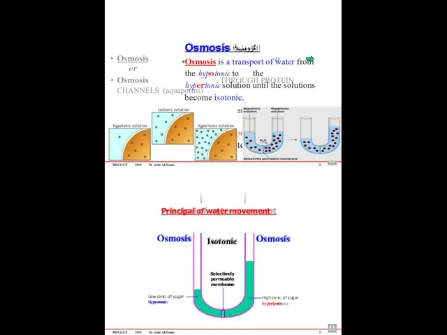
Osmosis ةٌزومسلأا
Osmosis is a transport of water from the
BIOLOGY
2019
Dr. Amin Al-Doaiss
Osmosis ةٌزومسلأا
Osmosis is a transport of water from the
BIOLOGY
2019
Dr. Amin Al-Doaiss
12
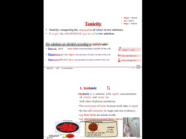
Tonicity
Tonicity: comparing the concentration of solute in two solutions.
Example:
BIOLOGY
2019
Dr. Amin Al-Doaiss
12
Tonicity
Tonicity: comparing the concentration of solute in two solutions.
Example:
BIOLOGY
2019
Dr. Amin Al-Doaiss
14
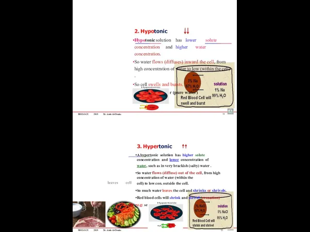
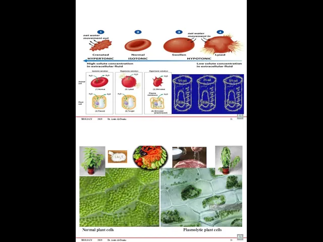
2. Hypotonic
Hypotonic solution has lower solute concentration and higher water concentration.
So water flows (diffuses) inward the cell,
BIOLOGY
2019
Dr. Amin Al-Doaiss
14
2. Hypotonic
Hypotonic solution has lower solute concentration and higher water concentration.
So water flows (diffuses) inward the cell,
BIOLOGY
2019
Dr. Amin Al-Doaiss
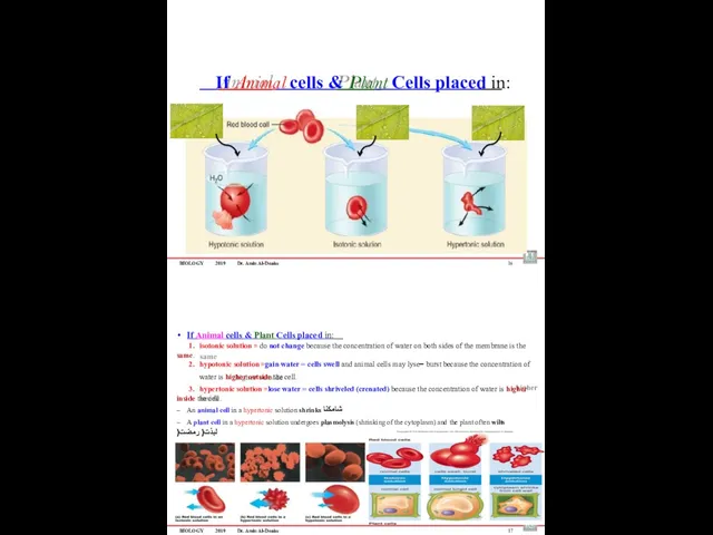
If Animal cells & Plant Cells placed in:
16
If Animal
BIOLOGY
2019
Dr. Amin Al-Doaiss
If Animal cells & Plant Cells placed in:
16
If Animal
BIOLOGY
2019
Dr. Amin Al-Doaiss
18
Normal plant cells
Plasmolytic plant cells
BIOLOGY
2019
Dr. Amin Al-Doaiss
19
BIOLOGY
2019
Dr. Amin Al-Doaiss
18
Normal plant cells
Plasmolytic plant cells
BIOLOGY
2019
Dr. Amin Al-Doaiss
19
Phagocytosis
Pinocytosis
Receptor-mediated
endocytosis
ةمعلبلا
Cellular eating
يولخلا برشلا
Cellular drinking
تلابقتسملا قٌرط نع يولخلا لاخدلاا
Selective eating
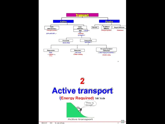
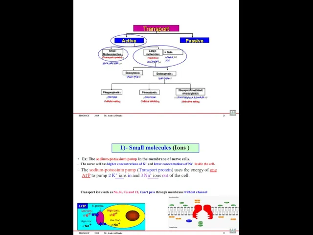
Transport
Passive
Active
Simple Diffusion
Facilitated
diffusion
Large molecules
Small
Phagocytosis
Pinocytosis
Receptor-mediated
endocytosis
ةمعلبلا
Cellular eating
يولخلا برشلا
Cellular drinking
تلابقتسملا قٌرط نع يولخلا لاخدلاا
Selective eating
Transport
Passive
Active
Simple Diffusion
Facilitated
diffusion
Large molecules
Small
BIOLOGY
2019
Dr. Amin Al-Doaiss
22
Small uncharged (non-polar) molecules enter or leave the cell
BIOLOGY
2019
Dr. Amin Al-Doaiss
22
Small uncharged (non-polar) molecules enter or leave the cell
BIOLOGY
2019
Dr. Amin Al-Doaiss
24
Phagocytosis
Pinocytosis
Receptor-mediated
endocytosis
يولخلا لكلاا
Cellular eating
يولخلا برشلا
Cellular drinking
تلابقتسملا قٌرط نع يولخلا لاخدلاا
Selective
BIOLOGY
2019
Dr. Amin Al-Doaiss
24
Phagocytosis
Pinocytosis
Receptor-mediated
endocytosis
يولخلا لكلاا
Cellular eating
يولخلا برشلا
Cellular drinking
تلابقتسملا قٌرط نع يولخلا لاخدلاا
Selective
BIOLOGY
2019
Dr. Amin Al-Doaiss
26
Large molecules are transported by small vesicles (Bulk Transport)
Materials
BIOLOGY
2019
Dr. Amin Al-Doaiss
26
Large molecules are transported by small vesicles (Bulk Transport)
Materials
BIOLOGY
2019
Dr. Amin Al-Doaiss
Types of Endocytosis
1. Phagocytosis – “cell eating” ىوللخا )لكلأا( علاتبلإا
Engulfs
BIOLOGY
2019
Dr. Amin Al-Doaiss
Types of Endocytosis
1. Phagocytosis – “cell eating” ىوللخا )لكلأا( علاتبلإا
Engulfs
BIOLOGY
2019
Dr. Amin Al-Doaiss
Dr. Amin Al-Doaiss
1
1-The nucleus
BIOLOGY
2019
Dr. Amin Al-Doaiss
2
BIOLOGY
2019
Dr. Amin Al-Doaiss
Dr. Amin Al-Doaiss
1
1-The nucleus
BIOLOGY
2019
Dr. Amin Al-Doaiss
2
BIOLOGY
2019
Dr. Amin Al-Doaiss
Structure of the nucleus
3
The Nucleus composed of:
Nuclear Envelop with
BIOLOGY
2019
Dr. Amin Al-Doaiss
Structure of the nucleus
3
The Nucleus composed of:
Nuclear Envelop with
BIOLOGY
2019
Dr. Amin Al-Doaiss
Chromatin
Is a nucleoprotein structure composed of DNA double helix
BIOLOGY
2019
Dr. Amin Al-Doaiss
Chromatin
Is a nucleoprotein structure composed of DNA double helix
BIOLOGY
2019
Dr. Amin Al-Doaiss
Types of Ribosomes
according their location & function
1) Free ribosomes ةرح
Location:
BIOLOGY
2019
Dr. Amin Al-Doaiss
Types of Ribosomes
according their location & function
1) Free ribosomes ةرح
Location:
BIOLOGY
2019
Dr. Amin Al-Doaiss
The Endomembrane system يلخادلا يئاشغلا زاهجلا
The endomembrane system: (endo-
BIOLOGY
2019
Dr. Amin Al-Doaiss
The Endomembrane system يلخادلا يئاشغلا زاهجلا
The endomembrane system: (endo-
BIOLOGY
2019
Dr. Amin Al-Doaiss
11
The cisternae of the (RER) they are flat membranes
BIOLOGY
2019
Dr. Amin Al-Doaiss
11
The cisternae of the (RER) they are flat membranes
BIOLOGY
2019
Dr. Amin Al-Doaiss
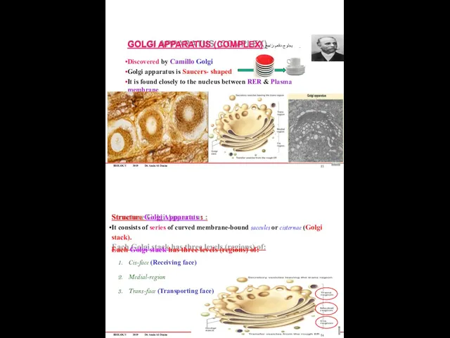
GOLGI APPARATUS (COMPLEX) يجلوج-دقعم-زاهج
Discovered by Camillo Golgi
Golgi apparatus
BIOLOGY
2019
Dr. Amin Al-Doaiss
GOLGI APPARATUS (COMPLEX) يجلوج-دقعم-زاهج
Discovered by Camillo Golgi
Golgi apparatus
BIOLOGY
2019
Dr. Amin Al-Doaiss
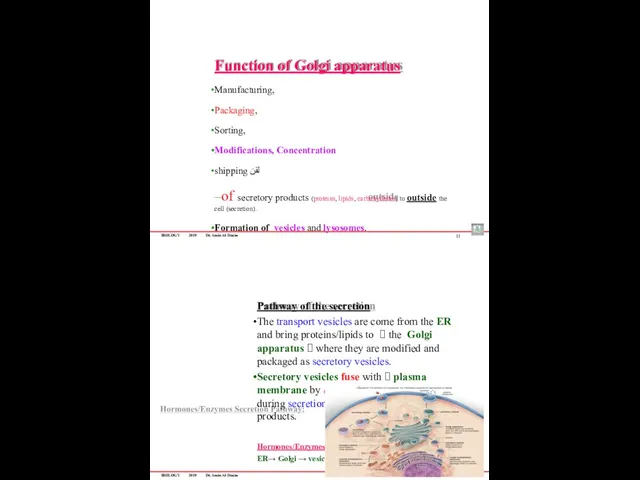
Function of Golgi apparatus
Manufacturing,
Packaging,
Sorting,
Modifications, Concentration
shipping لقن
–of secretory products (proteins,
BIOLOGY
2019
Dr. Amin Al-Doaiss
Function of Golgi apparatus
Manufacturing,
Packaging,
Sorting,
Modifications, Concentration
shipping لقن
–of secretory products (proteins,
BIOLOGY
2019
Dr. Amin Al-Doaiss
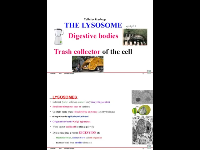
THE LYSOSOME
Digestive bodies
Trash collector of the cell
17
ة للحمُ لا
BIOLOGY
2019
Dr. Amin Al-Doaiss
THE LYSOSOME
Digestive bodies
Trash collector of the cell
17
ة للحمُ لا
BIOLOGY
2019
Dr. Amin Al-Doaiss
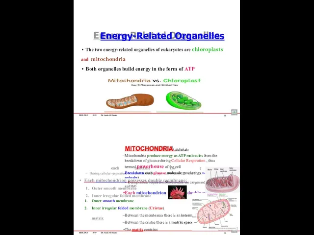
Energy-Related Organelles
The two energy-related organelles of eukaryotes are chloroplasts
and
BIOLOGY
2019
Dr. Amin Al-Doaiss
Energy-Related Organelles
The two energy-related organelles of eukaryotes are chloroplasts
and
BIOLOGY
2019
Dr. Amin Al-Doaiss
21
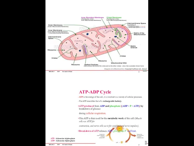
ATP-ADP Cycle
ATP is the energy of the cell, it
BIOLOGY
2019
Dr. Amin Al-Doaiss
21
ATP-ADP Cycle
ATP is the energy of the cell, it
BIOLOGY
2019
Dr. Amin Al-Doaiss
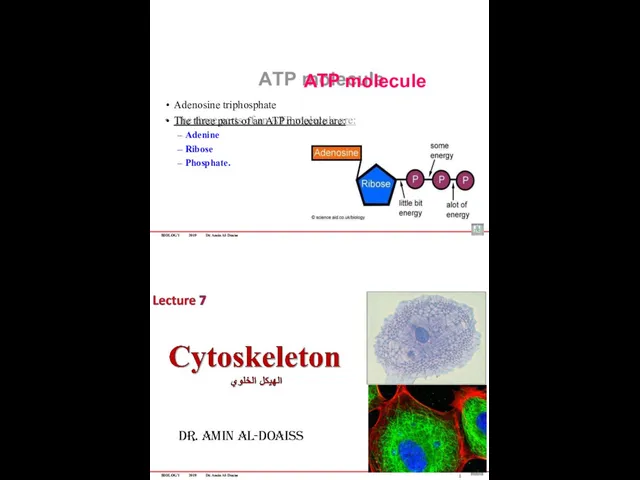
ATP molecule
Adenosine triphosphate
The three parts of an ATP molecule
BIOLOGY
2019
Dr. Amin Al-Doaiss
ATP molecule
Adenosine triphosphate
The three parts of an ATP molecule
BIOLOGY
2019
Dr. Amin Al-Doaiss
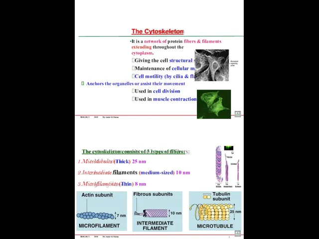
The Cytoskeleton
It is a network of protein fibers &
BIOLOGY
2019
Dr. Amin Al-Doaiss
The Cytoskeleton
It is a network of protein fibers &
BIOLOGY
2019
Dr. Amin Al-Doaiss
4
5
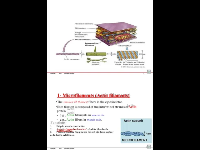
1- Microfilaments (Actin filaments)
The smallest & thinnest
BIOLOGY
2019
Dr. Amin Al-Doaiss
4
5
1- Microfilaments (Actin filaments)
The smallest & thinnest
6
BIOLOGY 2019 Dr. Amin Al-Doaiss
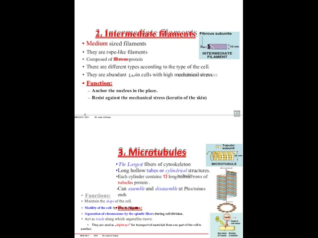
Medium sized filaments
They are rope-like filaments
Composed of
6
BIOLOGY 2019 Dr. Amin Al-Doaiss
Medium sized filaments
They are rope-like filaments
Composed of
BIOLOGY
2019
Dr. Amin Al-Doaiss
8
Microtubule
Motor Protein
P
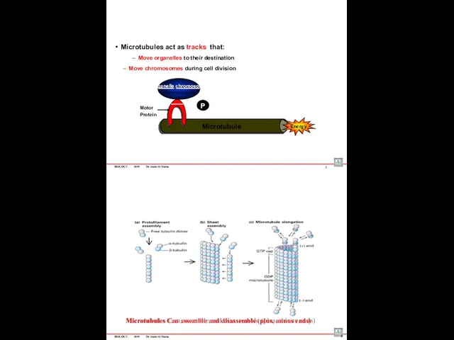
Microtubules act as tracks that:
Move organelles to their
BIOLOGY
2019
Dr. Amin Al-Doaiss
8
Microtubule
Motor Protein
P
Microtubules act as tracks that:
Move organelles to their
BIOLOGY
2019
Dr. Amin Al-Doaiss
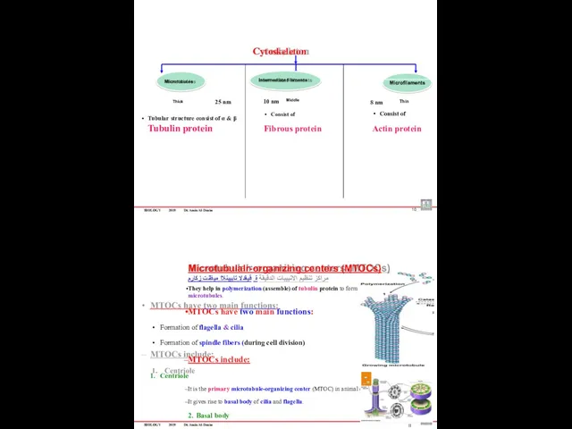
Cytoskeleton
Tubular structure consist of α & β
Tubulin protein
Consist of
Actin
BIOLOGY
2019
Dr. Amin Al-Doaiss
Cytoskeleton
Tubular structure consist of α & β
Tubulin protein
Consist of
Actin
BIOLOGY
2019
Dr. Amin Al-Doaiss
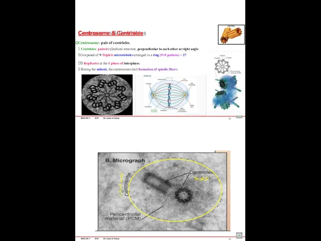
Centrosome & Centrioles
Centrosome: pair of centrioles
⮚ Centrioles: paired cylindrical
BIOLOGY
2019
Dr. Amin Al-Doaiss
Centrosome & Centrioles
Centrosome: pair of centrioles
⮚ Centrioles: paired cylindrical
BIOLOGY
2019
Dr. Amin Al-Doaiss
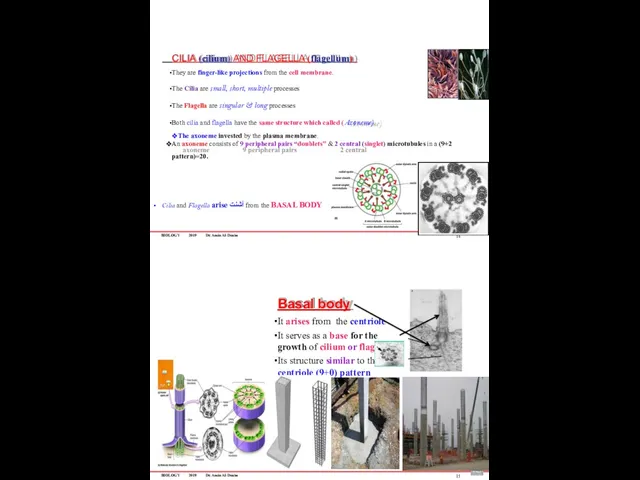
CILIA (cilium) AND FLAGELLA (flagellum)
They are finger-like projections from
BIOLOGY
2019
Dr. Amin Al-Doaiss
CILIA (cilium) AND FLAGELLA (flagellum)
They are finger-like projections from
BIOLOGY
2019
Dr. Amin Al-Doaiss
Dr. Amin Al-Doaiss
1
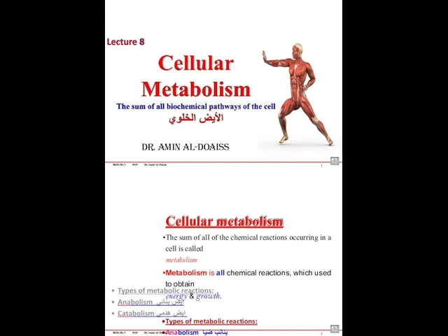
Cellular metabolism
The sum of all of the
BIOLOGY
2019
Dr. Amin Al-Doaiss
Dr. Amin Al-Doaiss
1
Cellular metabolism
The sum of all of the
BIOLOGY
2019
Dr. Amin Al-Doaiss
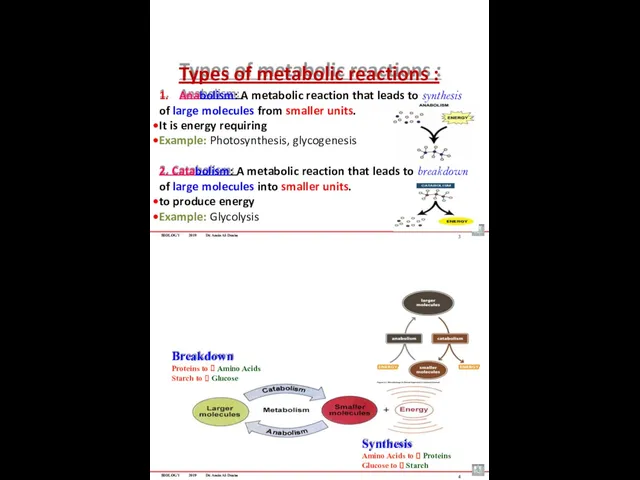
Types of metabolic reactions :
1. Anabolism: A metabolic reaction that
BIOLOGY
2019
Dr. Amin Al-Doaiss
Types of metabolic reactions :
1. Anabolism: A metabolic reaction that
BIOLOGY 2019 Dr. Amin Al-Doaiss
5
Cellular Metabolism
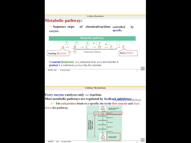
Metabolic pathway:
Sequence steps of chemical reactions
enzyme.
controlled by specific
1
2
3 4 5 6
Starting Reactant
End product
Intermediate reactants
Metabolic pathways:
A → B → C → D → E → F → G
A reactant
BIOLOGY 2019 Dr. Amin Al-Doaiss
5
Cellular Metabolism
Metabolic pathway:
Sequence steps of chemical reactions
enzyme.
controlled by specific
1
2
3 4 5 6
Starting Reactant
End product
Intermediate reactants
Metabolic pathways:
A → B → C → D → E → F → G
A reactant
BIOLOGY 2019 Dr. Amin Al-Doaiss
7
Cellular Metabolism
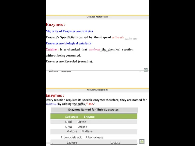
Enzymes :
Majority of Enzymes are proteins
Enzyme’s Specificity is
BIOLOGY 2019 Dr. Amin Al-Doaiss
7
Cellular Metabolism
Enzymes :
Majority of Enzymes are proteins
Enzyme’s Specificity is
BIOLOGY 2019 Dr. Amin Al-Doaiss
9
Cellular Metabolism
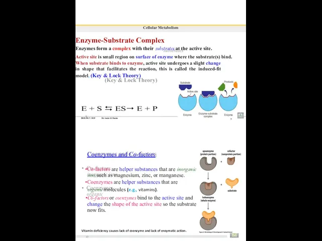
Enzyme-Substrate Complex
Enzymes form a complex with their substrates
BIOLOGY 2019 Dr. Amin Al-Doaiss
9
Cellular Metabolism
Enzyme-Substrate Complex
Enzymes form a complex with their substrates
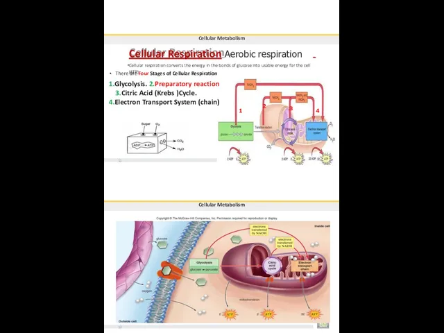
Cellular Respiration Aerobic respiration
Cellular respiration converts the energy in the bonds
Cellular Respiration Aerobic respiration
Cellular respiration converts the energy in the bonds
Cellular Metabolism
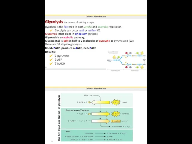
Glycolysis the process of splitting a sugar.
glycolysis is the first
Cellular Metabolism
Glycolysis the process of splitting a sugar.
glycolysis is the first
Cellular Metabolism
Preparatory (transition, link) reaction
Takes place in the matrix of the
Cellular Metabolism
Preparatory (transition, link) reaction
Takes place in the matrix of the
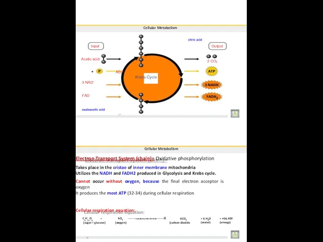
Cellular Metabolism
citric acid
Input
Acetic acid
ADP
Krebs Cycle
3 NAD+
FAD
oxaloacetic acid
17
Output
2 CO
2
C6H12O6
+
6O2 --------cytoplasm/mitochondria--------?
6CO2
+ 6 H2O
(water)
+ ≈36
Cellular Metabolism
citric acid
Input
Acetic acid
ADP
Krebs Cycle
3 NAD+
FAD
oxaloacetic acid
17
Output
2 CO
2
C6H12O6
+
6O2 --------cytoplasm/mitochondria--------?
6CO2
+ 6 H2O
(water)
+ ≈36
 Презентация к уроку биологии в 5 классе по теме: Живое и неживое: каковы особенности биологических систем? к учебнику В.А.Самкова, Д.И.Рокотова Биология 5 класс
 Царства живой природы
 Презентация к занятию по теме Аллергия
 Жасуша теориясы
 Презентация про ЗОЖ для 5классов.
 Russian desman
 Class Basidiomycetes. Class Deuteromyces
 Нейрофизиология соматосенсорной системы
 Зоофагтарды, гербифагтарды және микроорганизмдерді қолдану әдістері. (Лекция 10)
 Птичий калейдоскоп. Внеклассное мероприятие
 Поучительные рассказы о животных от профессора Колобкова
 Большие личности миниатюрных пород
 Значение гомологии и аналогии в живом мире
 What is the engine of our body machine
 Строение и функции кожи человека
 Тема навчального проекту. Біологія-це цікаво
 Тип Кишечнополостные
 Общий путь катаболизма. (Лекция 9)
 Организационно-экономические основы формирования и развития устойчивой системы селекции и семеноводства в регионе
 Құрамында антрацен туындылары бар дәрілік өсімдік шикізаттарын талдау
 Разнообразие растений
 Физиология кровообращения
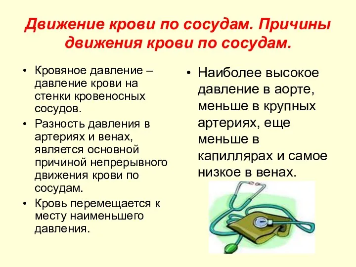 Движение крови по сосудам. Причины движения крови по сосудам
 Испарение воды растением
 Культура горох посевной
 Возникновение и развитие эволюционных представлений
 Методы исследования генетики человека
 Споровые растения