- Nucleic acids

Содержание
- 2. Plan. First isolation of DNA (deoxyribonucleic acid). Pirimidine and purine bases. Minor bases. Structure of nucleosides
- 3. DNA (deoxyribonucleic acid) was firstly isolated by the Swiss physician Friedrich Miescher in 1869. Actually it
- 4. The polymeric structure of DNA may be described in terms of monomeric units of increasing complexity.
- 9. Bases attached to a sugar is called nucleoside. Sugar + phosphate + base = nucleotide. DNA
- 10. deoxyribonucleotide
- 11. deoxyribonucleotide
- 12. deoxyribonucleotide
- 13. deoxyribonucleotide
- 14. Types of bonds in 2'-deoxycytidine-5'-diphosphate
- 16. 3 end
- 17. RNA are easily hydrolyzed under mild alkaline conditions to nucleotides which is cleaved in alkaline medium
- 19. According to the Watson-Crick model of a DNA molecule consists of two polynucleotide chains forming a
- 20. DNA double helix fragment in space-filling and wire-frame format
- 21. Base Pairing 3 Hydrogen bonds . . . . . . . . .
- 22. Base Pairing 2 Hydrogen bonds . . . . . .
- 23. A always pairs with T in DNA. C also pairs with G in DNA. The amount
- 24. Different levels of DNA structure to fit the enormously long DNA duplexes into the nuclei of
- 25. Replication — the process by which identical copies of DNA are made so that information can
- 28. Almost all the cells in our body have DNA with the exception of red blood cells.
- 30. Nicotinamide adenine dinucleotide (NAD) is one of the principal oxidation-reduction reagents in biological systems.
- 31. Bial’s test (pentose detection in products of nucleoprotein hydrolysis) When reacted with concentrated solution of H2SO4
- 32. Bial’s test Also they gave red products of condensation with thymol (2-isopropyl-5-methylphenol).
- 33. A mutation is an error in the base sequence of a gene. The end result can
- 34. Antiviral drugs DNA synthesis terminates whenever AZT is incorporated into the growing DNA strands in the
- 36. Скачать презентацию
 Строение и развитие мужских половых клеток
 Психофизиология эмоций. Возникновение и протекание эмоций. Виды эмоций
 Сон. Правила здорового сна
 Размножение живых организмов. Гаметогенез
 Защита овощных и цветочных культур в защищенном грунте препаратами ООО Сингента
 Биохимия соединительной ткани
 класс пресмыкающиеся
 Строение и работа сердца
 Покрытосеменные, или Цветковые
 Достижения и перспективы развития биотехнологии в Беларуси
 Жынысты және жыныссыз көбею және олардың көбею түрлері
 Сравнительная характеристика однодольных и двудольных растений
 Будова скелета людини
 Водоросли. Царство - растений
 Қалталылар отряды – Marsupiala
 Человеческий мозг
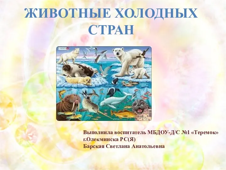 Животные холодных стран
 Дерево секвойя
 Healthy eating
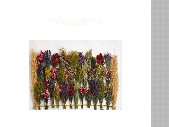 Сухоцветы. Составление зимних букетов
 День кошек
 Chondrichthyes. Хрящевые рыбы
 Відчуття. Види відчуттів
 урок по теме Строение цветка
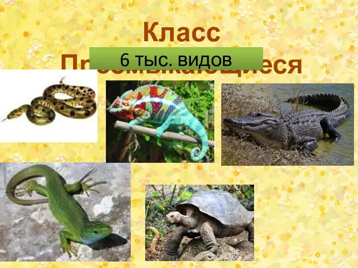 Класс Пресмыкающиеся
 Ядовитые растения Крыма
 Строение тела человека и его функциональные системы
 Растительный мир Дальнего Востока