- Phyllum plathyhelminthes

Содержание
- 2. QUESTIONS -Plathyhelmintes in general -Class Trematoda: main features and life cycles of some parasites -Class Cestodea:
- 3. Plathyhelminthes are known as flatworms, because they are much flattened dorso-ventrally. They are characterized by the
- 4. *Body dorsoventrally flattened, leaf like or tape like and segmented. *First animals to have organ system
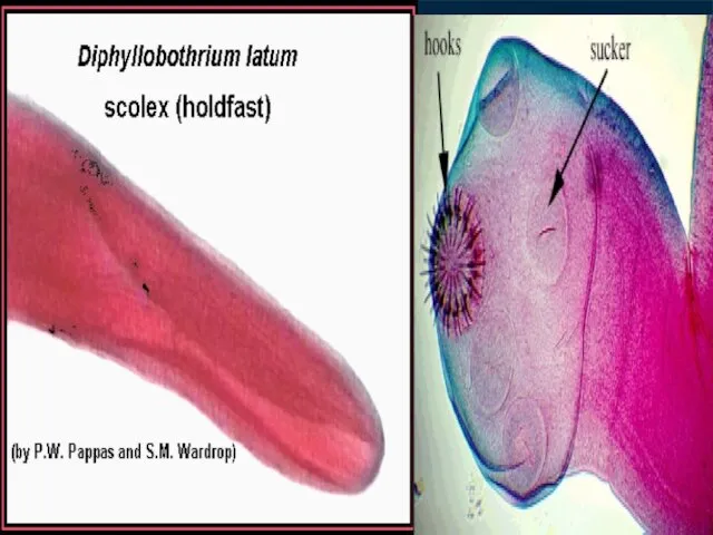
- 5. Classification of Plathyhelminthes Free – living, Planaria flukes: Fasciola, tapeworms: Taenia, Clonorchis, Echinoccocus, Schistosomes Diphillobothrium latum
- 6. Сlass Trematoda. General: Trematodes have flat and oval body, ranging from 1 mm to 3 cm
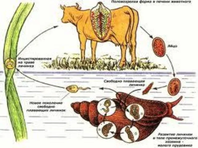
- 7. Fasciola hepatica
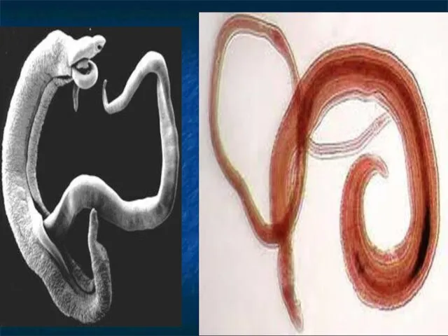
- 9. Schistosomes or Blood Flukes They are much differ from the other Trematodes: they are flukes with
- 11. Life – history of Schistosomes
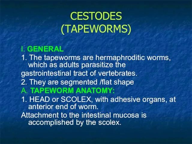
- 12. CESTODES (TAPEWORMS) I. GENERAL 1. The tapeworms are hermaphroditic worms, which as adults parasitize the gastrointestinal
- 13. 2. PROGLOTTIDS -the multiple, hermaphroditic, egg-producing units. These are the flattened segments of the worm body.
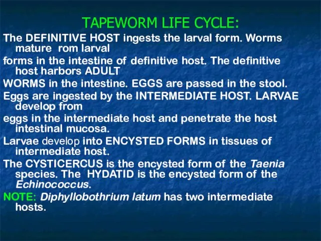
- 15. TAPEWORM LIFE CYCLE: The DEFINITIVE HOST ingests the larval form. Worms mature rom larval forms in
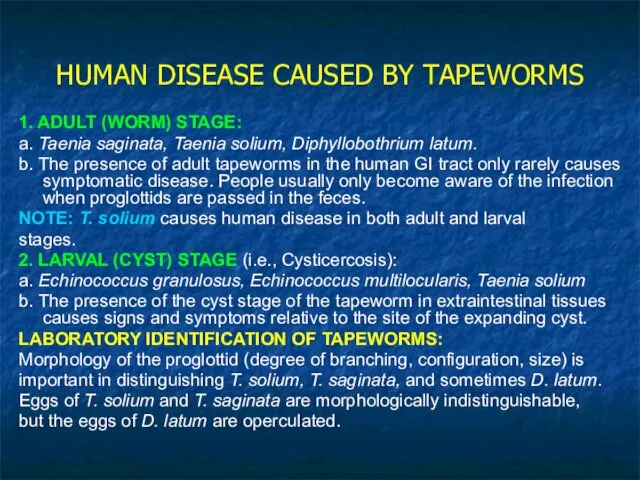
- 16. HUMAN DISEASE CAUSED BY TAPEWORMS 1. ADULT (WORM) STAGE: a. Taenia saginata, Taenia solium, Diphyllobothrium latum.
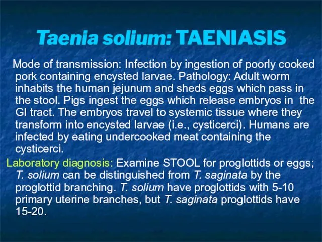
- 17. Taenia solium: TAENIASIS Mode of transmission: Infection by ingestion of poorly cooked pork containing encysted larvae.
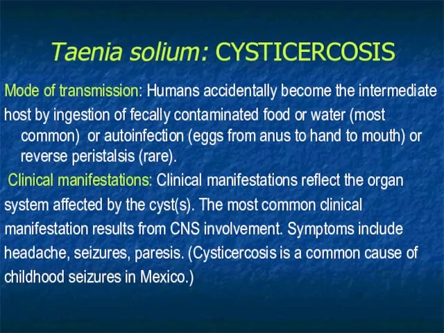
- 18. Taenia solium: CYSTICERCOSIS Mode of transmission: Humans accidentally become the intermediate host by ingestion of fecally
- 19. Pathology: Embryos emerge from the ingested egg and travel through the human body where cysticerci develop.
- 20. Echinococcus granulosus
- 21. Echinococcus granulosus: Epidemiology: people (e.g., herders or hunters) who have close contact with dogs that may
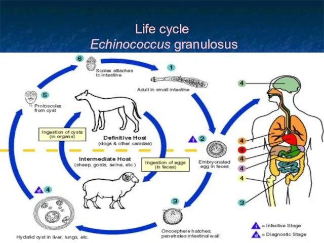
- 22. Life cycle Echinococcus granulosus
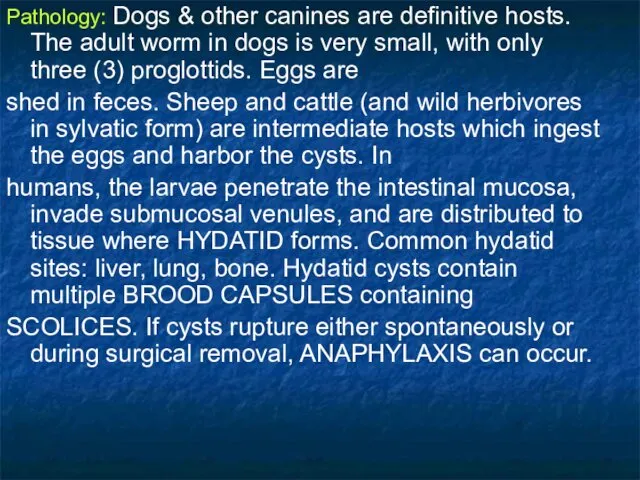
- 23. Pathology: Dogs & other canines are definitive hosts. The adult worm in dogs is very small,
- 25. Скачать презентацию
QUESTIONS
-Plathyhelmintes in general
-Class Trematoda: main features and life cycles of some
QUESTIONS
-Plathyhelmintes in general
-Class Trematoda: main features and life cycles of some
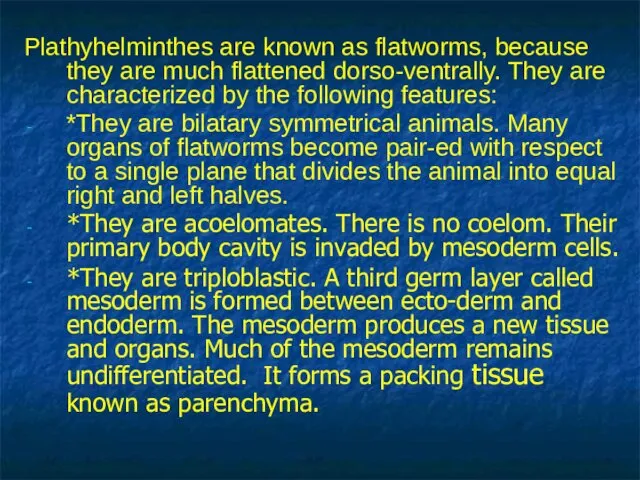
Plathyhelminthes are known as flatworms, because they are much flattened dorso-ventrally.
Plathyhelminthes are known as flatworms, because they are much flattened dorso-ventrally.
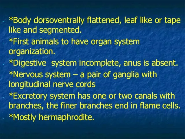
*Body dorsoventrally flattened, leaf like or tape like and segmented.
*First animals
*First animals
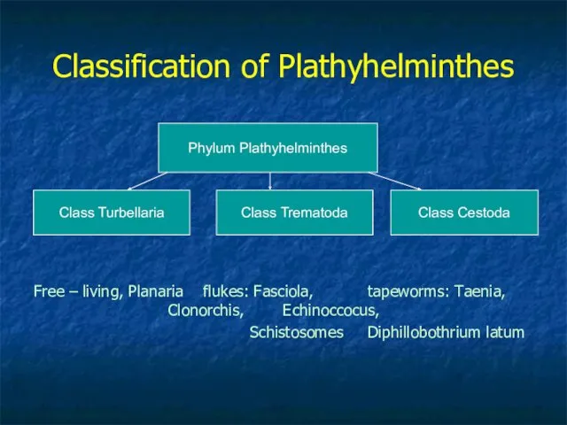
Classification of Plathyhelminthes
Free – living, Planaria flukes: Fasciola, tapeworms: Taenia, Clonorchis,
Classification of Plathyhelminthes
Free – living, Planaria flukes: Fasciola, tapeworms: Taenia, Clonorchis,
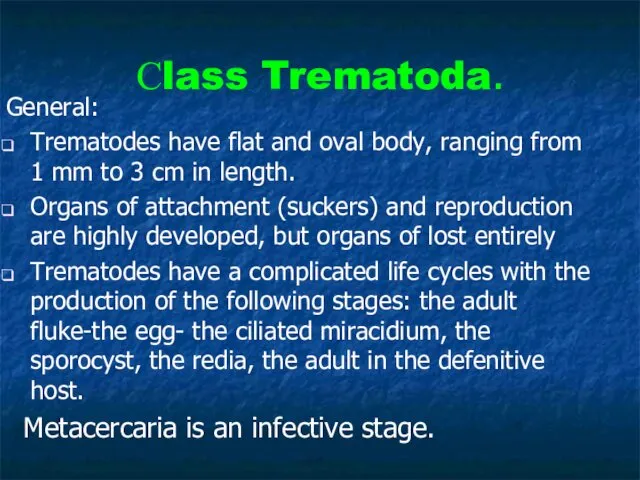
Сlass Trematoda.
General:
Trematodes have flat and oval body, ranging from 1 mm
Сlass Trematoda.
General:
Trematodes have flat and oval body, ranging from 1 mm
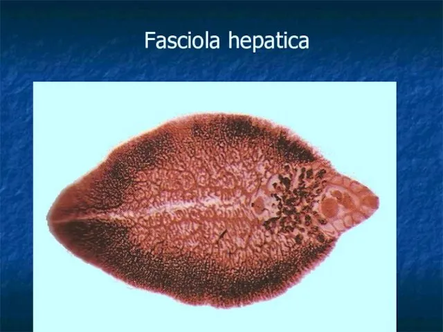
Fasciola hepatica
Fasciola hepatica
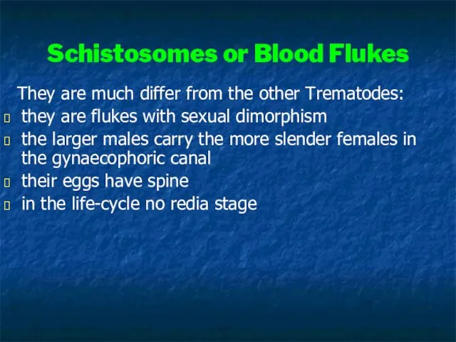
Schistosomes or Blood Flukes
They are much differ from the other
Schistosomes or Blood Flukes
They are much differ from the other
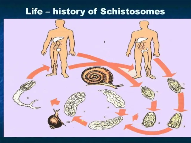
Life – history of Schistosomes
Life – history of Schistosomes
CESTODES
(TAPEWORMS)
I. GENERAL
1. The tapeworms are hermaphroditic worms, which as adults
CESTODES
(TAPEWORMS)
I. GENERAL
1. The tapeworms are hermaphroditic worms, which as adults
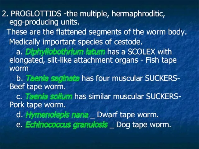
2. PROGLOTTIDS -the multiple, hermaphroditic, egg-producing units.
These are the flattened
2. PROGLOTTIDS -the multiple, hermaphroditic, egg-producing units.
These are the flattened
TAPEWORM LIFE CYCLE:
The DEFINITIVE HOST ingests the larval form. Worms
TAPEWORM LIFE CYCLE:
The DEFINITIVE HOST ingests the larval form. Worms
HUMAN DISEASE CAUSED BY TAPEWORMS
1. ADULT (WORM) STAGE:
a. Taenia saginata, Taenia
HUMAN DISEASE CAUSED BY TAPEWORMS
1. ADULT (WORM) STAGE:
a. Taenia saginata, Taenia
Taenia solium: TAENIASIS
Mode of transmission: Infection by ingestion of
Taenia solium: TAENIASIS
Mode of transmission: Infection by ingestion of
Taenia solium: CYSTICERCOSIS
Mode of transmission: Humans accidentally become the intermediate
host
Taenia solium: CYSTICERCOSIS
Mode of transmission: Humans accidentally become the intermediate
host
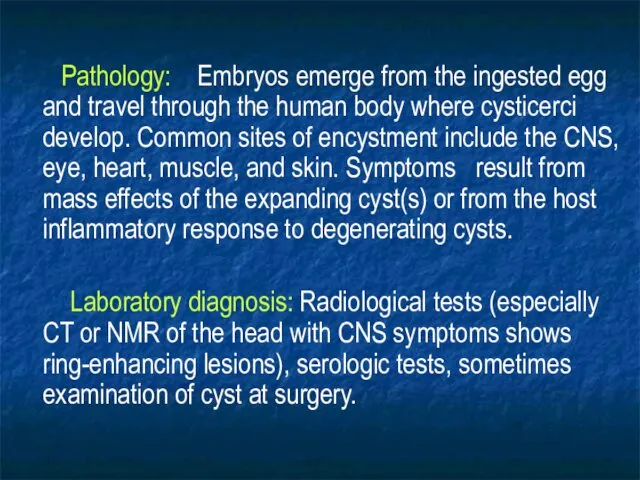
Pathology: Embryos emerge from the ingested egg and travel
Pathology: Embryos emerge from the ingested egg and travel
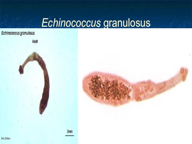
Echinococcus granulosus
Echinococcus granulosus
Echinococcus granulosus:
Epidemiology: people (e.g., herders or hunters) who have close
Echinococcus granulosus:
Epidemiology: people (e.g., herders or hunters) who have close
Life cycle
Echinococcus granulosus
Life cycle
Echinococcus granulosus
Pathology: Dogs & other canines are definitive hosts. The adult worm
Pathology: Dogs & other canines are definitive hosts. The adult worm
 Бактериофаги. Строение бактериофага
 Царства живой природы
 Цветок – самый красивый орган растения
 Бактерии в жизни человека. Польза и вред
 Ядовитые растения России
 Филогенетическое разнообразие грибоподобных организмов. Разнообразие. Систематика. Жизненные циклы
 Значение опорно-двигательной системы. 8 класс
 Животные (звери)
 Центры происхождения культурных растений
 Интересные зеленые. Игра для учащихся 6-х классов
 Пути и направления эволюции
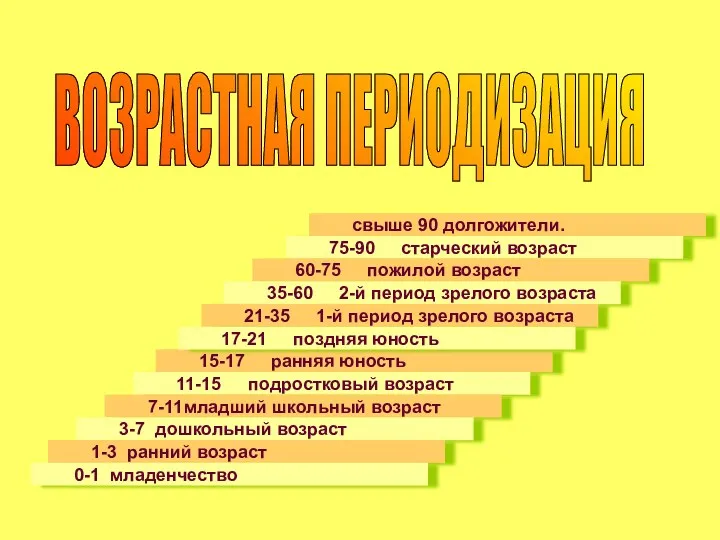 Возрастная периодизация
 Бөлме гүлдері - адамның досы
 Онтогенез. Лекция 14
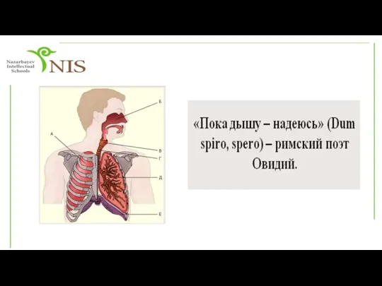 Жәндіктер, балықтар мен адамның тыныс алу мүшелері құрылысының ерекшеліктерін зерттеу және салыстыру
 Нервная ткань
 Культивирование и рост микроорганизмов
 Путешествие по страницам Красной книги
 Размножение живых организмов
 Комнатное растение альсобия
 Вода. Роль воды в жизни человека
 Генетика. Становление и роль в современном естествознании
 Эндокринді бездер, қызметі. Гормондар. Гуморальдық реттелу
 Витамины для зрения
 Полезный мед - натуральный мед
 Эмбриональное развитие организмов
 Развитие индюка от яйца до взрослой особи
 Универсальность строительных и функциональных блоков на молекулярном уровне организации биологических систем