- Physiology of eye. Physiology of vision

Содержание
- 2. Diapasone of the visible light
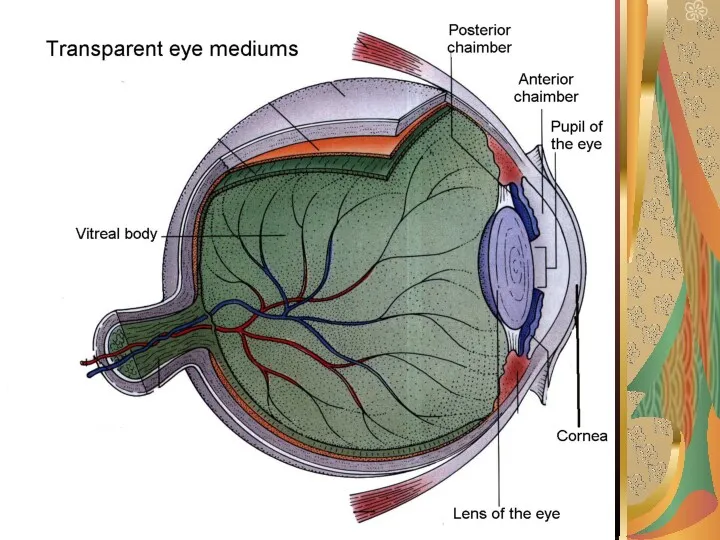
- 3. Optic system of eyeball Cornea allows light to enter the eyeball. Aqueous humor fills anterior and
- 5. Aqueous humor circulation Ciliary processes in posterior chamber secrete aqueous fluid. It flows between the ligament
- 6. Physical refraction and reduced eye Refraction is bending of light rays at surface between two media.
- 8. Clinical refraction All variants of refraction, which are observed in patients we call clinical refraction. These
- 9. Presbyopia occurs due to a lessening of flexibility of the crystalline lens, as well as to
- 10. Aberrations and astigmatism In spherical aberration light rays pass through peripheral parts of the eye lens
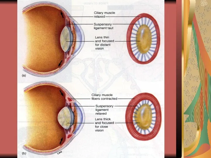
- 11. Accommodation and its regulation Accommodation is adjustment of eye lens for various distances. Relaxation of ciliary
- 13. Defensive mechanisms in eye Fibrous tunic of eyeball is composed by avascular connective tissue, which gives
- 14. Pupillary reflexes When light pass into eye, pupil contracts. In darkness pupil dilates. This is pupillary
- 15. Age peculiarities in eye structure In old age lens of eye loose elasticity. So this condition,
- 16. Development of refraction In newborn eye is hypermetropic. Eyeball grows with age, and normally gets spherical
- 17. Composition of retina Layers of retina from outside to inside: pigmented layer; layer of rods and
- 18. Physiological peculiarities of pigmented layer and photoreceptors. Light falls on retina on inner side i.e. on
- 19. Photochemical reactions in retina Outer segment of photoreceptors contain photochemicals. Inner segment contains nucleus, synaptic body
- 20. Central division of visual analyzer Impulses from retina pass to optic nerve – optic chiasm (fibers
- 21. Other connections of optic tract In addition to lateral genicular body, fibers from optic tract also
- 22. Light and dark adaptation . If a person remains in bright light for a long time,
- 23. Theories of color perception According to Jung-Helmgolc theory there are three types of cones for three
- 24. Disorders of color perception . There are three fundamental colors: red, which is marked “protos”; green
- 25. Visual acuity Ability of human eye to discriminate between point sources of light is called visual
- 26. Field of vision Field of vision is area that is seeing by an eye at a
- 28. Скачать презентацию
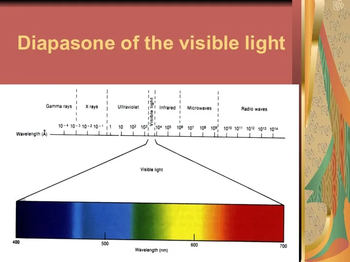
Diapasone of the visible light
Diapasone of the visible light
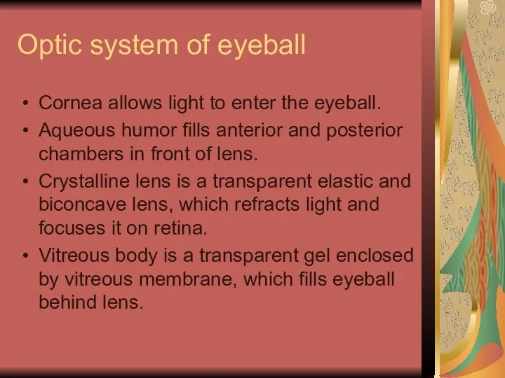
Optic system of eyeball
Cornea allows light to enter the eyeball.
Optic system of eyeball
Cornea allows light to enter the eyeball.
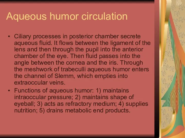
Aqueous humor circulation
Ciliary processes in posterior chamber secrete aqueous fluid.
Aqueous humor circulation
Ciliary processes in posterior chamber secrete aqueous fluid.
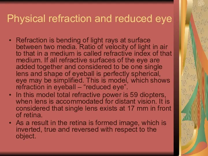
Physical refraction and reduced eye
Refraction is bending of light rays
Physical refraction and reduced eye
Refraction is bending of light rays
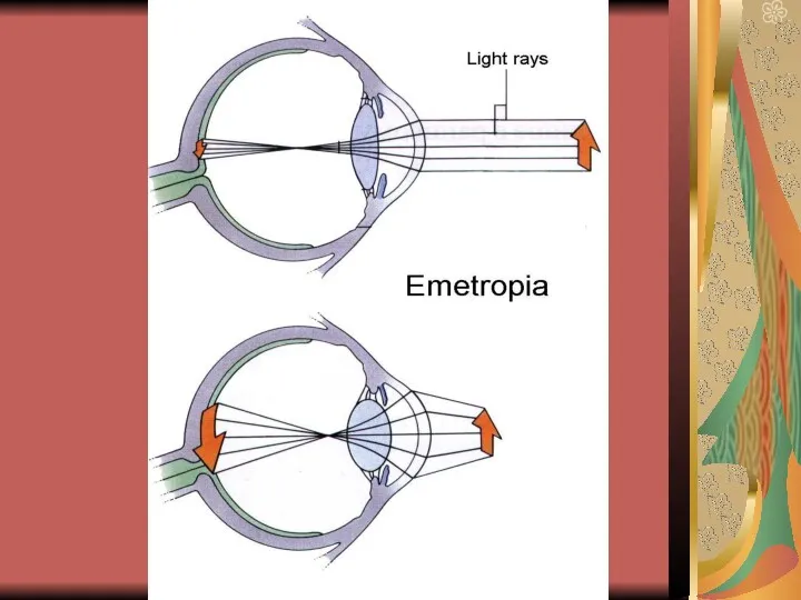
Clinical refraction
All variants of refraction, which are observed in patients
Clinical refraction
All variants of refraction, which are observed in patients
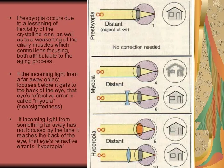
Presbyopia occurs due to a lessening of flexibility of the crystalline
Aberrations and astigmatism
In spherical aberration light rays pass through peripheral
Aberrations and astigmatism
In spherical aberration light rays pass through peripheral
Accommodation and its regulation
Accommodation is adjustment of eye lens for
Accommodation and its regulation
Accommodation is adjustment of eye lens for
Defensive mechanisms in eye
Fibrous tunic of eyeball is composed by
Defensive mechanisms in eye
Fibrous tunic of eyeball is composed by
Pupillary reflexes
When light pass into eye, pupil contracts. In darkness
Pupillary reflexes
When light pass into eye, pupil contracts. In darkness
Age peculiarities in eye structure
In old age lens of eye
Age peculiarities in eye structure
In old age lens of eye
Development of refraction
In newborn eye is hypermetropic. Eyeball grows with
Development of refraction
In newborn eye is hypermetropic. Eyeball grows with
Composition of retina
Layers of retina from outside to inside:
pigmented
Composition of retina
Layers of retina from outside to inside:
pigmented
Physiological peculiarities of pigmented layer and photoreceptors.
Light falls on retina
Physiological peculiarities of pigmented layer and photoreceptors.
Light falls on retina
Photochemical reactions in retina
Outer segment of photoreceptors contain photochemicals. Inner
Photochemical reactions in retina
Outer segment of photoreceptors contain photochemicals. Inner
Central division of visual analyzer
Impulses from retina pass to optic
Central division of visual analyzer
Impulses from retina pass to optic
Other connections of optic tract
In addition to lateral genicular
Other connections of optic tract
In addition to lateral genicular
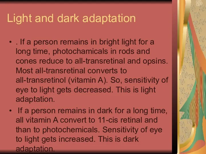
Light and dark adaptation
. If a person remains in bright
Light and dark adaptation
. If a person remains in bright
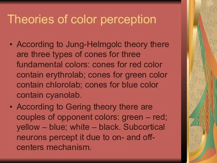
Theories of color perception
According to Jung-Helmgolc theory there are three
Theories of color perception
According to Jung-Helmgolc theory there are three
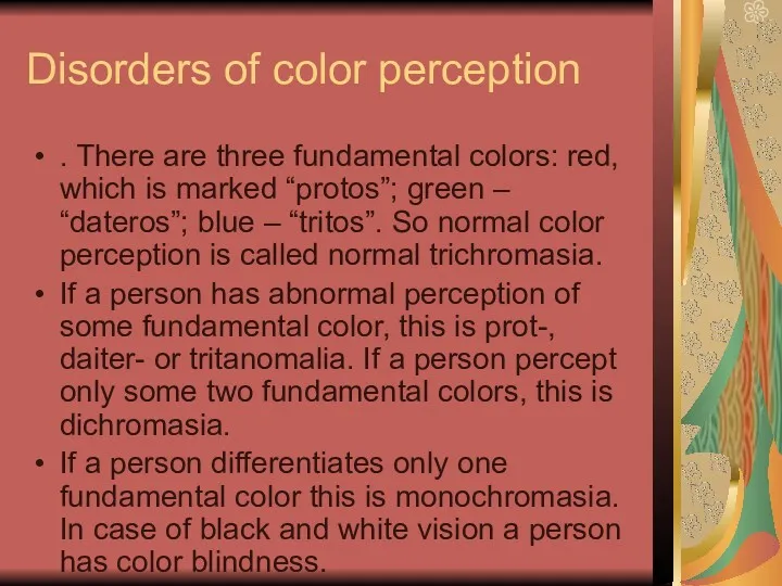
Disorders of color perception
. There are three fundamental colors: red,
Disorders of color perception
. There are three fundamental colors: red,
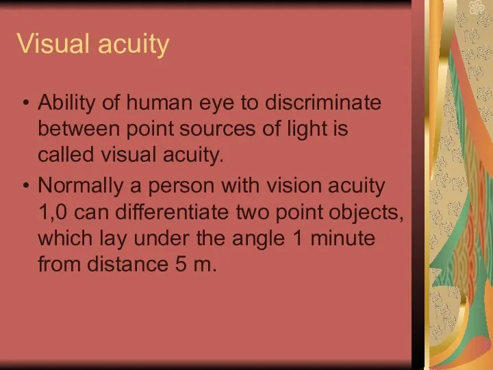
Visual acuity
Ability of human eye to discriminate between point sources
Visual acuity
Ability of human eye to discriminate between point sources
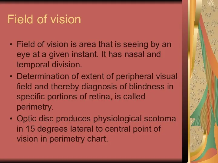
Field of vision
Field of vision is area that is seeing
Field of vision
Field of vision is area that is seeing
 Наука генетика и её связь с другими науками
 Анатомо-физиологические особенности человека в подростковом возрасте
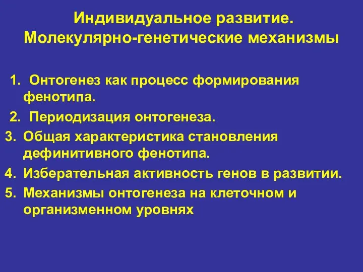 Индивидуальное развитие. Молекулярно-генетические механизмы
 Жизненные формы организмов. Лекция 8
 Тест Вода. Свойства воды, биология 6 класс
 Forest Animals
 Компания Хелатные БиоТехнологии. Органический биостимулятор для открытого и защищённого грунтов
 Секреты перелётных птиц
 Екологічні групи рослин по відношенню до вологи
 Слиновиділення і ферменти слини
 Газообмен в легких и тканях. Дыхательные движения и их регуляция
 Жизненный цикл клетки
 В гости к весне. (2 класс)
 Творчість Тараса Григоровича Шевченка
 Микробиология, как наука. Основные методы микробиологических исследований
 20231227_pishchevye_produkty
 Домашний питомец, кот Мурзик
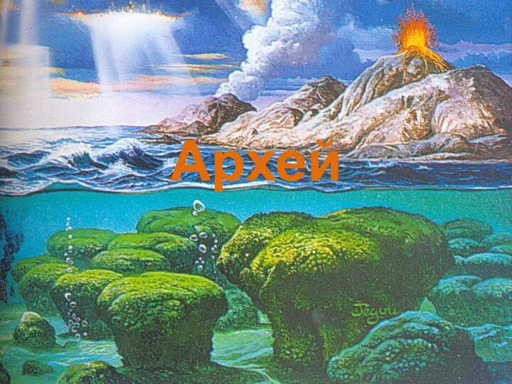 Архейская эра. Развитие органического мира
 Волокнистые соединительные ткани. Специализированные соединительные ткани
 Жүйке жүйесі. Жүйке жүйесінің мүшелерінің ПРе- және ПОстнатальды дамуы. Адам онтогенезінде функционалды жүйелер туралы түсінік
 Потребности клеток в питательных веществах. Принципы составления питательных сред. Сырье для биотехнологической промышленности
 Подводный мир!
 Человек, общество, природа
 Красная книга России
 Микробиологическое производство лекарственных средств, регуляторов роста и средств защиты растений
 Факторы, негативно влияющие на дыхательную систему
 Домашні рослини
 Сравнительное наблюдение за прорастанием семян, ростом растений