- Protein structure at action: bind transform release

Содержание
- 2. BIND: repressors α- turn - α
- 3. DNA & RNA BINDING Zn- fingers Leu-zipper
- 4. -BINDING-INDUCED DEFORMATION MAKES REPRESSOR ACTIVE, and IT BINDS TO DNA BIND ? ? RELEASE: REPRESSOR
- 5. Immunoglobulin
- 6. Standard positions of active sites in protein folds
- 7. There are some with catalytic (Ser-protease) site
- 8. Preferential binding of TS: RIGID enzyme Catalysis: stabilization of the transition state (TS) Theory: Pauling &
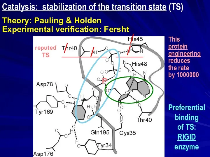
- 9. Catalysis: stabilization of the transition state (TS) Theory: Pauling & Holden Experimental verification: Fersht ______ __________
- 10. Catalysis: stabilization of the transition state (TS) Theory: Pauling & Holden Experimental verification: Fersht ______ __________
- 11. Catalytic antibodies ABZYM = AntyBody enZYM Antibodies are selected to TS-like molecule Transition state (TS ‡)
- 12. BIND ? TRANSFORM ? RELEASE: ENZYME Note: small active site chymotrypsin
- 13. Sometimes: Different folds with the same active site: the same biochemical function
- 14. POST-TRANSLATIONAL MODIFICATION Sometimes, only the CHAIN CUT-INDUCED DEFORMATION MAKES THE ENZYME ACTIVE READY Chymotripsinogen active cat.
- 15. Chymotrypsin catalyses hydrolysis of a peptide Spontaneous hydrolysis: very slow
- 16. SER-protease: catalysis
- 17. CHYMOTRYPSIN ACTIVE SITE with INHIBITOR
- 18. Preferential binding of TS: RIGID enzyme F = k1x1 = - k2x2 Ei = (ki /2)(xi)2
- 19. PROTEIN STRUCTURE AT ACTION: BIND ? TRANSFORM ? RELEASE RIGID CATALITIC SITE INDEPENDENT ON OVERALL CHAIN
- 20. Induced fit model for enzyme catalysis. Daniel Edward Koshland, Jr. (1920 – 2007) Hermann Emil Louis
- 21. MOTIONS
- 22. Double sieve: movement of substrate from one active site to another ⇑ tRNAIle Fersht A.R., Dingwall
- 23. Movement in two-domain enzyme: One conformation for binding (and release), another for catalysis Induced fit
- 24. Two-domain dehydrogenases: Universal NAD-binding domain; Individual substrate-binding domain
- 25. Movement in quaternary structure: Hemoglobin vs. myoglobin non-covalent bonding of O2 move of O2 to and
- 26. Kinesin : Linear cyclic motor the simplest one-direction walking machine with cyclic ligand-induced conformational changes and
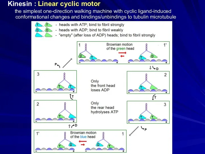
- 27. Kinesin : Linear cyclic motor the simplest one-direction walking machine with cyclic ligand-induced conformational changes and
- 28. Sir Andrew Fielding Huxley (1917 – 2012) Nobel Prize 1963 Myosin "cross-bridges"
- 29. Механохимический цикл Миозин Актин АТФ → АДФ + Ф 15 ккал/моль в клеточных условиях
- 30. Mechanochemical cycle Myosin Actin
- 31. structure from the X-ray data: Junge, Sielaff, Engelbrecht, Nature, 459, 364 (2009) Rotary motor F0F1-ATP synthase
- 32. Engelbrecht & Junge, FEBS Lett. 414, 485 (1997) Elston, Wang, Oster, Nature, 391, 510 (1998) F0-machine:
- 33. Rotary motor F0F1-ATP synthase ⎯ working cycle of the H+-turbine
- 34. H3O+ binding in Bacillus pseudofirmus Ion binding to the rotor ring of F0F1-ATP synthase H+ binding
- 35. SUMMARY of the course
- 36. PROTEIN PHYSICS Interactions Structures Selection States & transitions
- 37. Intermediates & nuclei Structure prediction & bioinformatics Protein engineering & design Functioning
- 38. Благодарю за внимание … товарищи офизевшие биологи!
- 40. Скачать презентацию
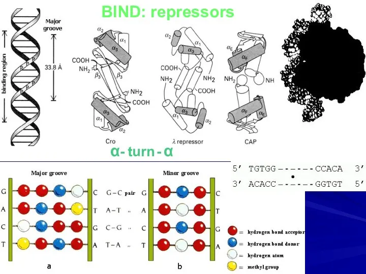
BIND: repressors
α- turn - α
BIND: repressors
α- turn - α
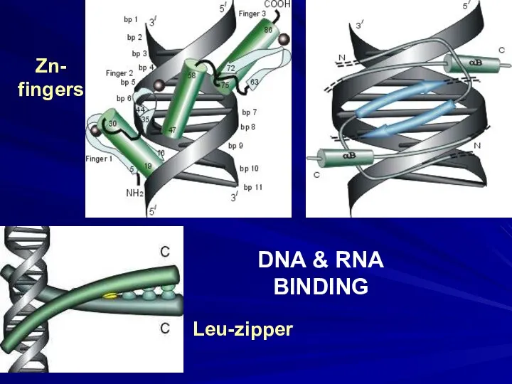
DNA & RNA
BINDING
Zn-
fingers
Leu-zipper
DNA & RNA
BINDING
Zn-
fingers
Leu-zipper
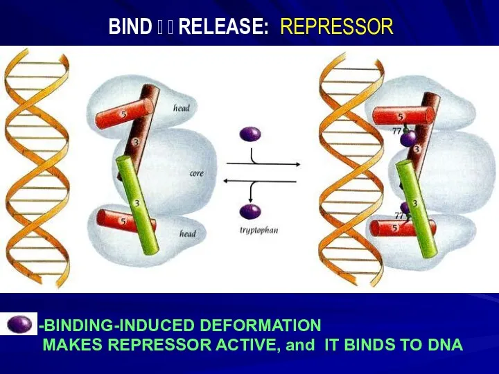
-BINDING-INDUCED DEFORMATION
MAKES REPRESSOR ACTIVE, and IT BINDS TO DNA
BIND
-BINDING-INDUCED DEFORMATION
MAKES REPRESSOR ACTIVE, and IT BINDS TO DNA
BIND
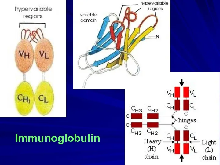
Immunoglobulin
Immunoglobulin
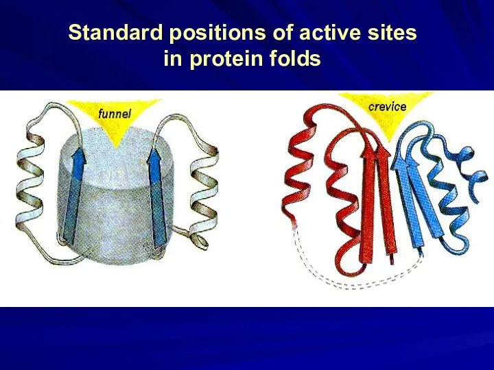
Standard positions of active sites in protein folds
Standard positions of active sites in protein folds

There are some
with catalytic
(Ser-protease) site
There are some
with catalytic
(Ser-protease) site

Preferential binding of TS: RIGID enzyme
Catalysis: stabilization of the transition state
Preferential binding of TS: RIGID enzyme
Catalysis: stabilization of the transition state
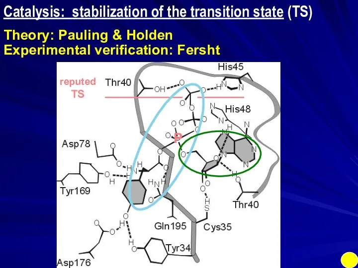
Catalysis: stabilization of the transition state (TS)
Theory: Pauling & Holden
Experimental verification:
Catalysis: stabilization of the transition state (TS)
Theory: Pauling & Holden
Experimental verification:
Catalysis: stabilization of the transition state (TS)
Theory: Pauling & Holden
Experimental verification:
Catalysis: stabilization of the transition state (TS)
Theory: Pauling & Holden
Experimental verification:
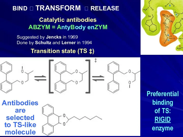
Catalytic antibodies
ABZYM = AntyBody enZYM
Antibodies
are
selected
to TS-like
molecule
Transition state (TS
Catalytic antibodies
ABZYM = AntyBody enZYM
Antibodies
are
selected
to TS-like
molecule
Transition state (TS

BIND ? TRANSFORM ? RELEASE: ENZYME
Note:
small
active
site
chymotrypsin
BIND ? TRANSFORM ? RELEASE: ENZYME
Note:
small
active
site
chymotrypsin
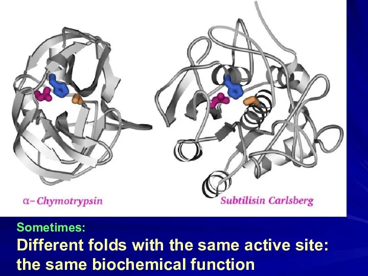
Sometimes:
Different folds with the same active site:
the same biochemical function
Sometimes:
Different folds with the same active site:
the same biochemical function
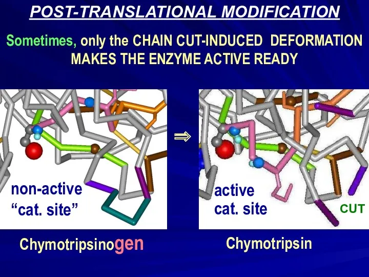
POST-TRANSLATIONAL MODIFICATION
Sometimes, only the CHAIN CUT-INDUCED DEFORMATION
MAKES THE ENZYME ACTIVE
POST-TRANSLATIONAL MODIFICATION
Sometimes, only the CHAIN CUT-INDUCED DEFORMATION
MAKES THE ENZYME ACTIVE
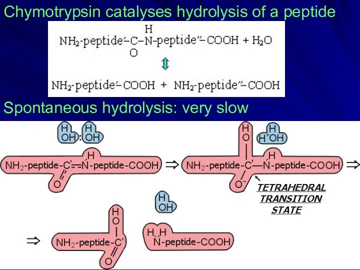
Chymotrypsin catalyses hydrolysis of a peptide
Spontaneous hydrolysis: very slow
Chymotrypsin catalyses hydrolysis of a peptide
Spontaneous hydrolysis: very slow

SER-protease:
catalysis
SER-protease:
catalysis
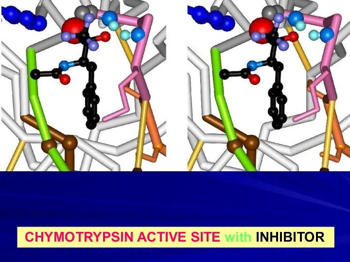
CHYMOTRYPSIN ACTIVE SITE with INHIBITOR
CHYMOTRYPSIN ACTIVE SITE with INHIBITOR
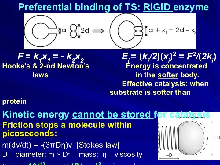
Preferential binding of TS: RIGID enzyme
F = k1x1 = -
Preferential binding of TS: RIGID enzyme
F = k1x1 = -
PROTEIN STRUCTURE AT ACTION:
BIND ? TRANSFORM ? RELEASE
RIGID CATALITIC SITE
INDEPENDENT
PROTEIN STRUCTURE AT ACTION:
BIND ? TRANSFORM ? RELEASE
RIGID CATALITIC SITE
INDEPENDENT
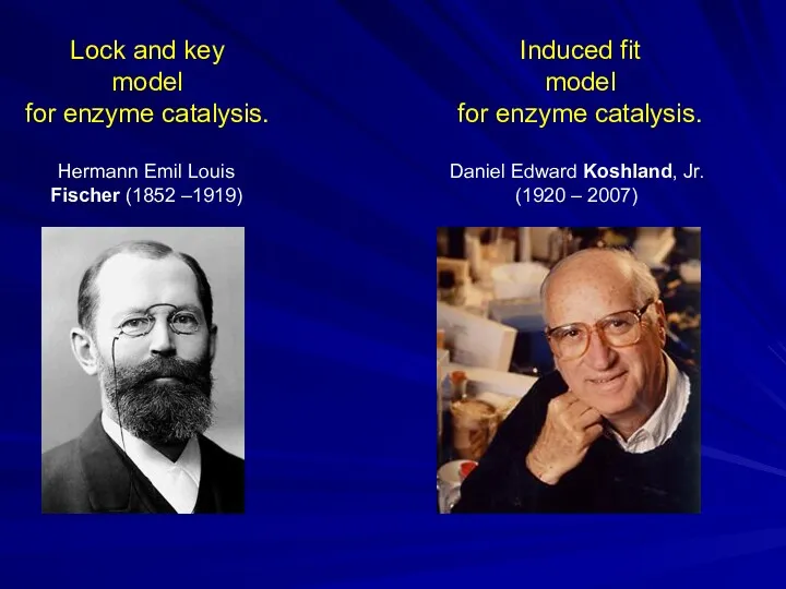
Induced fit
model
for enzyme catalysis.
Daniel Edward Koshland, Jr.
(1920 – 2007)
Hermann Emil Louis
Induced fit
model
for enzyme catalysis.
Daniel Edward Koshland, Jr.
(1920 – 2007)
Hermann Emil Louis

MOTIONS
MOTIONS

Double sieve:
movement of substrate
from one active site to another
⇑
tRNAIle
Fersht A.R., Dingwall C.
Double sieve:
movement of substrate
from one active site to another
⇑
tRNAIle
Fersht A.R., Dingwall C.
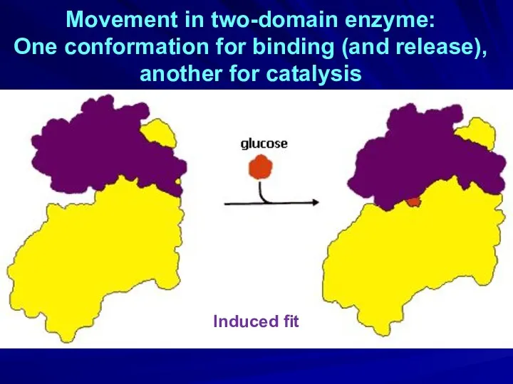
Movement in two-domain enzyme:
One conformation for binding (and release),
another for
Movement in two-domain enzyme:
One conformation for binding (and release),
another for
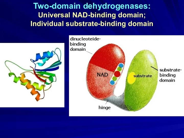
Two-domain dehydrogenases:
Universal NAD-binding domain;
Individual substrate-binding domain
Two-domain dehydrogenases:
Universal NAD-binding domain;
Individual substrate-binding domain
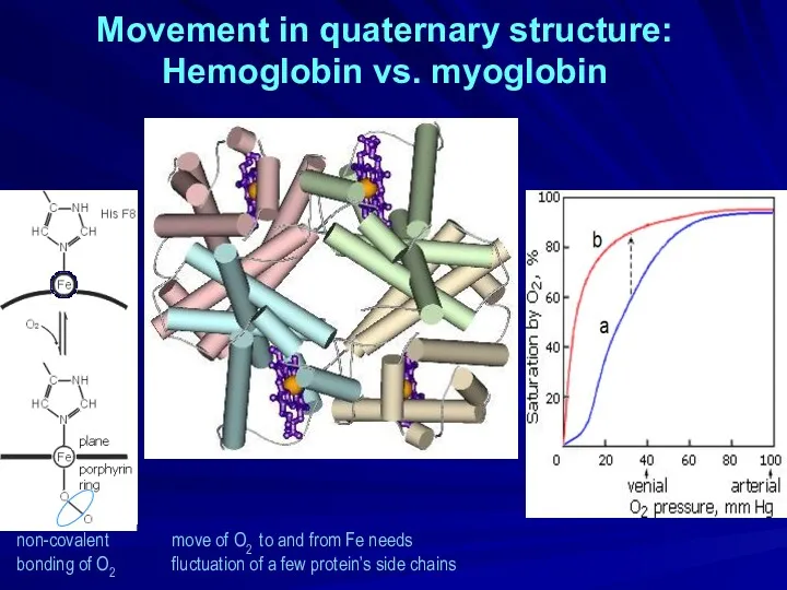
Movement in quaternary structure:
Hemoglobin vs. myoglobin
non-covalent
bonding of O2
move of
Movement in quaternary structure:
Hemoglobin vs. myoglobin
non-covalent
bonding of O2
move of
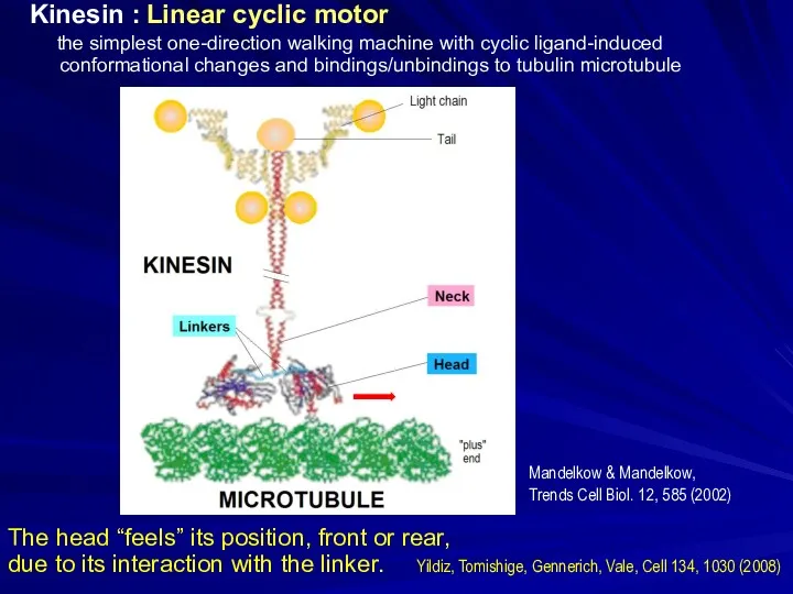
Kinesin : Linear cyclic motor
the simplest one-direction walking machine
Kinesin : Linear cyclic motor
the simplest one-direction walking machine
Kinesin : Linear cyclic motor
the simplest one-direction walking machine
Kinesin : Linear cyclic motor
the simplest one-direction walking machine

Sir Andrew Fielding Huxley
(1917 – 2012)
Nobel Prize 1963
Myosin "cross-bridges"
Sir Andrew Fielding Huxley
(1917 – 2012)
Nobel Prize 1963
Myosin "cross-bridges"

Механохимический цикл
Миозин Актин
АТФ → АДФ + Ф
15 ккал/моль
в клеточных условиях
Механохимический цикл
Миозин Актин
АТФ → АДФ + Ф
15 ккал/моль
в клеточных условиях
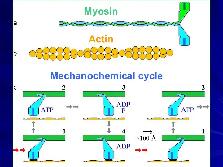
Mechanochemical cycle
Myosin
Actin
Mechanochemical cycle
Myosin
Actin

structure from the X-ray data: Junge, Sielaff, Engelbrecht, Nature, 459, 364
structure from the X-ray data: Junge, Sielaff, Engelbrecht, Nature, 459, 364

Engelbrecht & Junge, FEBS Lett. 414, 485 (1997)
Elston, Wang, Oster, Nature,
Engelbrecht & Junge, FEBS Lett. 414, 485 (1997)
Elston, Wang, Oster, Nature,
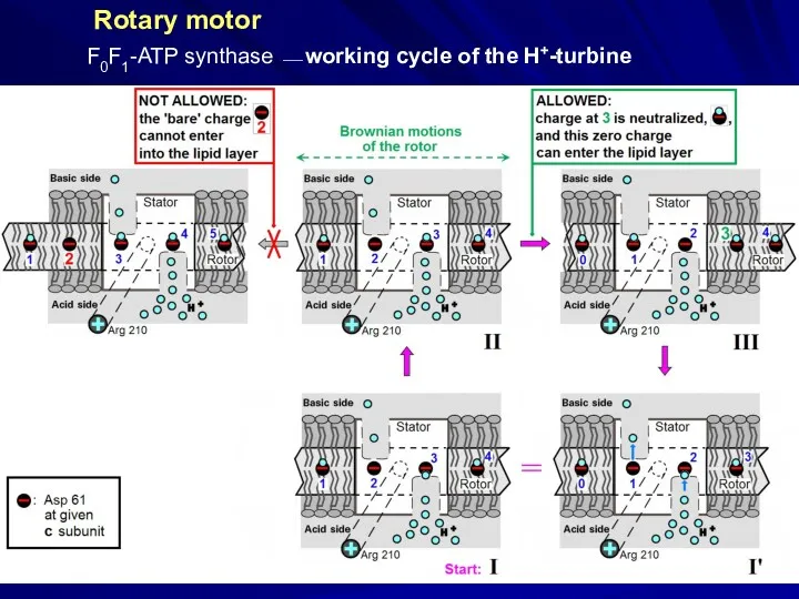
Rotary motor
F0F1-ATP synthase ⎯ working cycle of the H+-turbine
Rotary motor
F0F1-ATP synthase ⎯ working cycle of the H+-turbine

H3O+ binding in Bacillus pseudofirmus
Ion binding
to the rotor
H3O+ binding in Bacillus pseudofirmus
Ion binding
to the rotor
SUMMARY
of the course
SUMMARY
of the course
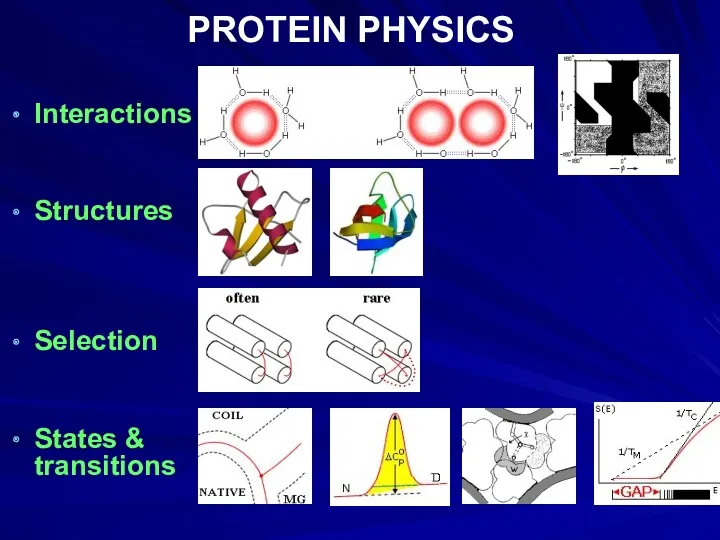
PROTEIN PHYSICS
Interactions
Structures
Selection
States & transitions
PROTEIN PHYSICS
Interactions
Structures
Selection
States & transitions

Intermediates & nuclei
Structure prediction & bioinformatics
Protein engineering & design
Functioning
Structure prediction & bioinformatics
Protein engineering & design
Functioning
Благодарю за внимание
…
товарищи офизевшие биологи!
Благодарю за внимание
…
товарищи офизевшие биологи!
 f611e845-2141-4c61-a9b2-34e2049b0658
 Морские свинки
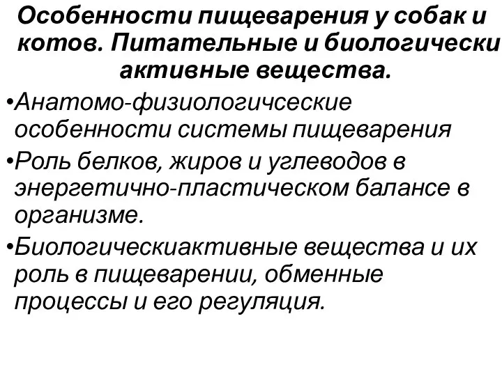 Особенности пищеварения у собак и котов. Питательные и биологически активные вещества
 Лекция 1. Растениеводство
 Этимология названий лекарственных растений. Мифы и легенды
 Почки растений
 Турбота про потомство
 Путешествие в страну Легумия. Систематика покрытосеменных. 6 класс
 Нейропрорывы-2017/2018
 Презентация по биологии для учащихся 7 класса по теме Класс Пресмыкающиеся
 Вітаміни. Їх роль у життєдіяльності людини
 Размножение - свойство живых организмов
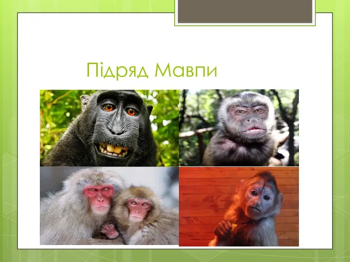 Підряд Мавпи
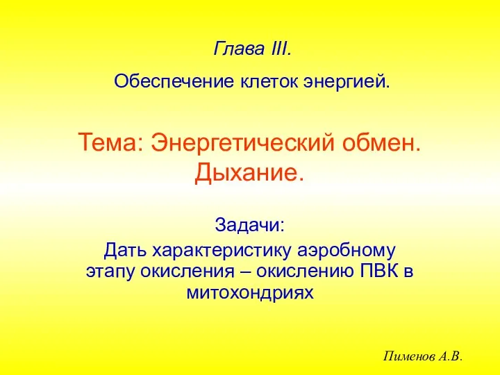 Энергетический обмен. Дыхание
 Цікаві факти про риб
 Талдағыштар физиологиясы. Талдағыштардың жалпы қасиеттері. Көру талдағыштары
 Структура и функции клетки
 Размножение и развитие человека
 Покрытосеменные, или Цветковые
 Самые страшные животные мира
 Рост и развитие растений
 Освоение суши растениями
 Хромосомная теория наследственностию. Генетика пола
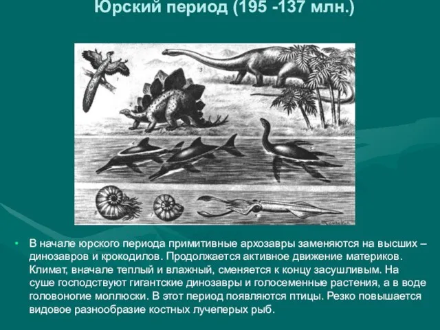 Юрский период (195 -137 млн.)
 Презентация Великаны и карлики в животном мире
 Насекомые. Отряд равнокрылые
 Биотические факторы
 Становление наук о человеке. (8 класс)