- The First Three Weeks of Human Embryogenesis

Содержание
- 2. Week 1 1. Fertilization – is the fusion of the sperm and ovum (male and female
- 3. Week 1 Zygote – 1 cell embryo – starts to divide: 2. Cleavage – is the
- 4. zygote Fertilization zygote Fertilization Cleavage 2 cells stage 3-5 cells stage Morula Blastocyst Implantation Day 6
- 5. At the end of cleavage blastula is formed. Human blastula is called blastocyst (has cavity -cyst)
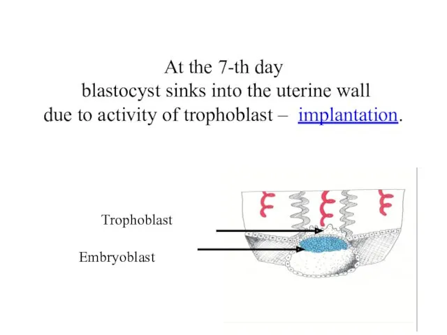
- 6. Embryoblast At the 7-th day blastocyst sinks into the uterine wall due to activity of trophoblast
- 7. Week 2: Beginning of 3. Gastrulation – formation of 3 germ layers Early Gastrulation take place
- 8. Result of early Delamination Embryonic disc:
- 9. Late gastrulation – formation of mesoderm – 3-d germ layer – take place by cell migration:
- 10. 1-st appear extraembryonic mesoderm: it surrounds upper and lower sacs, and underly trophoblast
- 11. Trophoblast Extraembryonic Mesoderm * Ectoderm Endoderm Extraembryonic Mesoderm *
- 12. As a result appear so-called extraembryonic organs - amnion, yolk sac and chorion
- 13. Chorion Amnion Yolk Sac
- 14. Migration of cells within the embryonic disc leads to formation of the embryonic mesoderm and axial
- 15. Migration of cells within embryonic disc leads to formation of temporal cellular assemblage between ectoderm and
- 16. Transverse section Ectoderm Endoderm Amniotic Cavity Primitive streak Yolk Sac
- 17. In front of primitive streak appears primitive knot. Cells of Primitive Streak begin to move laterally.
- 18. ectoderm endoderm mesoderm Notochord appears by the primitive knot invagination. Mesoderm appears by migration of cells
- 19. 3-2.(next step): Development of the Neural Tube - future nerve system - by the invagination of
- 20. Neural plate in Surface Ectoderm forms Neural groove
- 21. Then - Neural Tube
- 23. Development of the Neural Tube Surface Ectoderm Neural Crest Neural Tube
- 24. Neural tube formation
- 25. Neural tube formation
- 26. Gastrulation is finished with the formation of axial organs – neural tube, notochord, somites (mesoderm) Somite
- 27. 4. Formation of the embryo body (20-th day) by: - body flexion, - head and tail
- 28. Body flexion
- 29. Differentiation of GERM LAYERS: 1. Differentiation of Ectoderm A. Surface Ectoderm B. Neural Tube 2. Differentiation
- 30. Surface Ectoderm differentiates to epithelium of skin, and its derivatives, oral cavity epithelium, rectal epithelium, outer
- 31. Neural tube (neuroectoderm) --- brain, spinal cord, and the retina Neural crests --- Peripheral Nervous system,
- 32. Endoderm differentiates to epithelium of stomach, intestine, liver, pancreas, respiratory system
- 33. Mesoderm Notochord Endoderm Ectoderm Yolk Sac Amniotic Cavity Somite Intermediate mesoderm (nephrotome) Lateral plate mesoderm (somatopleuric,
- 34. Mesoderm Nephrotome urogenital system including kydneys, gonads, ducts, and accessory glands Somite dermatome - dermis of
- 35. Late embryonic stages Histogenesis Organogenesis
- 37. Скачать презентацию
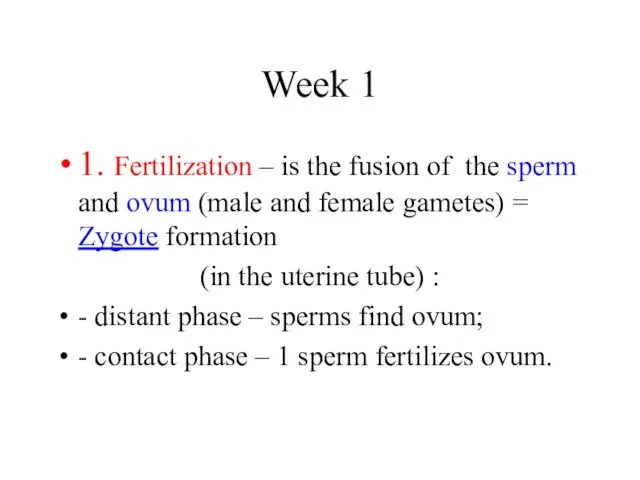
Week 1
1. Fertilization – is the fusion of the sperm and
Week 1
1. Fertilization – is the fusion of the sperm and
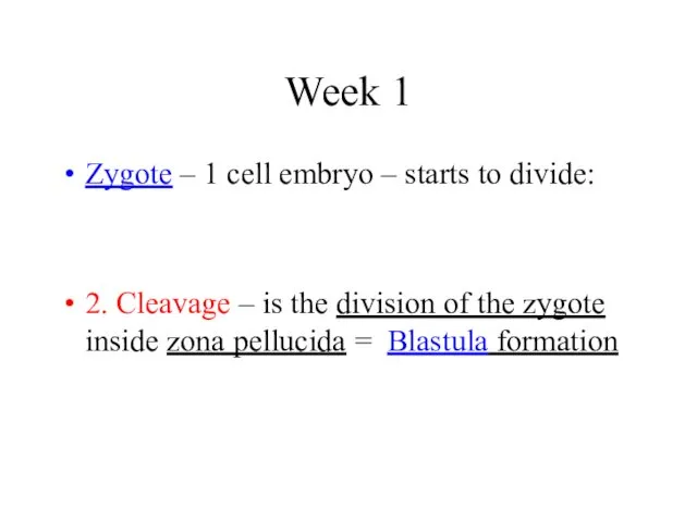
Week 1
Zygote – 1 cell embryo – starts to divide:
2. Cleavage
Week 1
Zygote – 1 cell embryo – starts to divide:
2. Cleavage
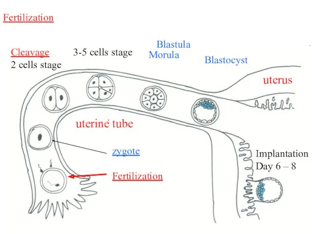
zygote
Fertilization
zygote
Fertilization
Cleavage
2 cells stage
3-5 cells stage
Morula
Blastocyst
Implantation
Day 6 – 8
uterus
uterine
zygote
Fertilization
zygote
Fertilization
Cleavage
2 cells stage
3-5 cells stage
Morula
Blastocyst
Implantation
Day 6 – 8
uterus
uterine
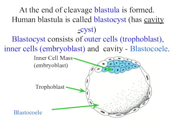
At the end of cleavage blastula is formed. Human blastula is
At the end of cleavage blastula is formed. Human blastula is
Embryoblast
At the 7-th day blastocyst sinks into the uterine wall
due
Embryoblast
At the 7-th day blastocyst sinks into the uterine wall due

Week 2:
Beginning of
3. Gastrulation – formation of 3 germ
Week 2: Beginning of 3. Gastrulation – formation of 3 germ
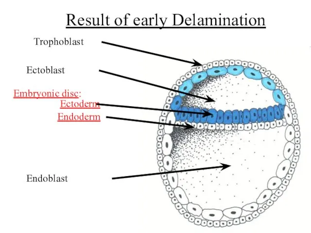
Result of early Delamination
Embryonic disc:
Result of early Delamination
Embryonic disc:
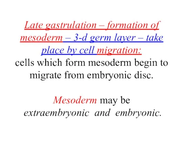
Late gastrulation – formation of mesoderm – 3-d germ layer –
Late gastrulation – formation of mesoderm – 3-d germ layer –
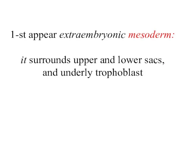
1-st appear extraembryonic mesoderm:
it surrounds upper and lower sacs,
and underly
1-st appear extraembryonic mesoderm: it surrounds upper and lower sacs, and underly
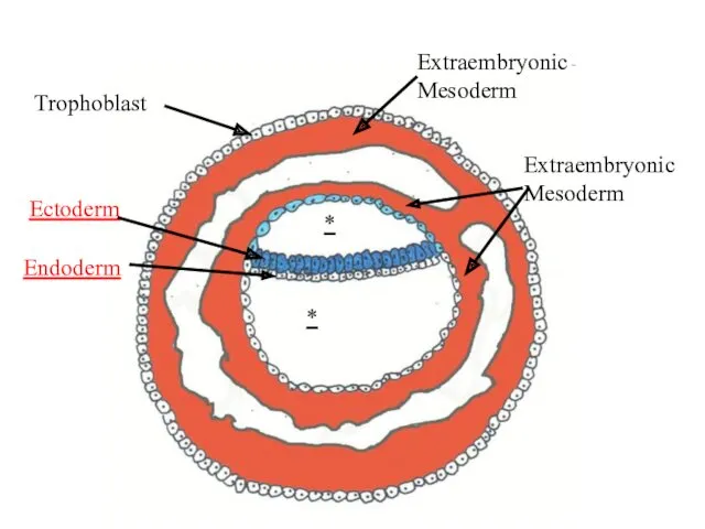
Trophoblast
Extraembryonic
Mesoderm
*
Ectoderm
Endoderm
Extraembryonic
Mesoderm
*
Trophoblast
Extraembryonic
Mesoderm
*
Ectoderm
Endoderm
Extraembryonic
Mesoderm
*
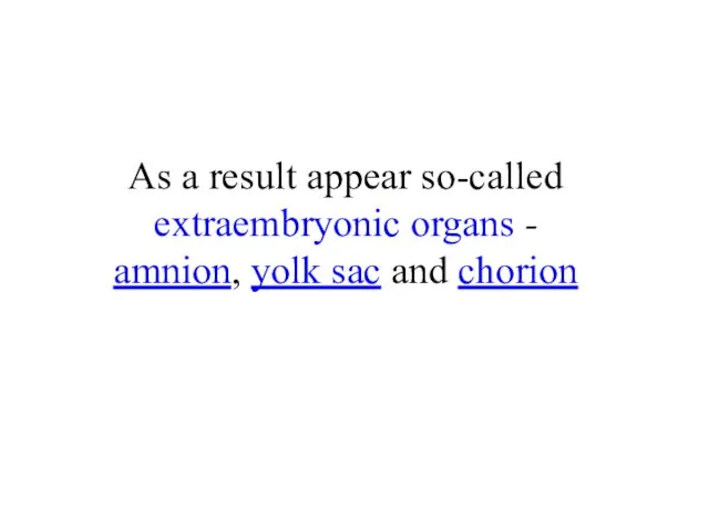
As a result appear so-called extraembryonic organs -
amnion, yolk sac
As a result appear so-called extraembryonic organs - amnion, yolk sac
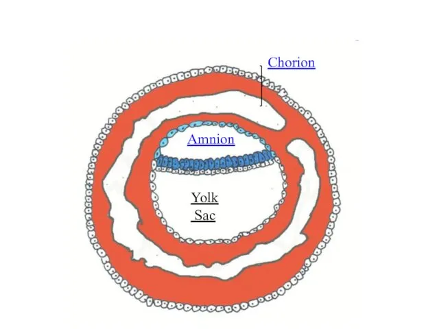
Chorion
Amnion
Yolk
Sac
Chorion
Amnion
Yolk
Sac
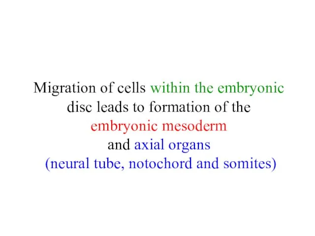
Migration of cells within the embryonic disc leads to formation of
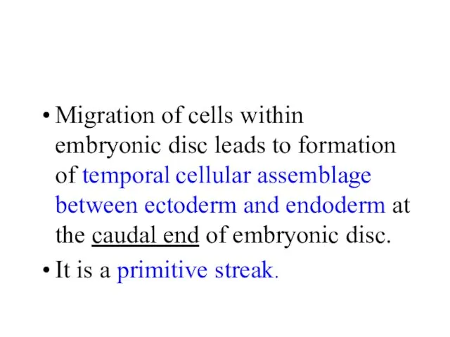
Migration of cells within embryonic disc leads to formation of temporal
Migration of cells within embryonic disc leads to formation of temporal
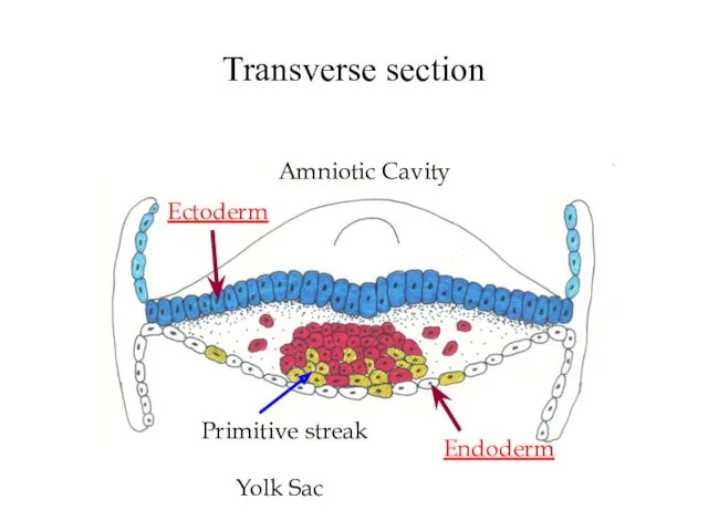
Transverse section
Ectoderm
Endoderm
Amniotic Cavity
Primitive streak
Yolk Sac
Transverse section
Ectoderm
Endoderm
Amniotic Cavity
Primitive streak
Yolk Sac
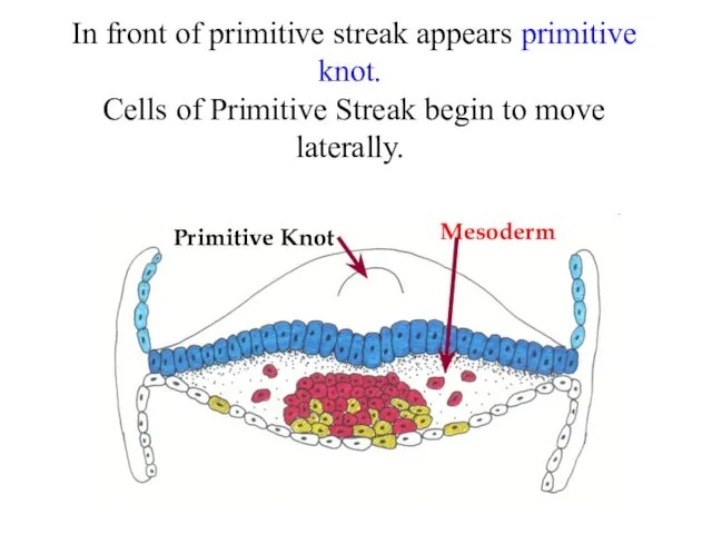
In front of primitive streak appears primitive knot.
Cells of
In front of primitive streak appears primitive knot. Cells of
ectoderm
endoderm
mesoderm
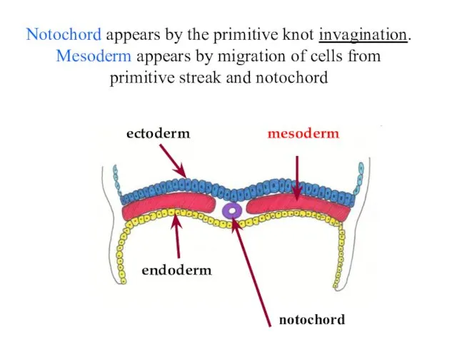
Notochord appears by the primitive knot invagination. Mesoderm appears by migration
ectoderm
endoderm
mesoderm
Notochord appears by the primitive knot invagination. Mesoderm appears by migration
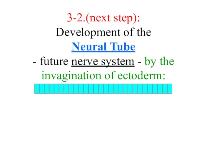
3-2.(next step):
Development of the
Neural Tube
- future nerve system - by
3-2.(next step): Development of the Neural Tube - future nerve system - by

Neural plate in Surface Ectoderm forms Neural groove
Neural plate in Surface Ectoderm forms Neural groove
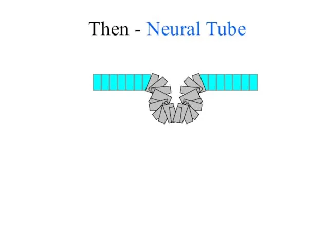
Then - Neural Tube
Then - Neural Tube
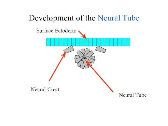
Development of the Neural Tube
Surface Ectoderm
Neural Crest
Neural Tube
Development of the Neural Tube
Surface Ectoderm
Neural Crest
Neural Tube
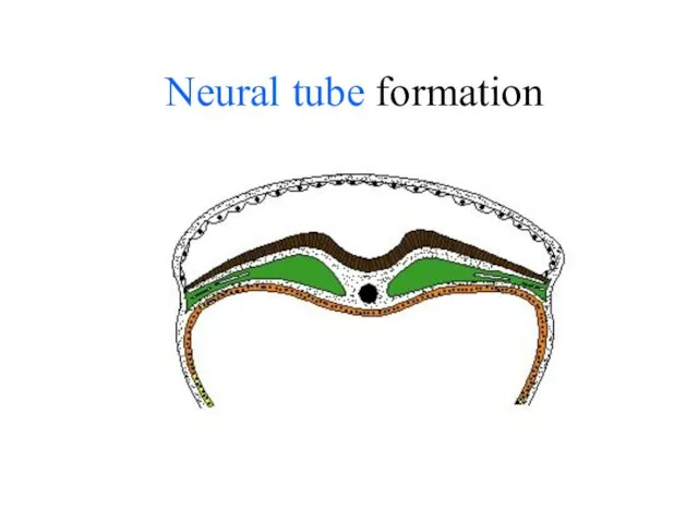
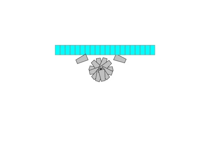
Neural tube formation
Neural tube formation
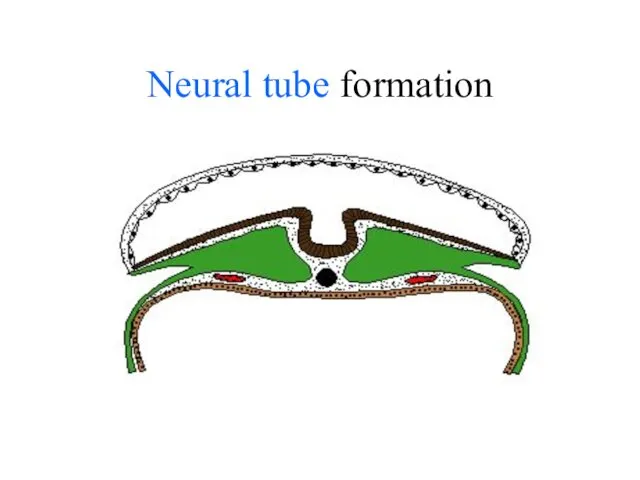
Neural tube formation
Neural tube formation
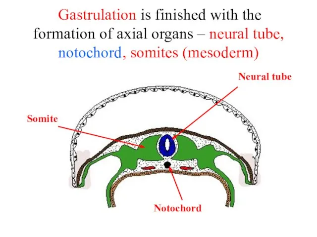
Gastrulation is finished with the formation of axial organs –
Gastrulation is finished with the formation of axial organs –
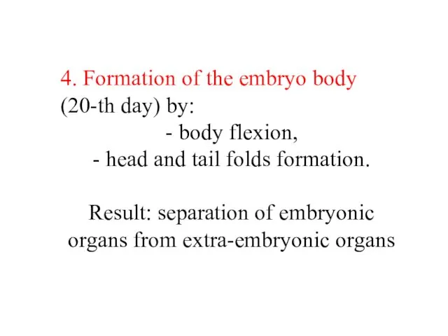
4. Formation of the embryo body
(20-th day) by:
- body flexion,
4. Formation of the embryo body
(20-th day) by:
- body flexion,
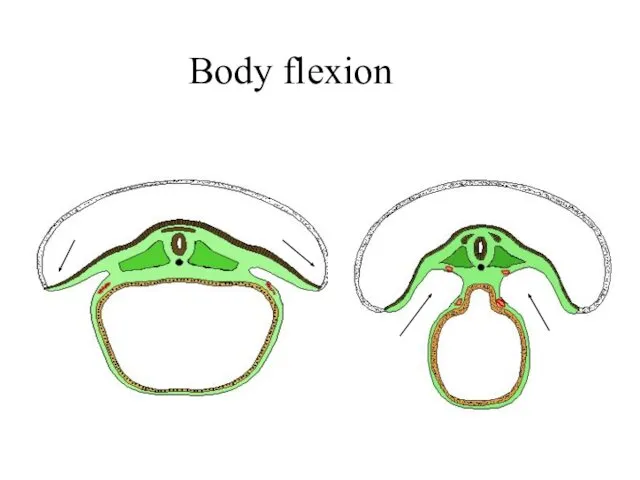
Body flexion
Body flexion
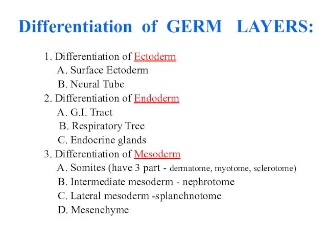
Differentiation of GERM LAYERS:
1. Differentiation of Ectoderm
A. Surface Ectoderm
Differentiation of GERM LAYERS:
1. Differentiation of Ectoderm
A. Surface Ectoderm
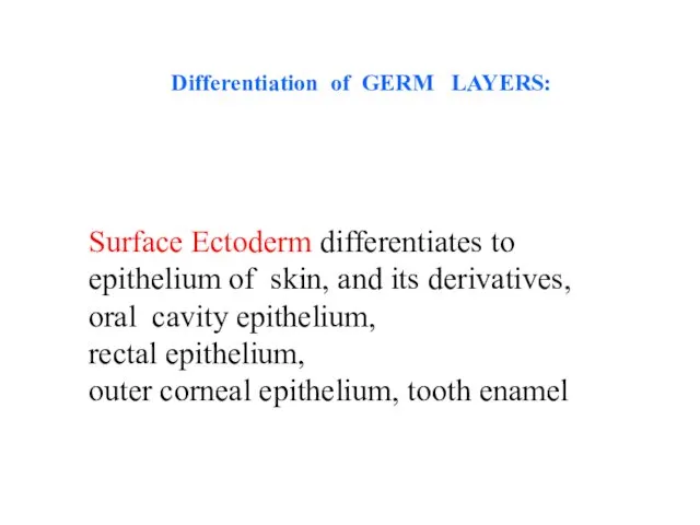
Surface Ectoderm differentiates to
epithelium of skin, and its derivatives,
oral
Surface Ectoderm differentiates to
epithelium of skin, and its derivatives,
oral
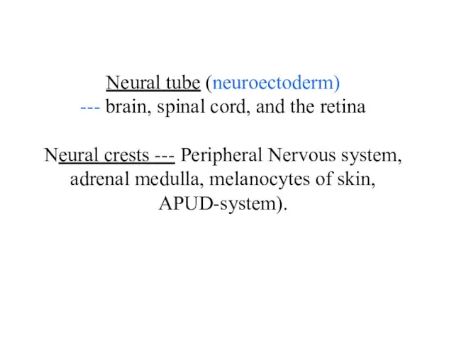
Neural tube (neuroectoderm)
--- brain, spinal cord, and the retina
Neural crests ---
Neural tube (neuroectoderm) --- brain, spinal cord, and the retina Neural crests ---
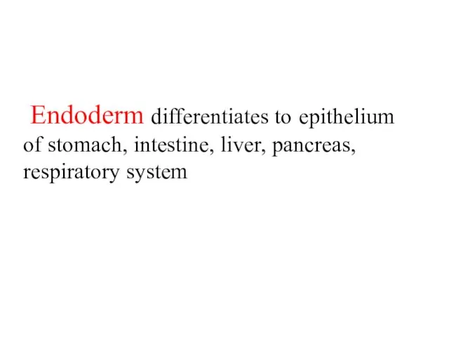
Endoderm differentiates to epithelium of stomach, intestine, liver, pancreas, respiratory
Endoderm differentiates to epithelium of stomach, intestine, liver, pancreas, respiratory

Mesoderm
Notochord
Endoderm
Ectoderm
Yolk Sac
Amniotic Cavity
Somite
Intermediate mesoderm
(nephrotome)
Lateral plate mesoderm
(somatopleuric,
splanchnopleuric
mesoderm)
Mesoderm
Notochord
Endoderm
Ectoderm
Yolk Sac
Amniotic Cavity
Somite
Intermediate mesoderm
(nephrotome)
Lateral plate mesoderm
(somatopleuric,
splanchnopleuric
mesoderm)
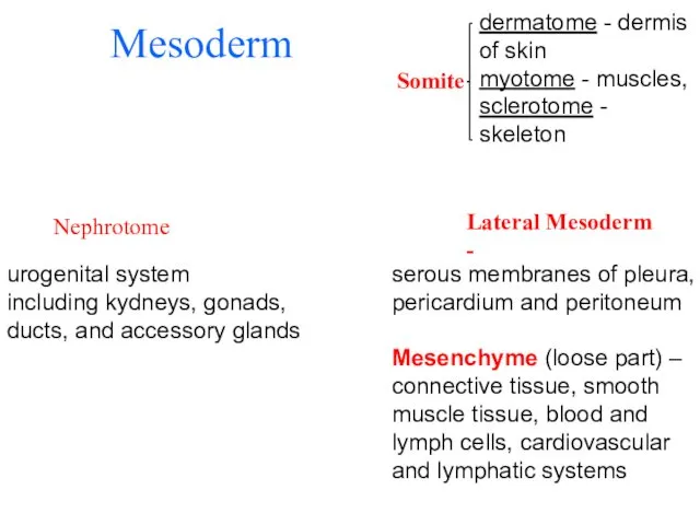
Mesoderm
Nephrotome
urogenital system
including kydneys, gonads,
ducts, and accessory glands
Somite
dermatome - dermis of
Mesoderm
Nephrotome
urogenital system
including kydneys, gonads,
ducts, and accessory glands
Somite
dermatome - dermis of

Late embryonic stages
Histogenesis
Organogenesis
Late embryonic stages
Histogenesis
Organogenesis
 Этические проблемы использования животных
 Пищеварение 1.1
 Общие признаки животных. 5 класс
 Ретикулярная формация. Лимбическая система. Оболочки мозга
 Хемосинтез. Хемосинтезирующие организмы
 Визначення типу шкіри на різних ділянках обличчя та складання правил догляду за власною шкірою
 Прокариоты на службе по защите почвенной среды жизни от тяжёлых металлов
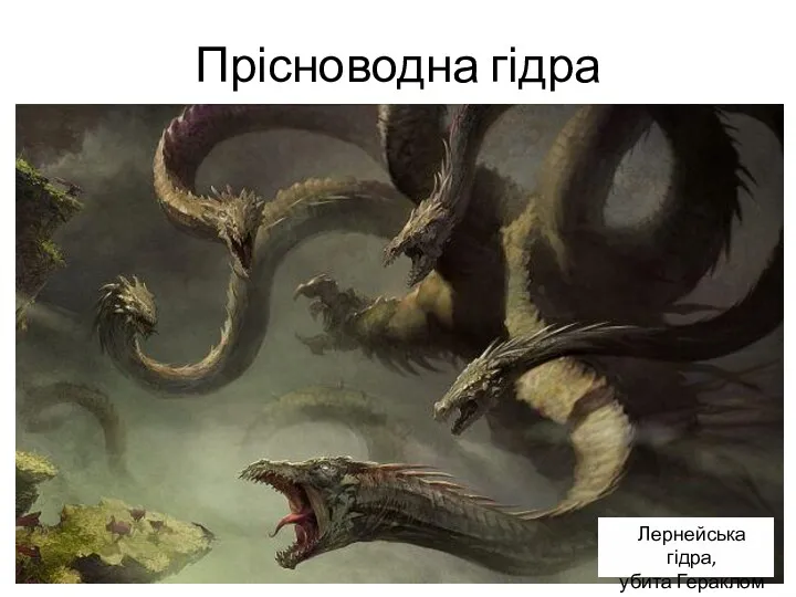 Прісноводна гідра
 Zebra is a striped member of the horse family
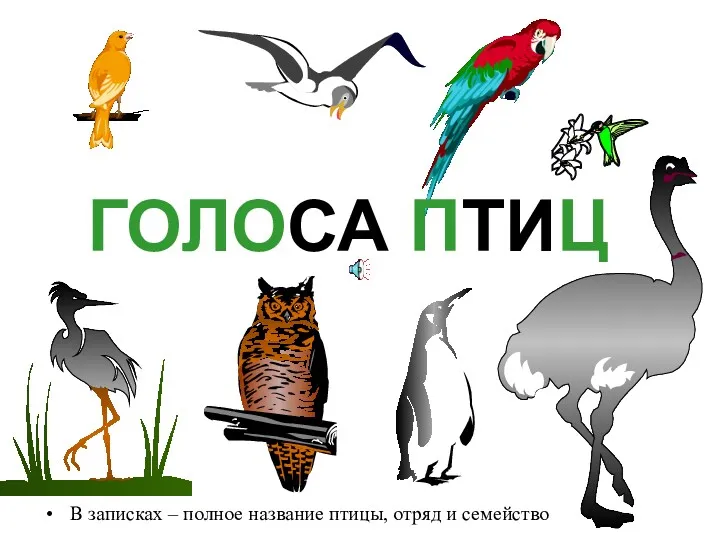 Голоса птиц
 Імунітет рослин до інфекційних хвороб
 Морфологическое описание растения
 Презентация по биологии для учащихся 9 класса по теме Клеточная мембрана
 Про кошек
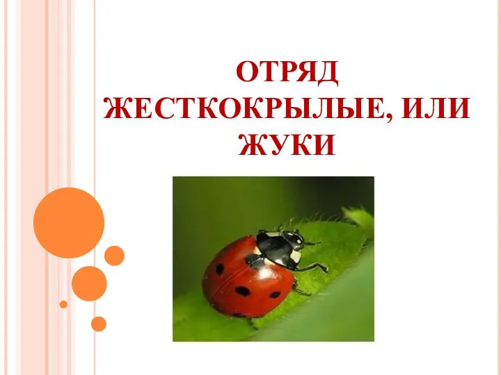 Презентация к уроку биологии в 7 классе Отряд Жесткокрылые, или Жуки
 Транскрипция и трансляция. Генетическая трансформация
 Китообразные и ластоногие
 Породы кошек
 Классификация и характеристика рыб
 Строение клеток прокариот и эукариот
 Органы и системы органов
 Мед - пчелиная заслуга
 Микробиология - наука о микроорганизмах
 Карл Линней - основоположник биологической систематики
 Движение крови в организме. Органы кровообращения
 Приглашение на семинар по управлению инкубаторием компании Пас Реформ
 Витамины красоты
 Внеклассное мероприятие Красная книга Оренбургской области