- Structures of water-soluble globular proteins

Содержание
- 2. Fibrous H-bonds & hydrophobics Membrane ____ Globular proteins (water-soluble)
- 3. Hermann Emil Louis Fischer (1852 –1919) Nobel Prize 1902 Protein chain Protein sequence Frederick Sanger (1918
- 4. domain 1 domain 2 ← single-domain globular protein fold stack
- 5. Secondary structures (α-helices, β-strands) are the most rigid and conserved details of proteins; they are determined
- 6. Max Ferdinand Perutz (1914 –2002) Nobel Prize 1962 X-ray 3D protein structure Kurt Wüthrich, 1938 Nobel
- 7. Homologous proteins have similar folds. True, but trivial. NON-trivial: Many NON-homologous proteins have similar folds. Hemo-
- 8. β-proteins β-sheets: usually, twisted (usually, right-) ↑ H-bonds: within sheets Hydrophobics: between sheets ____
- 9. Orthogonal packing Aligned packing of β-sheets of β-sheets sandwiches & cylinders
- 10. orthogonal packing of one rolled β-sheet Retinol-binding protein
- 11. Trypsin-like SER-protease Acid-protease orthogonal packings of β-sheets 2 1 4 5 5’ 6 3 2’ 2
- 12. IG-fold: aligned packing of β-sheets Greek key 2::5 Greek key 3::6 1 2 3 4 5
- 13. β-sandwich Interlocked pairs: center of sandwich Greek key: edge of sandwich Hydrophobic surfaces of sheets of
- 14. aligned packings of β-sheets a) different: only topologies b) equal: even topology 6 5 8 3
- 15. aligned packing of β-sheets 6-bladed propeller neuraminidase
- 16. UNusual LEFT-HANDED chain turns (AND NO β−TWIST!) Left-handed β-prism: Acyl transferase Right-handed β-prism: Pectate lyase Usual
- 17. α-proteins H-bonds: within helices & Hydrophobics: between helices
- 18. Quasi-cylindrical core (in fibrous) Quasi-flat core Quasi-spherical core MOST COMMON
- 19. Orthogonal packing Similar to orthogonal of LONG α-helices packing of β-sheets
- 20. Aligned packing Similar to aligned of LONG α-helices packing of β-sheets
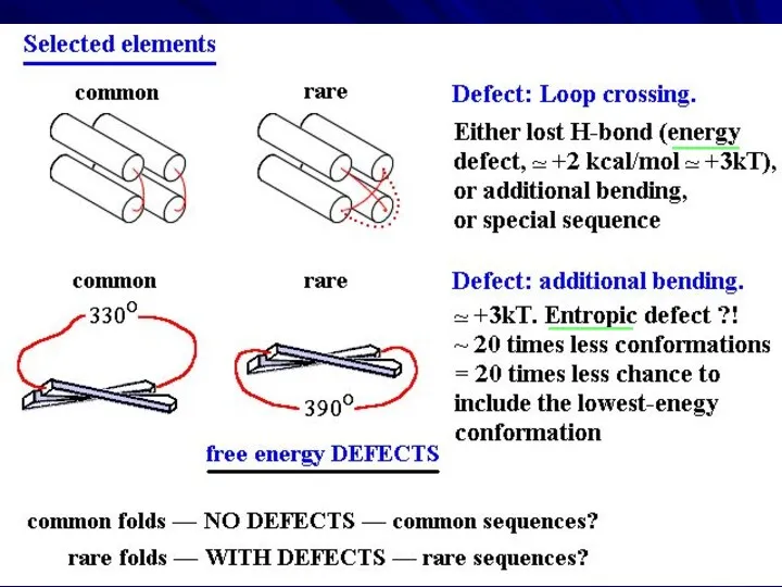
- 21. Quasi-spherical polyhedra Quasi- spherical core: MOST COMMON no loop turns of ~360o no loop crossings
- 22. Packing of ridges: “0-4” & “0-4”: -500 “0-4” & “1-4”: +200 IDEAL POLYHEDRA -600 ≈ -500
- 23. α/β proteins H-bonds: within helices & sheets Hydrophobics: between helices & sheets
- 24. TIM barrel Rossmann fold
- 25. α and β layers right-handed superhelices Regular secondary structure sequence: β − α − β −
- 26. Classification of β-barrels: “share number” S and strand number N. Here: S=8, N=8 Standard active site
- 27. α+β proteins H-bonds: within helices & sheets Hydrophobics: between helices & sheets
- 28. α+β: a) A kind of regularity in the secondary structure sequence: β − α − β
- 29. α+β: b) Secondary structure sequence: composed of irregular blocks, e.g.: β − β − β −
- 30. TYPICAL FOLDING PATTERNS (1977) Jane Shelby Richardson, 1941
- 31. EMPIRICAL RULES separate α and β layers right-handed superhelices no large, ~360o turns no loop crossings
- 32. RESULT: NARROW SET OF PREDOMINANT FOLDING PATTERNS these are those that have no ‘defects’
- 33. ALSO, these are “natively disordered proteins”, which form a definite structure only when bound to some
- 34. Globular domains C A T H ≈ S C O P
- 35. Алексей Григорьевич Мурзин, 1956 Dame Janet Maureen Thornton, 1949 Cyrus Homi Chothia, 1942 CATH SCOP Classification
- 36. Efimov’s “trees”
- 37. 80/20 LAW:
- 38. EMPIRICAL RULES for FREQUENT FOLDS α and β structures, right-handed separate α and β layers superhelices
- 39. Unusual fold (no α, almost no β structure: bad for stability) - BUT: very special sequence
- 40. Unusual fold (GFP): helix inside Usual folds: helices outside
- 41. What is more usual: sequence providing α inside or β β inside? α β β N>150
- 42. _____ ____
- 43. Miller, Janin, Chothia 1984 Example: Small protein details
- 44. THEORY Closed system: energy E = const CONSIDER: 1 state of “small part” with ε &
- 45. Protein structure is stable, if its free energy is below some threshold For example: below that
- 46. More stable detail – more random sequences Less stable detail – less random sequences What's good
- 47. “Multitude principle” for physical selection of folds of globular proteins (now: “designability”): the more sequences fit
- 48. Globular domains C A T H ≈ S C O P RATIONAL STRUCTURAL CLASSIFICATION OF PROTEINS
- 50. Скачать презентацию
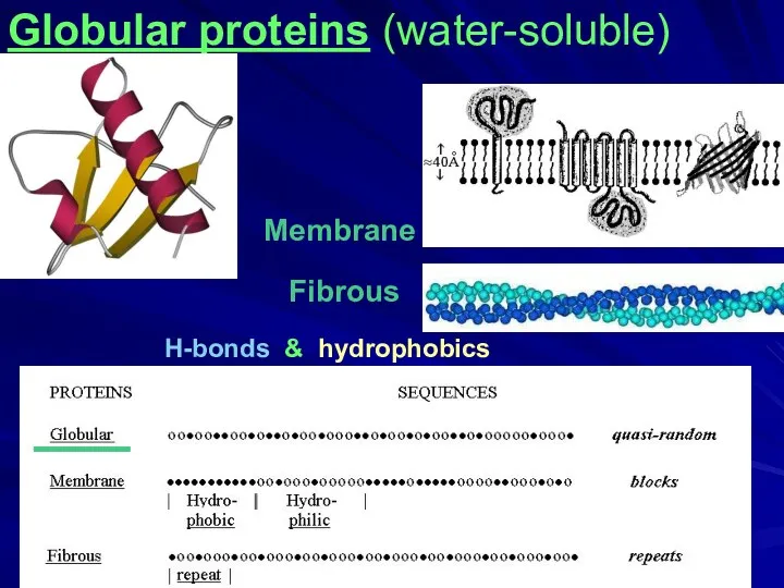
Fibrous
H-bonds & hydrophobics
Membrane
____
Globular proteins (water-soluble)
Fibrous
H-bonds & hydrophobics
Membrane
____
Globular proteins (water-soluble)
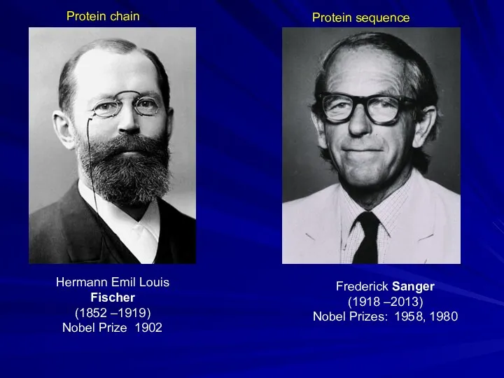
Hermann Emil Louis Fischer
(1852 –1919)
Nobel Prize 1902
Protein chain
Protein sequence
Frederick Sanger
(1918 –2013)
Nobel
Hermann Emil Louis Fischer
(1852 –1919)
Nobel Prize 1902
Protein chain
Protein sequence
Frederick Sanger
(1918 –2013)
Nobel
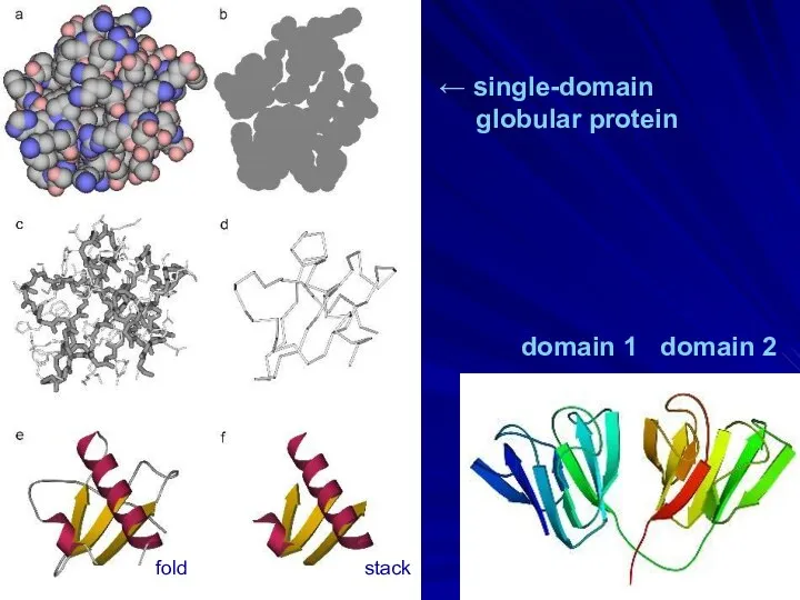
domain 1 domain 2
← single-domain
globular protein
fold stack
domain 1 domain 2
← single-domain
globular protein
fold stack
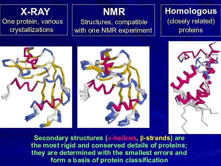
Secondary structures (α-helices, β-strands) are
the most rigid and conserved details
Secondary structures (α-helices, β-strands) are
the most rigid and conserved details
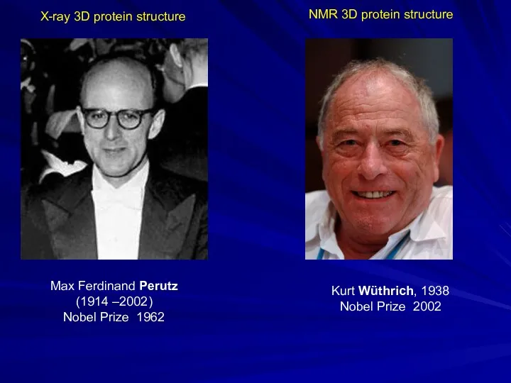
Max Ferdinand Perutz
(1914 –2002)
Nobel Prize 1962
X-ray 3D protein structure
Kurt Wüthrich, 1938
Nobel Prize
Max Ferdinand Perutz
(1914 –2002)
Nobel Prize 1962
X-ray 3D protein structure
Kurt Wüthrich, 1938
Nobel Prize
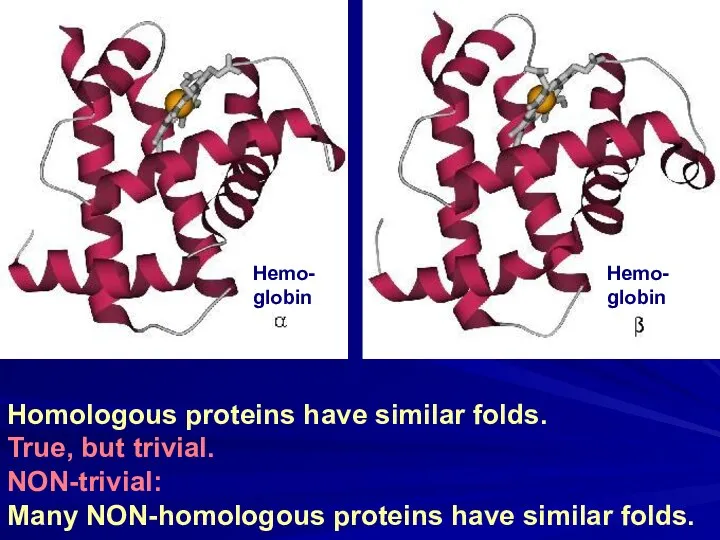
Homologous proteins have similar folds.
True, but trivial.
NON-trivial:
Many NON-homologous proteins have
Homologous proteins have similar folds.
True, but trivial.
NON-trivial:
Many NON-homologous proteins have
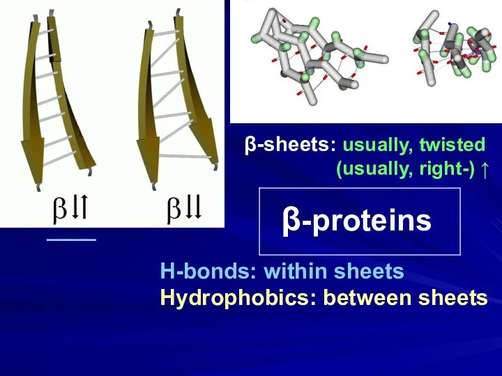
β-proteins
β-sheets: usually, twisted
(usually, right-) ↑
H-bonds: within sheets
Hydrophobics: between
β-proteins
β-sheets: usually, twisted
(usually, right-) ↑
H-bonds: within sheets
Hydrophobics: between
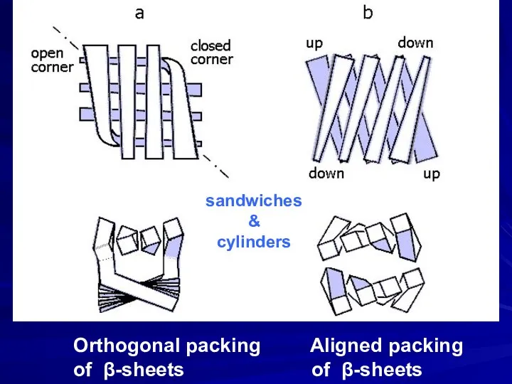
Orthogonal packing Aligned packing
of β-sheets of β-sheets
sandwiches
&
cylinders
Orthogonal packing Aligned packing
of β-sheets of β-sheets
sandwiches
&
cylinders
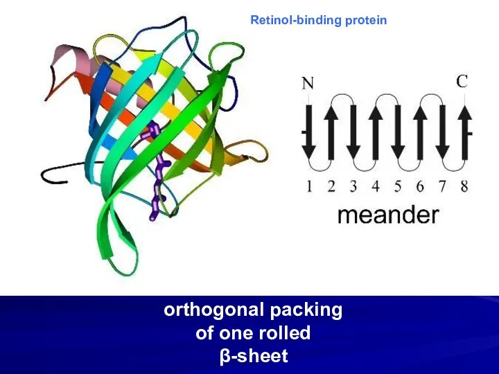
orthogonal packing
of one rolled β-sheet
Retinol-binding protein
orthogonal packing
of one rolled β-sheet
Retinol-binding protein
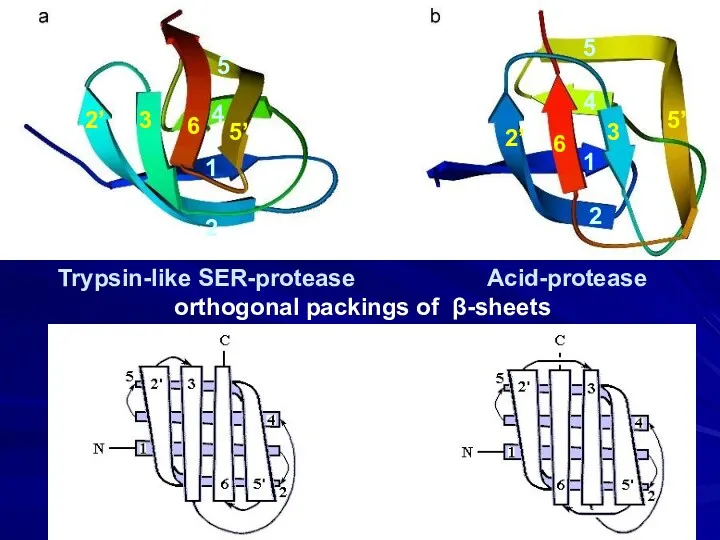
Trypsin-like SER-protease Acid-protease
orthogonal packings of β-sheets
2
1
4
5
5’
6
3
2’
2
1
4
5
6
2’
3
5’
Trypsin-like SER-protease Acid-protease
orthogonal packings of β-sheets
2
1
4
5
5’
6
3
2’
2
1
4
5
6
2’
3
5’
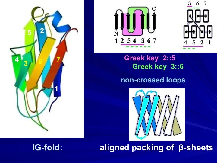
IG-fold: aligned packing of β-sheets
Greek key 2::5
Greek key 3::6
1
2
3
4
5
6
7
non-crossed
IG-fold: aligned packing of β-sheets
Greek key 2::5
Greek key 3::6
1
2
3
4
5
6
7
non-crossed
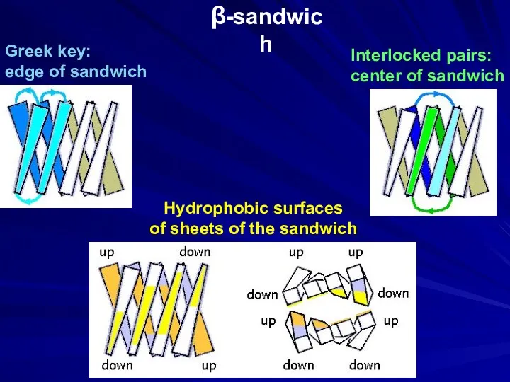
β-sandwich
Interlocked pairs:
center of sandwich
Greek key:
edge of sandwich
Hydrophobic surfaces
of sheets
β-sandwich
Interlocked pairs:
center of sandwich
Greek key:
edge of sandwich
Hydrophobic surfaces
of sheets
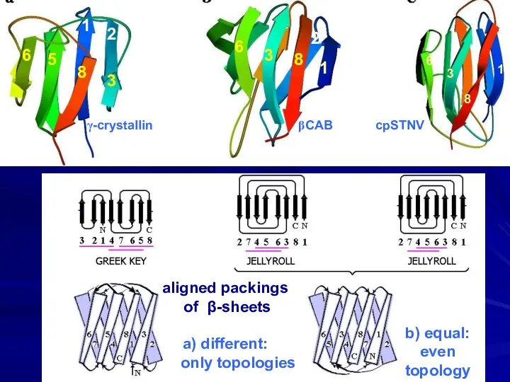
aligned packings
of β-sheets
a) different: only topologies
b) equal: even topology
6
5
8
3
2
1
6
3
8
1
2
6
3
8
1
γ-crystallin
aligned packings
of β-sheets
a) different: only topologies
b) equal: even topology
6
5
8
3
2
1
6
3
8
1
2
6
3
8
1
γ-crystallin
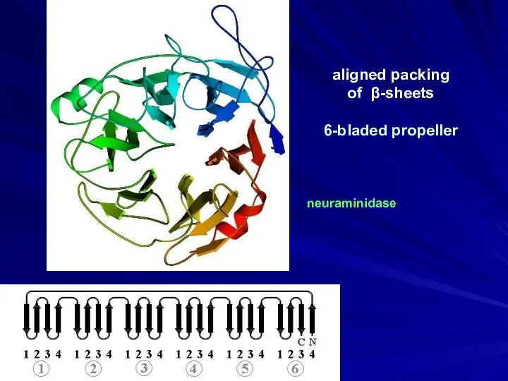
aligned packing
of β-sheets
6-bladed propeller
neuraminidase
aligned packing
of β-sheets
6-bladed propeller
neuraminidase
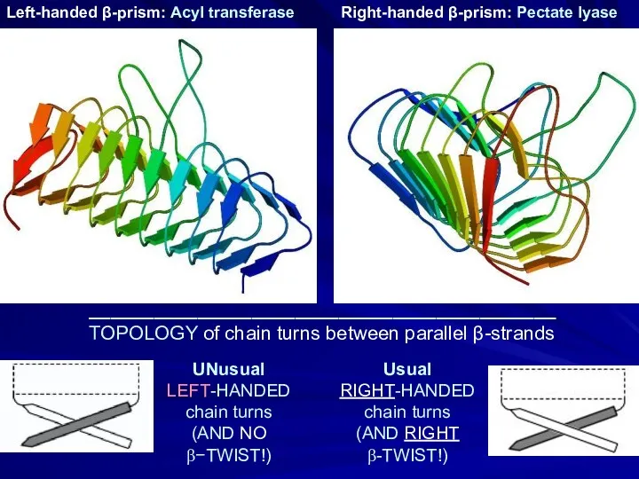
UNusual
LEFT-HANDED
chain turns
(AND NO
β−TWIST!)
Left-handed β-prism: Acyl transferase
Right-handed
UNusual
LEFT-HANDED
chain turns
(AND NO
β−TWIST!)
Left-handed β-prism: Acyl transferase
Right-handed
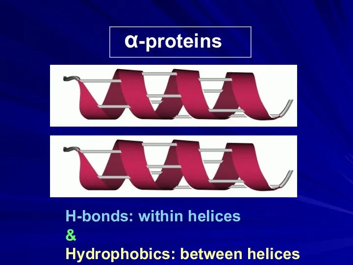
α-proteins
H-bonds: within helices
&
Hydrophobics: between helices
α-proteins
H-bonds: within helices
&
Hydrophobics: between helices
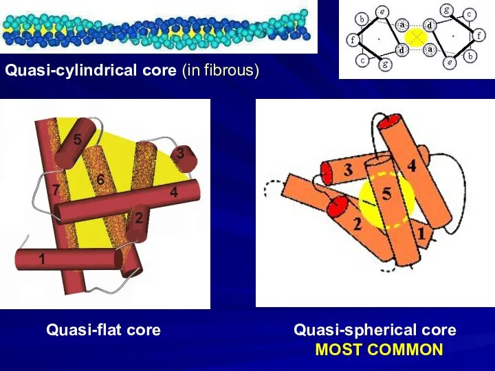
Quasi-cylindrical core (in fibrous)
Quasi-flat core
Quasi-spherical core
MOST COMMON
Quasi-cylindrical core (in fibrous)
Quasi-flat core
Quasi-spherical core
MOST COMMON
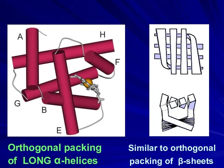
Orthogonal packing Similar to orthogonal
of LONG α-helices packing of β-sheets
Orthogonal packing Similar to orthogonal
of LONG α-helices packing of β-sheets
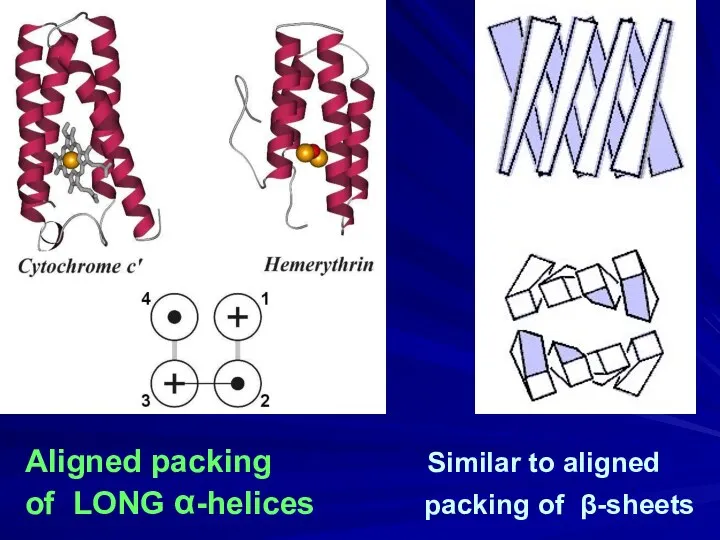
Aligned packing Similar to aligned
of LONG α-helices packing of β-sheets
Aligned packing Similar to aligned
of LONG α-helices packing of β-sheets
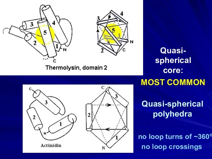
Quasi-spherical
polyhedra
Quasi-
spherical
core:
MOST COMMON
no loop turns of ~360o
no loop crossings
Quasi-spherical
polyhedra
Quasi-
spherical
core:
MOST COMMON
no loop turns of ~360o
no loop crossings
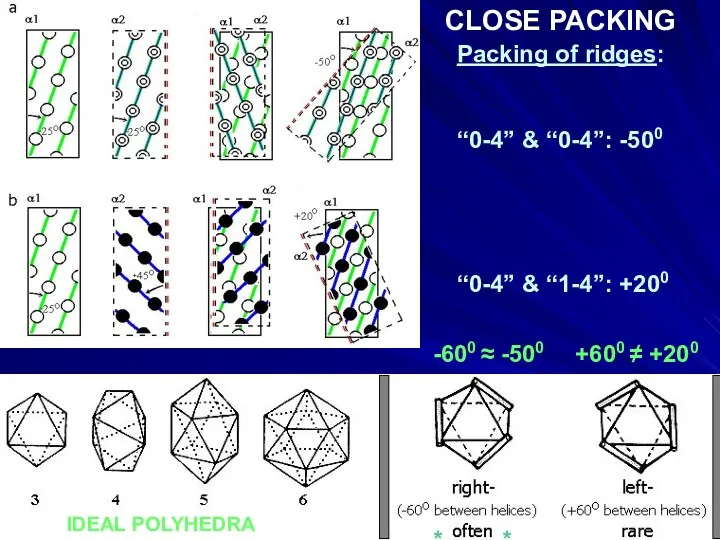
Packing of ridges:
“0-4” & “0-4”: -500
“0-4” & “1-4”: +200
IDEAL POLYHEDRA
-600
Packing of ridges:
“0-4” & “0-4”: -500
“0-4” & “1-4”: +200
IDEAL POLYHEDRA
-600
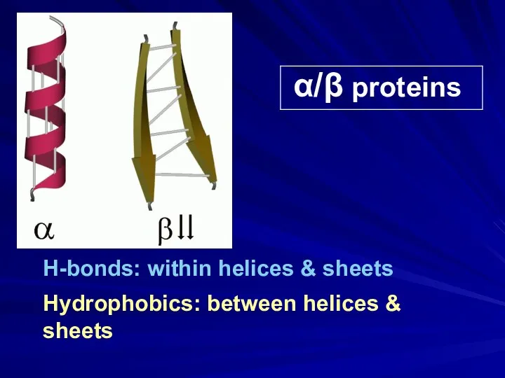
α/β proteins
H-bonds: within helices & sheets
Hydrophobics: between helices & sheets
α/β proteins
H-bonds: within helices & sheets
Hydrophobics: between helices & sheets
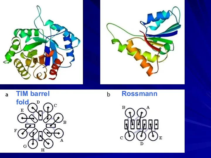
TIM barrel Rossmann fold
TIM barrel Rossmann fold
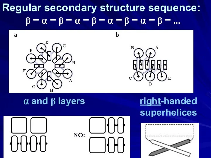
α and β layers right-handed
superhelices
Regular secondary structure sequence:
β −
α and β layers right-handed
superhelices
Regular secondary structure sequence:
β −
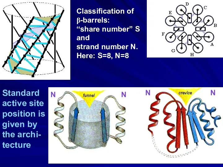
Classification of
β-barrels:
“share number” S
and
strand number N.
Here: S=8, N=8
Standard
active site
position is
Classification of
β-barrels:
“share number” S
and
strand number N.
Here: S=8, N=8
Standard
active site
position is
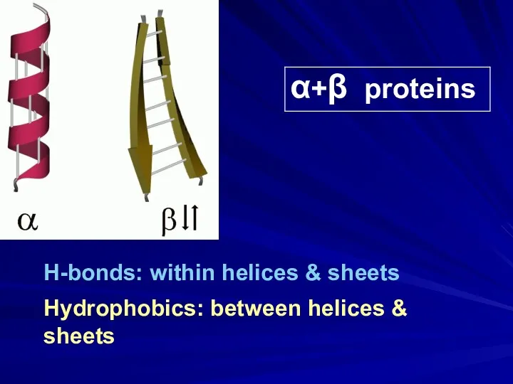
α+β proteins
H-bonds: within helices & sheets
Hydrophobics: between helices & sheets
α+β proteins
H-bonds: within helices & sheets
Hydrophobics: between helices & sheets
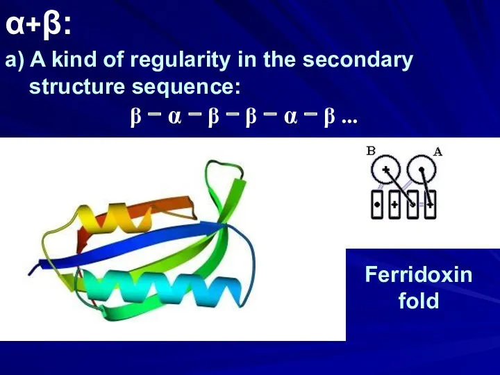
α+β:
a) A kind of regularity in the secondary
structure sequence:
β
α+β:
a) A kind of regularity in the secondary
structure sequence:
β
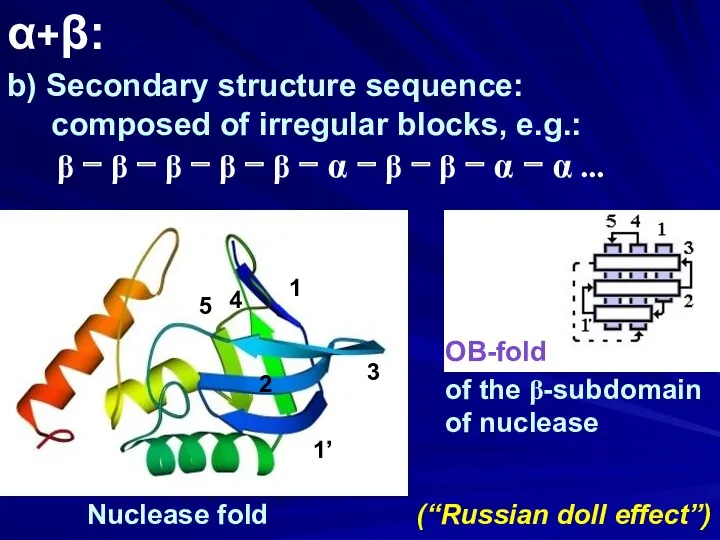
α+β:
b) Secondary structure sequence:
composed of irregular blocks, e.g.:
α+β:
b) Secondary structure sequence:
composed of irregular blocks, e.g.:
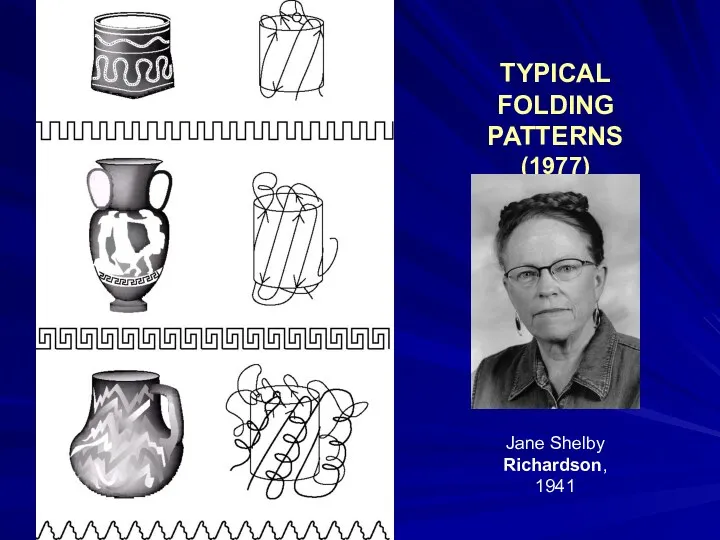
TYPICAL
FOLDING PATTERNS
(1977)
Jane Shelby
Richardson,
1941
TYPICAL
FOLDING PATTERNS
(1977)
Jane Shelby
Richardson,
1941
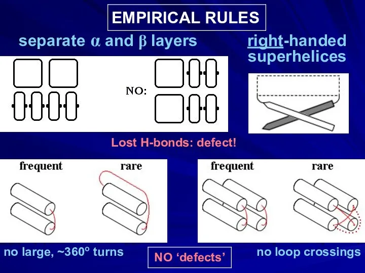
EMPIRICAL RULES
separate α and β layers right-handed
superhelices
no large, ~360o turns
no
EMPIRICAL RULES
separate α and β layers right-handed
superhelices
no large, ~360o turns
no
RESULT:
NARROW SET
OF PREDOMINANT FOLDING PATTERNS
these are those that have no
RESULT:
NARROW SET
OF PREDOMINANT FOLDING PATTERNS
these are those that have no
ALSO,
these are “natively disordered proteins”,
which form a definite structure
only when
ALSO,
these are “natively disordered proteins”,
which form a definite structure
only when
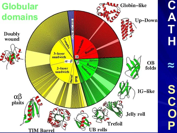
Globular
domains
C
A
T
H
≈
S
C
O
P
Globular
domains
C
A
T
H
≈
S
C
O
P
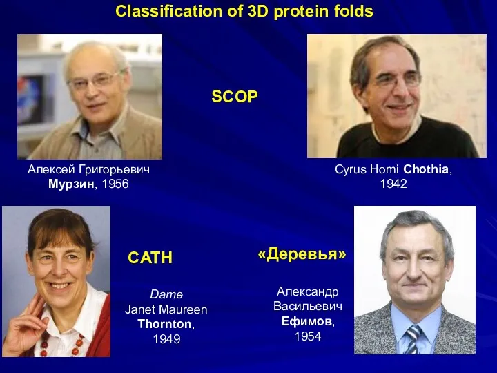
Алексей Григорьевич Мурзин, 1956
Dame
Janet Maureen Thornton,
1949
Cyrus Homi Chothia,
1942
CATH
SCOP
Classification of
Алексей Григорьевич Мурзин, 1956
Dame
Janet Maureen Thornton,
1949
Cyrus Homi Chothia,
1942
CATH
SCOP
Classification of
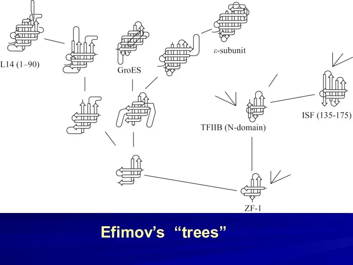
Efimov’s “trees”
Efimov’s “trees”
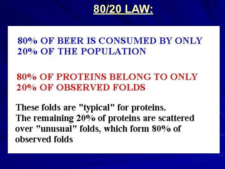
80/20 LAW:
80/20 LAW:
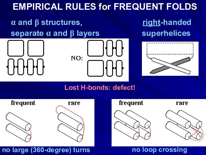
EMPIRICAL RULES for FREQUENT FOLDS
α and β structures, right-handed
separate α and
EMPIRICAL RULES for FREQUENT FOLDS
α and β structures, right-handed
separate α and
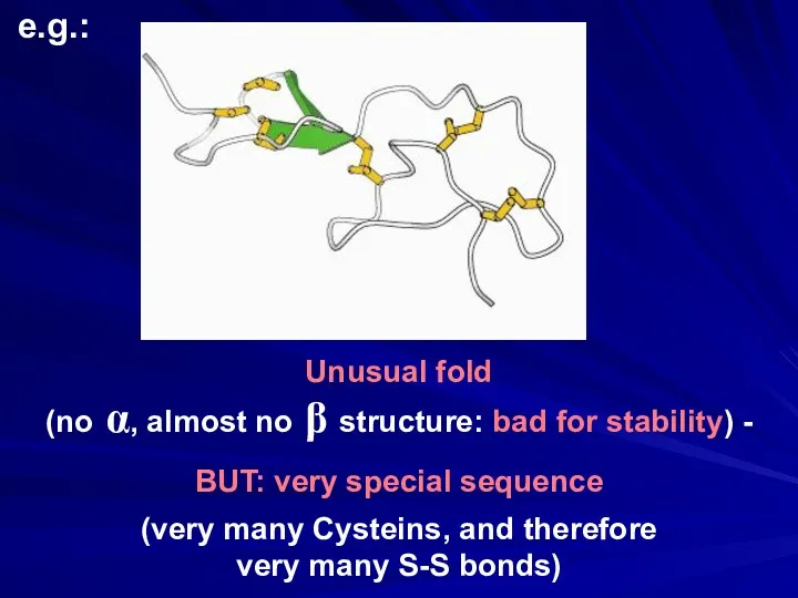
Unusual fold
(no α, almost no β structure: bad for stability) -
BUT:
Unusual fold
(no α, almost no β structure: bad for stability) -
BUT:
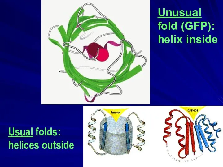
Unusual
fold (GFP):
helix inside
Usual folds:
helices outside
Unusual
fold (GFP):
helix inside
Usual folds:
helices outside
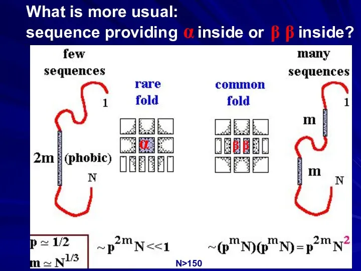
What is more usual:
sequence providing α inside or β β inside?
α
β
What is more usual:
sequence providing α inside or β β inside?
α
β
_____
____
_____
____
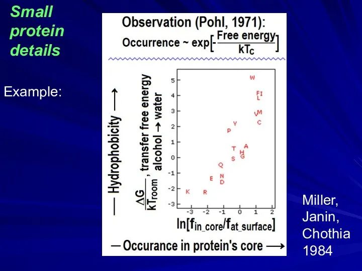
Miller,
Janin,
Chothia
1984
Example:
Small
protein
details
Miller,
Janin,
Chothia
1984
Example:
Small
protein
details
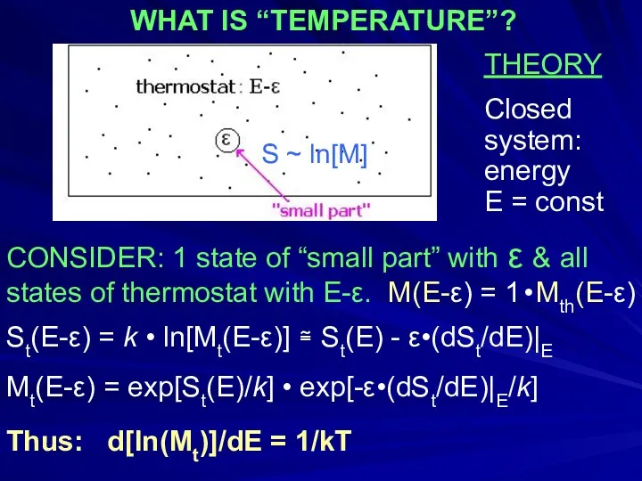
THEORY
Closed
system:
energy
E = const
CONSIDER: 1 state of “small part” with ε
THEORY
Closed
system:
energy
E = const
CONSIDER: 1 state of “small part” with ε
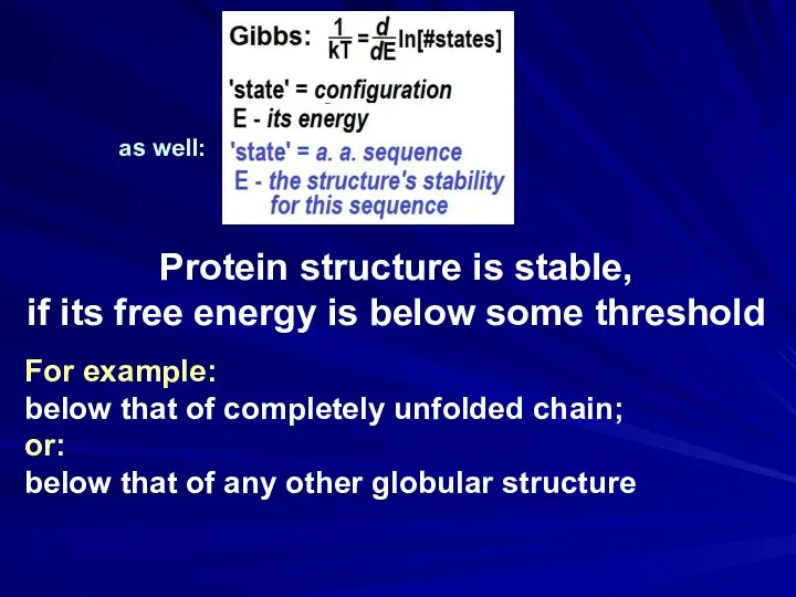
Protein structure is stable,
if its free energy is below some
Protein structure is stable,
if its free energy is below some
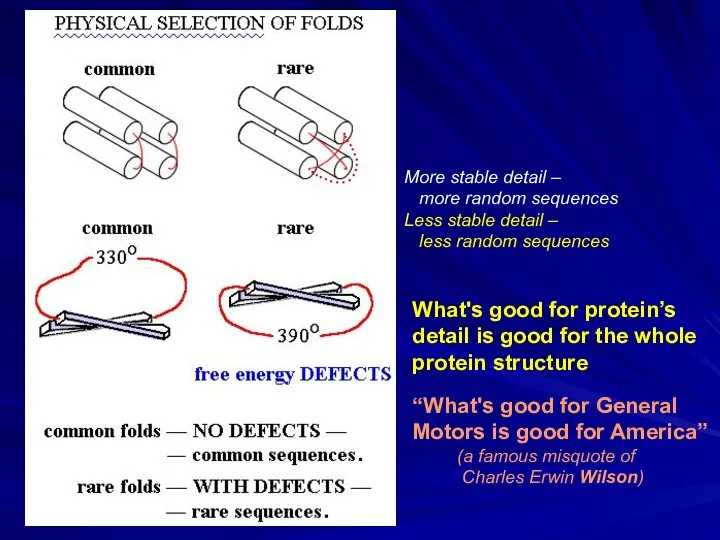
More stable detail –
more random sequences
Less stable detail –
More stable detail –
more random sequences
Less stable detail –
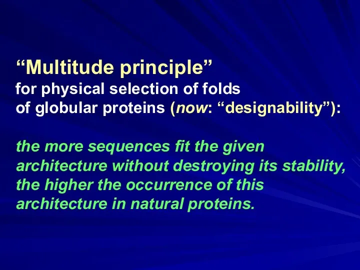
“Multitude principle”
for physical selection of folds
of globular proteins (now:
“Multitude principle”
for physical selection of folds
of globular proteins (now:
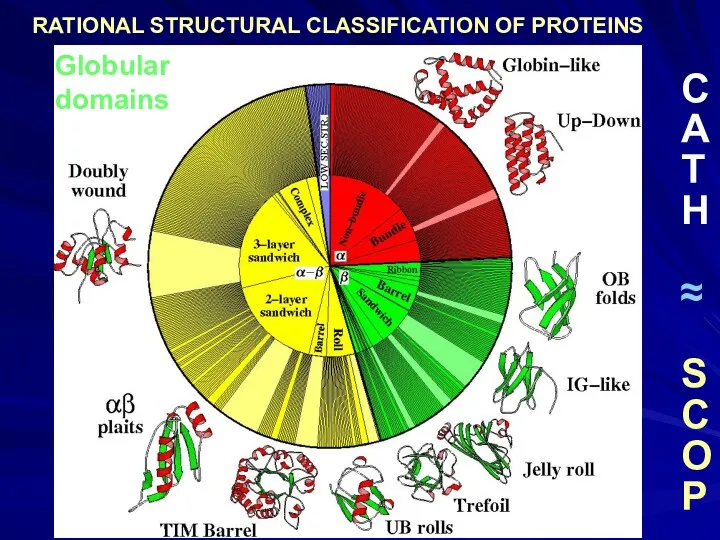
Globular
domains
C
A
T
H
≈
S
C
O
P
RATIONAL STRUCTURAL CLASSIFICATION OF PROTEINS
Globular
domains
C
A
T
H
≈
S
C
O
P
RATIONAL STRUCTURAL CLASSIFICATION OF PROTEINS
 Интерференция света. (Лекция 11)
 Происхождение элементов
 Кроссворд
 Молекулярно-кинетическая теория строения вещества (МКТ)
 Кинематика тела. Лекция 7
 тест по состояниям вещества 8 класс
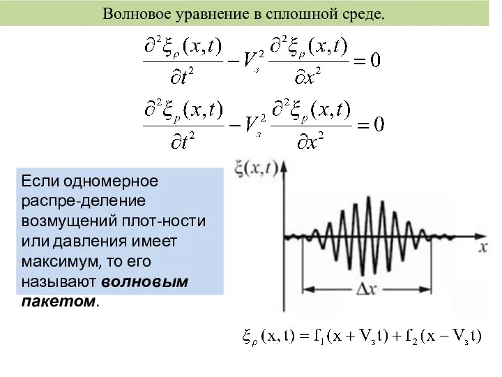 Волновое уравнение в сплошной среде
 Электромагнитные волны
 Методы исследования наносистем и наноматериалов. Классификация физико-химических методов исследования
 Презентация к уроку физики в 8 классе: Обобщение по теме Агрегатные состояния вещества.
 Магнитные материалы. Магнитное поле в веществе
 Урок физики 8 класс Испарение и конденсация
 Аеродинамічні характеристики літака. Аеродинамічне компонування літака. (Лекція 7.4.1)
 03.21г. Типы спектров. Спектральный анализ
 Фундаментальные взаимодействия
 Презентация к уроку Водород, 5 класс, предмет Физика и химия
 Закон Гука
 Лабораторная работа №2. Измерение массы тела на рычажных весах
 Электризация тел. Два рода зарядов
 Поляризация поперечных волн. Поляризация света. Дисперсия света. Виды спектров
 Батарейка своими руками
 Качество электроэнергии
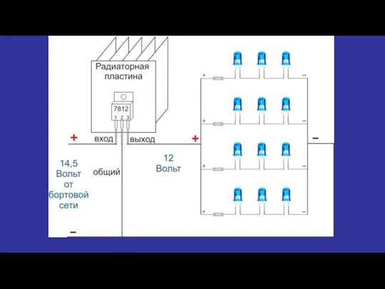 Электрическая схема светильника с регулировкой мощности
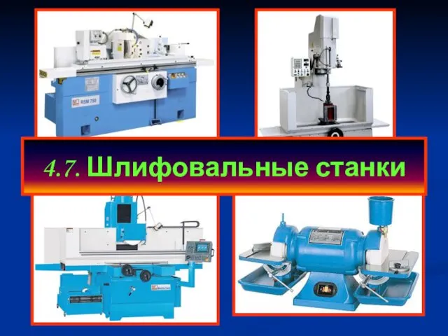 Шлифовальные станки. (Тема 7)
 Электромагнитная картина мира
 Курсовая работа по теоретической механике “Динамика кулисного механизма”
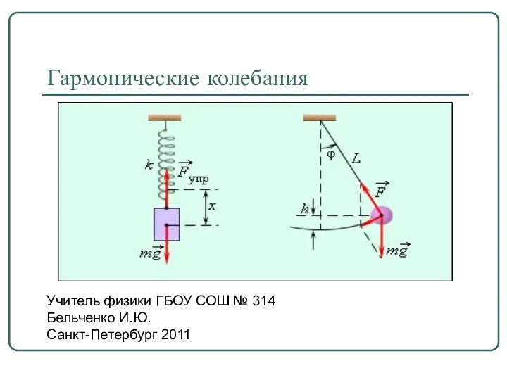 Гармонические колебания
 Работа силы Ампера. Магнитный поток. Явление электромагнитной индукции. Закон электромагнитной индукции. Правило Ленца