- Abdominal Wall Hernias

Содержание
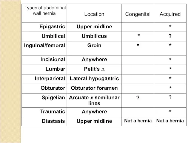
- 2. Hernia: The protrusion of tissue through a defect in fascial and/or muscular layer(s) that normally contain
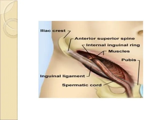
- 4. Semilunar line Arcuate line Basic Anatomy
- 5. Very common In midline between umbilicus and xiphoid May be multiple Small fascial defect ( Tongue
- 6. Umbilical Hernia Common in infancy Reacquired during adulthood Peritoneal sac Small ones of no significance Large
- 7. Typical Umbilical Hernia
- 8. Umbilical hernia Umbilical hernia is often found in childhood. However, it often manifests in adulthood.
- 9. MAYO REPAIR SAPEJKO REPAIR
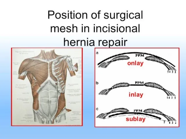
- 10. TERMINOLOGY OF MESH POSITIONS DURING VENTRAL HERNIA REPAIR
- 11. Umbilical & Inguinal Hernias
- 12. Most common Congenital ~ indirect Acquired ~ direct or indirect Indirect Hernia has peritoneal sac lateral
- 13. 16th century hernia repair Scrotal hernia, 1682 Hernia strap, 1758 Mention of hernias in painting
- 15. Typical scrotal hernia
- 16. Giant scrotal hernia Note scaphoid abdomen
- 17. The basic feature of all hernias Occur at a weak spot . Reduce on lying down
- 18. A hernia consist of 3 parts: 1. Sac: consist of a diverticulum of peritoneum. 2. Contents:
- 19. Complications of hernias Irreducible the hernia contents cannot be manipulated back into the abdominal cavity. Incarcerated
- 20. Sliding Hernia Hernia consisting of retroperitoneal fat and/or large bowel (cecum on the right, sigmoid on
- 21. Hernia surgery Circa 1300 ~1600 17th century ~1497
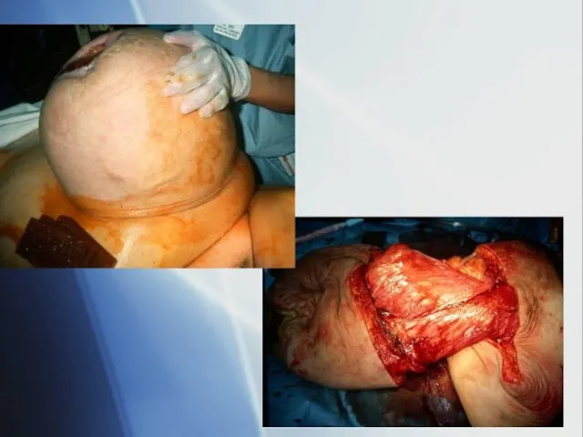
- 22. Giant Scrotal Hernia (1/2 of small bowel + right colon)
- 23. Incarcerated Inguinal Hernia with Bowel Obstruction
- 24. More typical inguinal hernia
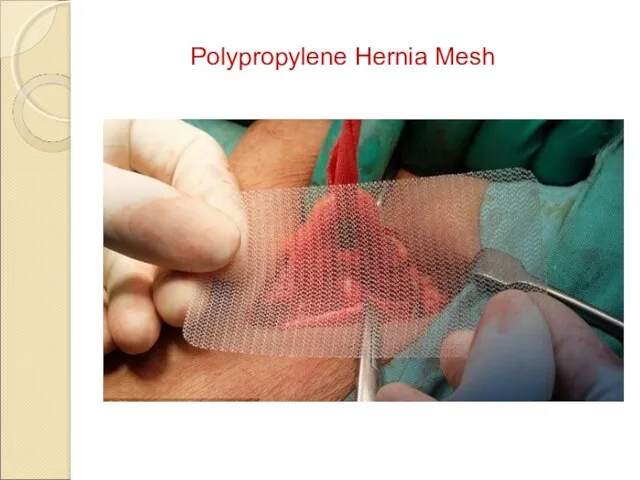
- 26. Polypropylene Hernia Mesh
- 27. Polypropylene Hernia System
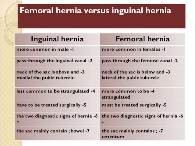
- 28. Femoral Hernia Develops in femoral canal, medial to femoral vein, below the inguinal ligament Occurs mainly
- 30. Femoral hernia versus inguinal hernia
- 31. Incarcerated Femoral Hernia causing obstruction
- 32. Bassini Repair Non-Mesh Hernia Repairs
- 34. Polypropylene Hernia Mesh
- 35. Can occur ANYWHERE an incision has been made, no matter how small. Incisional hernia
- 36. Incisional Hernia Can develop in the original incision site because of dehiscence or failure of wound
- 37. Incarcerated incisional hernia
- 38. Causes of Incisional Hernia Technical failure or fascial dehiscence: Sutures rip through, are placed improperly, or
- 39. Incisional Hernia Pressure on skin can cause ulceration
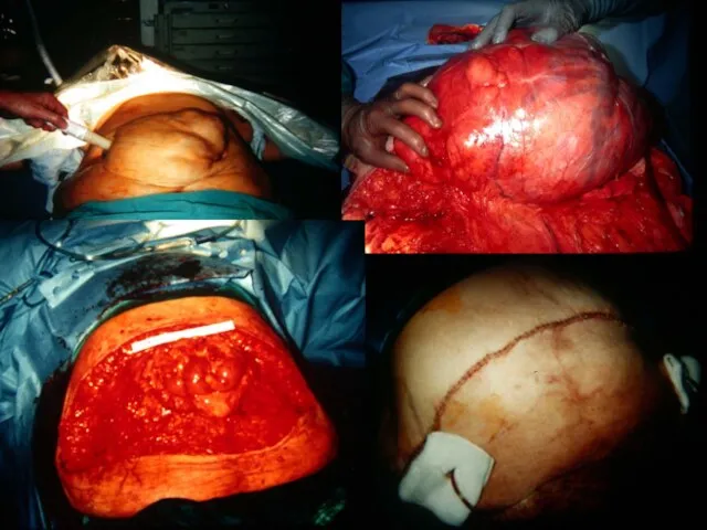
- 40. Incisional Hernia with Evisceration Note ulceration and spontaneous evisceration Cover with moist dressing. Take to operating
- 43. Incisional hernia with ‘peau d’orange’ (lymphedema)
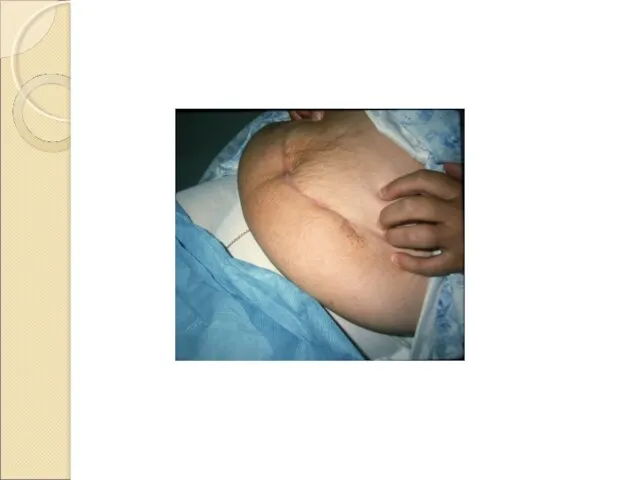
- 44. Large panniculus Small hernia
- 46. Interparietal hernia Very rare Between the layers of the abdominal wall Lateral to inguinal canal
- 47. Interparietal hernia Beneath external aponeurosis, coming through internal oblique muscle.
- 48. Left lower quadrant abdominal wall hernia outside inguinal canal containing sigmoid colon
- 49. Obturator Hernia Very rare Seen in elderly, emaciated patients Develops in obturator fossa Not visible or
- 50. Bowel obstruction from incarcerated obturator hernia
- 51. Obturator Hernia Causing Small Bowel Obstruction Site of obstruction deep in pelvis
- 52. Infarcted small bowel from obturator hernia
- 53. Spigelian Hernia Very rare, difficult to diagnose. Develops at or near intersection of arcuate and semilunar
- 54. Spigelian Hernia Laparoscopic view
- 55. Lumbar Hernia Develops at Petit’s Triangle Between abdominal and back muscles Fascia in this region is
- 56. Diastasis recti Not a hernia! Seen when there is wide separation of rectus muscle in epigastrium
- 59. Скачать презентацию

Hernia: The protrusion of tissue through a defect in fascial and/or
Hernia: The protrusion of tissue through a defect in fascial and/or

Semilunar line
Arcuate line
Basic Anatomy
Semilunar line
Arcuate line
Basic Anatomy
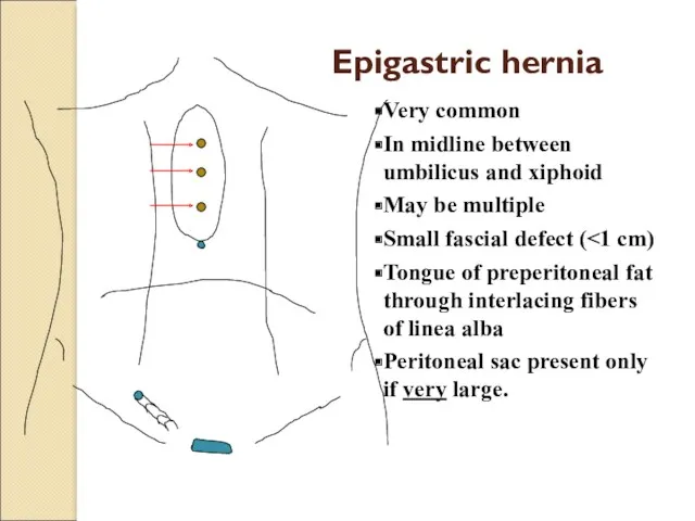
Very common
In midline between umbilicus and xiphoid
May be multiple
Small fascial defect
Very common
In midline between umbilicus and xiphoid
May be multiple
Small fascial defect
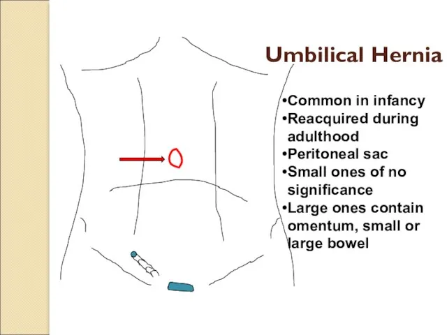
Umbilical Hernia
Common in infancy
Reacquired during adulthood
Peritoneal sac
Small ones of no
Umbilical Hernia
Common in infancy
Reacquired during adulthood
Peritoneal sac
Small ones of no
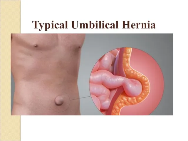
Typical Umbilical Hernia
Typical Umbilical Hernia
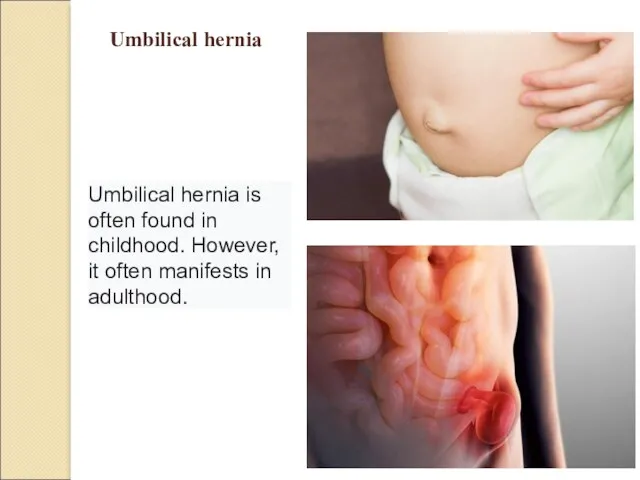
Umbilical hernia
Umbilical hernia is often found in childhood. However, it
Umbilical hernia
Umbilical hernia is often found in childhood. However, it
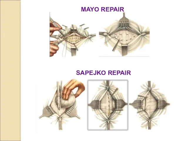
MAYO REPAIR
SAPEJKO REPAIR
MAYO REPAIR
SAPEJKO REPAIR
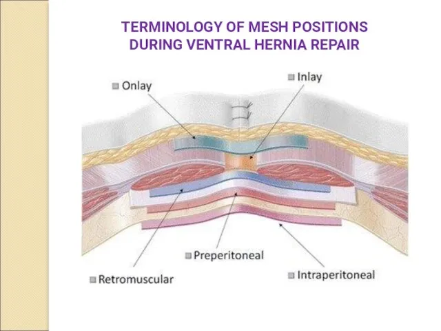
TERMINOLOGY OF MESH POSITIONS DURING VENTRAL HERNIA REPAIR
TERMINOLOGY OF MESH POSITIONS DURING VENTRAL HERNIA REPAIR

Umbilical & Inguinal Hernias
Umbilical & Inguinal Hernias

Most common
Congenital ~ indirect
Acquired ~ direct or indirect
Indirect Hernia
has peritoneal sac
lateral
Most common
Congenital ~ indirect
Acquired ~ direct or indirect
Indirect Hernia
has peritoneal sac
lateral
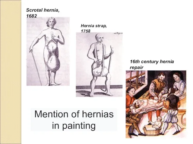
16th century hernia repair
Scrotal hernia, 1682
Hernia strap, 1758
Mention of hernias in
16th century hernia repair
Scrotal hernia, 1682
Hernia strap, 1758
Mention of hernias in
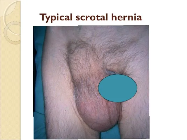
Typical scrotal hernia
Typical scrotal hernia

Giant scrotal hernia
Note scaphoid abdomen
Giant scrotal hernia
Note scaphoid abdomen
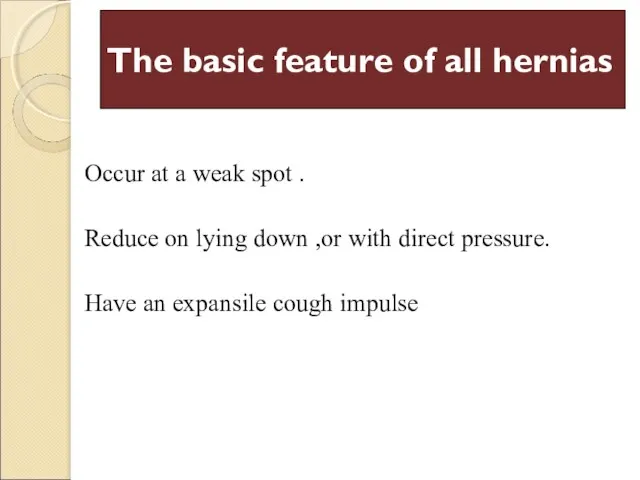
The basic feature of all hernias
Occur at a weak spot
The basic feature of all hernias
Occur at a weak spot
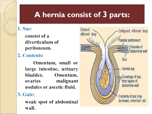
A hernia consist of 3 parts:
1. Sac:
consist of a diverticulum
A hernia consist of 3 parts:
1. Sac:
consist of a diverticulum
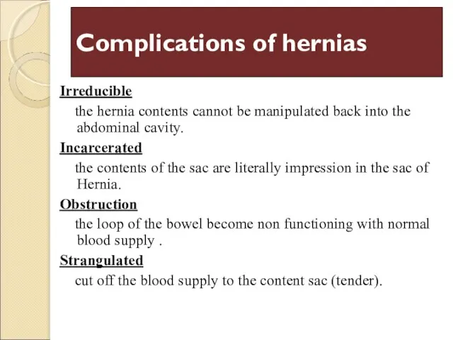
Complications of hernias
Irreducible
the hernia contents cannot be manipulated back
Complications of hernias
Irreducible
the hernia contents cannot be manipulated back
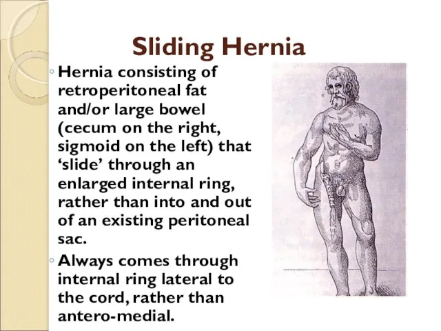
Sliding Hernia
Hernia consisting of retroperitoneal fat and/or large bowel (cecum on
Sliding Hernia
Hernia consisting of retroperitoneal fat and/or large bowel (cecum on
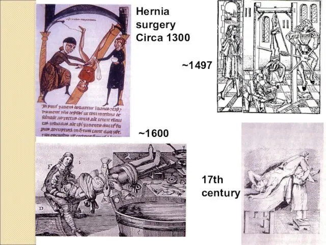
Hernia surgery
Circa 1300
~1600
17th century
~1497
Hernia surgery
Circa 1300
~1600
17th century
~1497

Giant Scrotal Hernia (1/2 of small bowel + right colon)
Giant Scrotal Hernia (1/2 of small bowel + right colon)
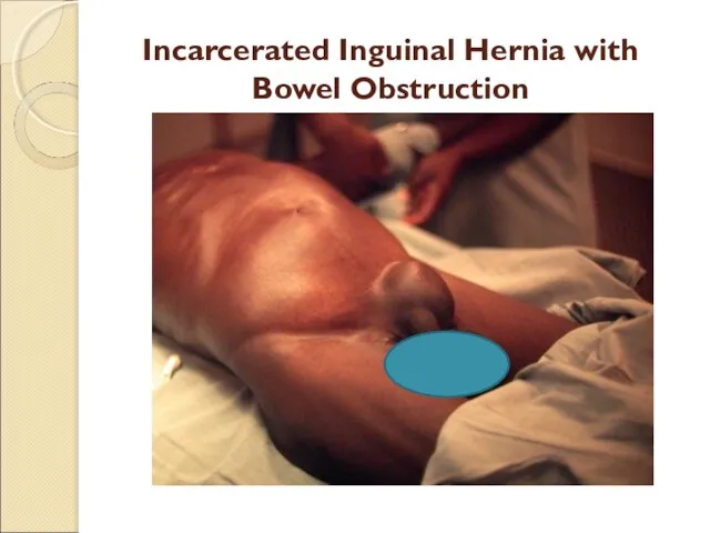
Incarcerated Inguinal Hernia with Bowel Obstruction
Incarcerated Inguinal Hernia with Bowel Obstruction

More typical inguinal hernia
More typical inguinal hernia
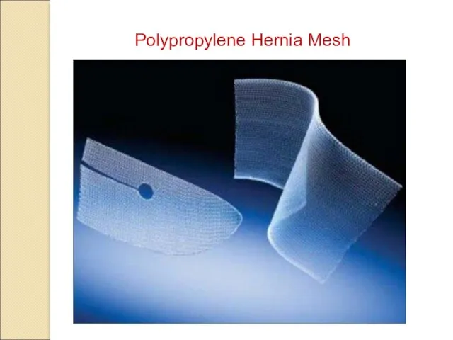
Polypropylene Hernia Mesh
Polypropylene Hernia Mesh

Polypropylene Hernia System
Polypropylene Hernia System
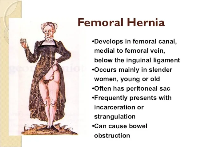
Femoral Hernia
Develops in femoral canal, medial to femoral vein, below the
Femoral Hernia
Develops in femoral canal, medial to femoral vein, below the
Femoral hernia versus inguinal hernia
Femoral hernia versus inguinal hernia
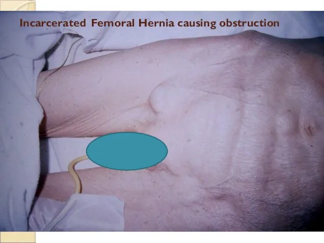
Incarcerated Femoral Hernia causing obstruction
Incarcerated Femoral Hernia causing obstruction
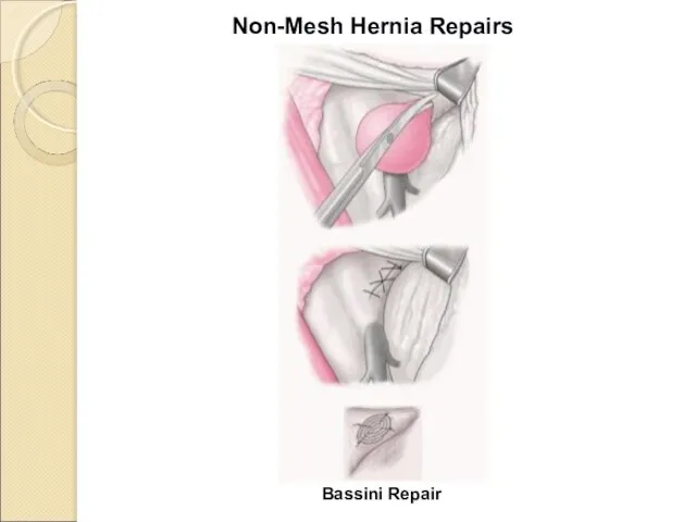
Bassini Repair
Non-Mesh Hernia Repairs
Bassini Repair
Non-Mesh Hernia Repairs
Polypropylene Hernia Mesh
Polypropylene Hernia Mesh

Can occur ANYWHERE an incision has been made, no matter how
Can occur ANYWHERE an incision has been made, no matter how
Incisional Hernia
Can develop in the original incision site because of dehiscence
Incisional Hernia
Can develop in the original incision site because of dehiscence
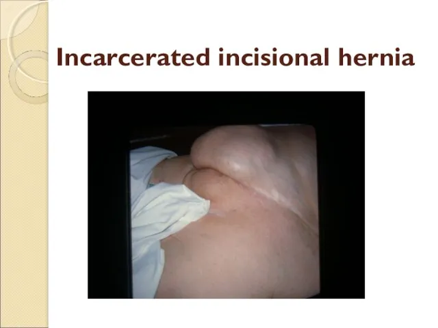
Incarcerated incisional hernia
Incarcerated incisional hernia
Causes of Incisional Hernia
Technical failure or fascial dehiscence:
Sutures rip through, are
Causes of Incisional Hernia
Technical failure or fascial dehiscence:
Sutures rip through, are
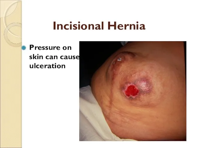
Incisional Hernia
Pressure on skin can cause ulceration
Incisional Hernia
Pressure on skin can cause ulceration
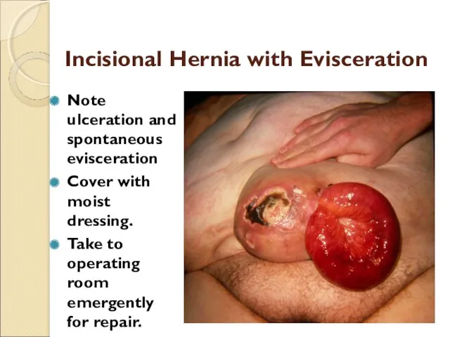
Incisional Hernia with Evisceration
Note ulceration and spontaneous evisceration
Cover with moist dressing.
Take
Incisional Hernia with Evisceration
Note ulceration and spontaneous evisceration
Cover with moist dressing.
Take
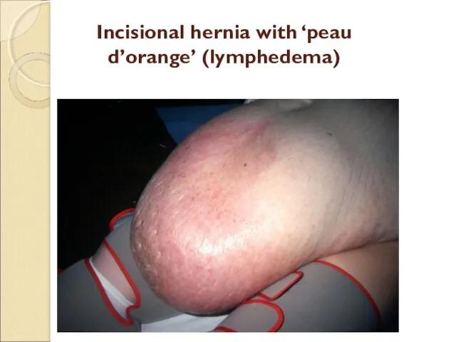
Incisional hernia with ‘peau d’orange’ (lymphedema)
Incisional hernia with ‘peau d’orange’ (lymphedema)
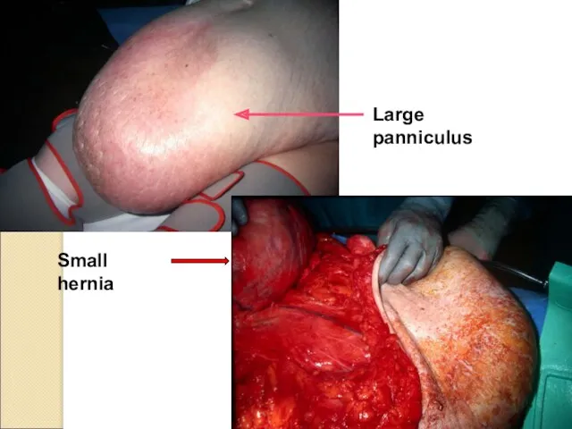
Large panniculus
Small hernia
Large panniculus
Small hernia
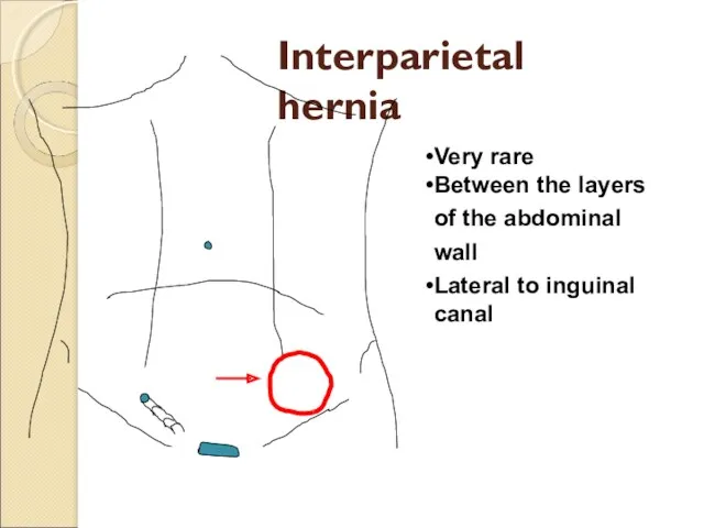
Interparietal hernia
Very rare
Between the layers of the abdominal wall
Lateral to inguinal
Interparietal hernia
Very rare
Between the layers of the abdominal wall
Lateral to inguinal
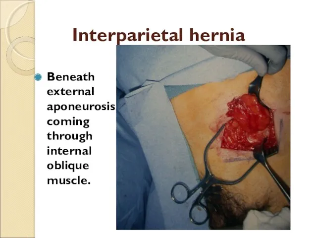
Interparietal hernia
Beneath external aponeurosis, coming through internal oblique muscle.
Interparietal hernia
Beneath external aponeurosis, coming through internal oblique muscle.
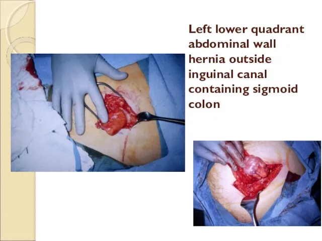
Left lower quadrant abdominal wall hernia outside inguinal canal
containing sigmoid colon
Left lower quadrant abdominal wall hernia outside inguinal canal
containing sigmoid colon
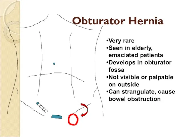
Obturator Hernia
Very rare
Seen in elderly, emaciated patients
Develops in obturator fossa
Not visible
Obturator Hernia
Very rare
Seen in elderly, emaciated patients
Develops in obturator fossa
Not visible
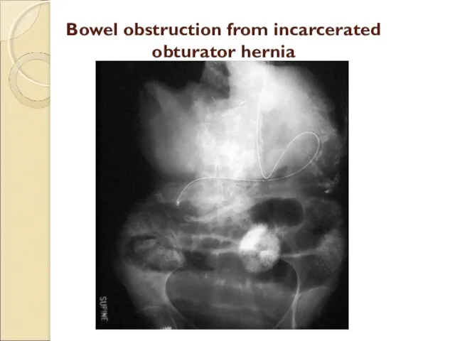
Bowel obstruction from incarcerated obturator hernia
Bowel obstruction from incarcerated obturator hernia

Obturator Hernia Causing Small
Bowel Obstruction
Site of obstruction deep in pelvis
Obturator Hernia Causing Small
Bowel Obstruction
Site of obstruction deep in pelvis
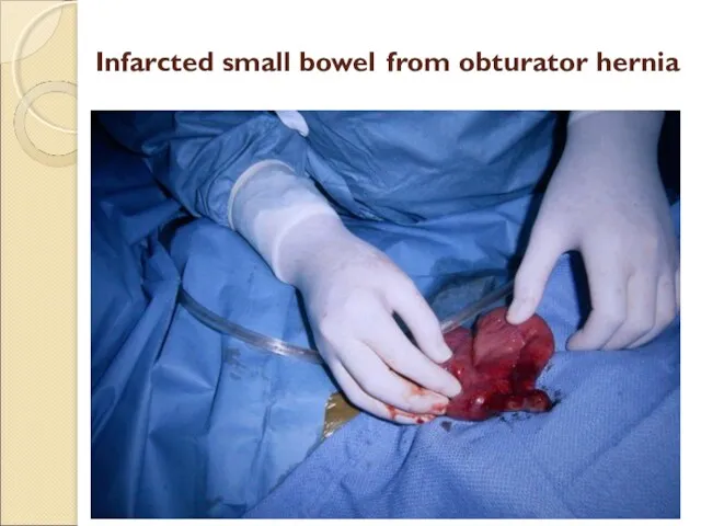
Infarcted small bowel from obturator hernia
Infarcted small bowel from obturator hernia

Spigelian Hernia
Very rare, difficult to diagnose.
Develops at or near intersection
Spigelian Hernia
Very rare, difficult to diagnose.
Develops at or near intersection
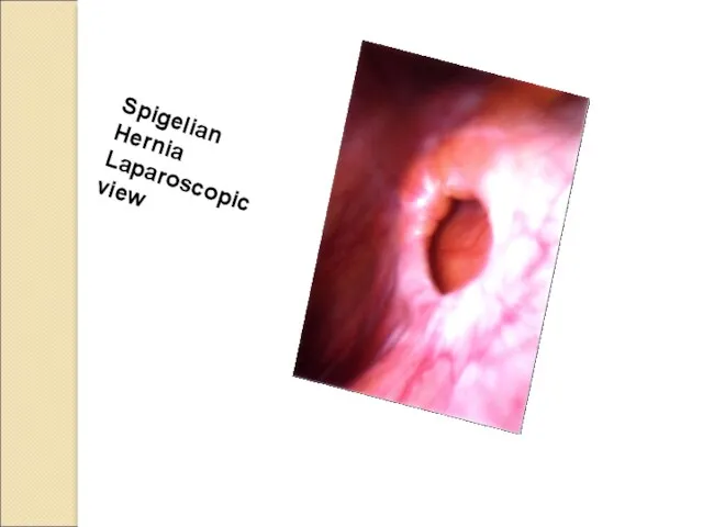
Spigelian
Hernia Laparoscopic view
Spigelian
Hernia Laparoscopic view
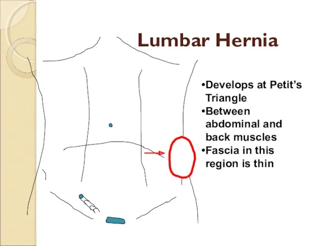
Lumbar Hernia
Develops at Petit’s Triangle
Between abdominal and back muscles
Fascia in this
Lumbar Hernia
Develops at Petit’s Triangle
Between abdominal and back muscles
Fascia in this
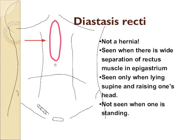
Diastasis recti
Not a hernia!
Seen when there is wide separation of
Diastasis recti
Not a hernia!
Seen when there is wide separation of
 Көпіршікті дерматоздар (пемфигус)
 Психологические аспекты работы в системе Медицинский регистратор – пациент
 Лабораторная диагностика паразитарных болезней
 Нәрестелердің іріңді септикалық аурулары. ГСЗ новорожденных
 Трансплантационный иммунитет
 Расспрос, осмотр больных с заболеваниями органов кровообращения. Пальпация и перкуссия области серца
 Особые формы плече-лопаточного периартрита
 Факторы иммунитета. Новый мир COVID-19. Инфекции и прививки
 Бронхиальная астма
 Анатомо-физиологические и психологические особенности лиц пожилого возраста
 АФО нервной системы и органов чувств ребенка. НПР детей раннего детского возраста
 Чувствительность и её нарушения
 Шеміршек тіні
 Реактивті артрит
 Эффект двойной дозы осельтамивира на лечение детей и взрослых госпитализированых с тяжелыми формами гриппа
 Проблема обеспеченности медицинской помощью жителей Бурятии
 Порядок выдачи и оформления листков нетрудоспособности
 Анестезиология и реаниматология – что это такое
 Острые лейкозы у детей
 Информационные технологии в медицине на примере практического занятия студентов
 Клинический разбор
 ВКР: Особенности деятельности медицинской сестры при обучении родителей уходу за новорожденным ребенком
 ВОП планирование семьи и репродуктивное сохранение здоровья. Пренатальная генетическая оценка и рекомендации
 Распространенные заболевания в терапии
 Групи крові. Переливання крові. Пересаджування кісткового мозку
 Блуждающий нерв (n.vagus). Анатомия. Клиника поражения
 Вакцинация
 Основы медицинской сортировки