- Chronic pancreatitis and pancreonecro sis

Содержание
- 2. ETIOLOGY Primary pancreatitis : Misuse of alcohol (70-80% of all diagnostic cases ) the systematic eating
- 3. THE PATHOGENESIS OF CHRONIC PANCREATITIS The main pathogenetic mechanism of the development of chronic pancreatitis is
- 4. DURATION OF CHRONIC PANCREATITIS IS DIVIDED INTO 3 PHASES : initial stage(1-5 years) – the most
- 5. ULTRASOUND INVESTIGATION. CHRONIC PANCREATITIS The pancreas might appear atrophic, calcified or fibrotic (advanced stages). Findings that
- 6. а) calcificates in the head of pancreas; б) Virsungov’s duct; в) pseudocyst of pancreas; г) increase
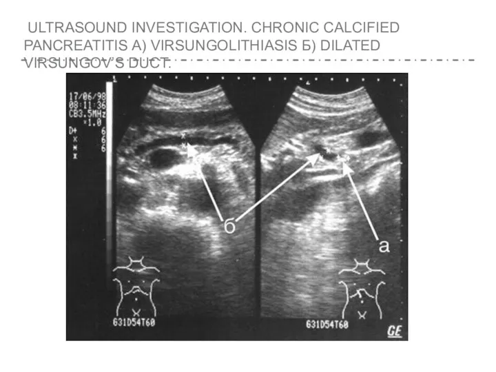
- 7. ULTRASOUND INVESTIGATION. CHRONIC CALCIFIED PANCREATITIS А) VIRSUNGOLITHIASIS Б) DILATED VIRSUNGOV’S DUCT.
- 8. ENDOSCOPIC ULTRASOUND has a vital diagnostic role because it is extremely sensitive in detecting the early
- 9. AN ENDOSCOPIC ULTRASOUND IMAGE DEMONSTRATING A DILATED PANCREATIC DUCT (MARKERS) IN A PATIENT WITH ADVANCED CHRONIC
- 10. COMPUTER TOMOGRAMPHY The diagnostic information similar to ultrasound, is indicated for suspected tumors and cysts of
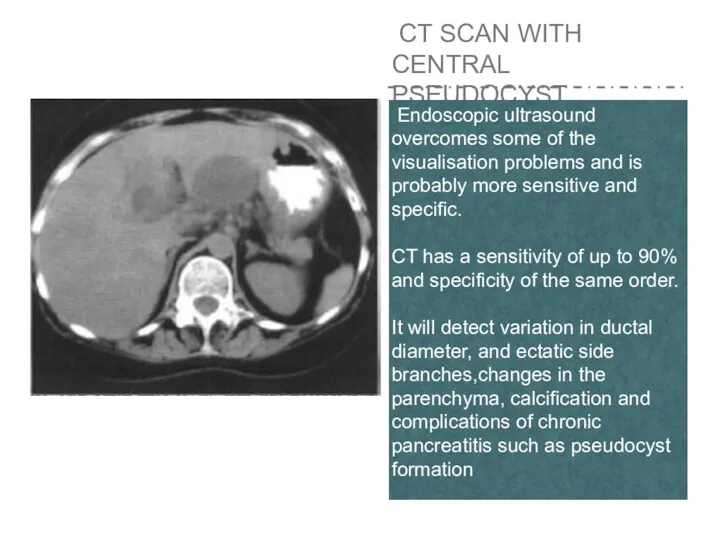
- 11. CT SCAN WITH CENTRAL PSEUDOCYST Endoscopic ultrasound overcomes some of the visualisation problems and is probably
- 12. ENDOSCOPIC RETROGRADE CHOLANGYIOPANKREATO GRAPHY reveals impaired patency of the main and secondary ducts. “Chain of lakes"
- 13. An endoscopic retrograde cholangiopancreatography image demonstrating minimal pancreatic duct abnormalities in a patient with painful small-duct
- 14. An endoscopic retrograde cholangiopancreatography image demonstrating massive pancreatic duct dilatation in a patient with bigduct chronic
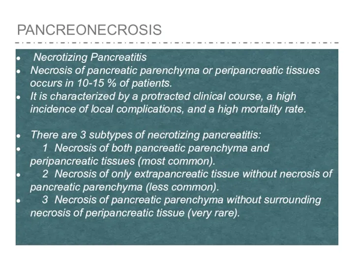
- 15. PANCREONECROSIS Necrotizing Pancreatitis Necrosis of pancreatic parenchyma or peripancreatic tissues occurs in 10-15 % of patients.
- 16. Necrosis of the pancreatic parenchyma can be diagnozed on a contrast-enhanced CT ⩾ 72 hours. Necrosis
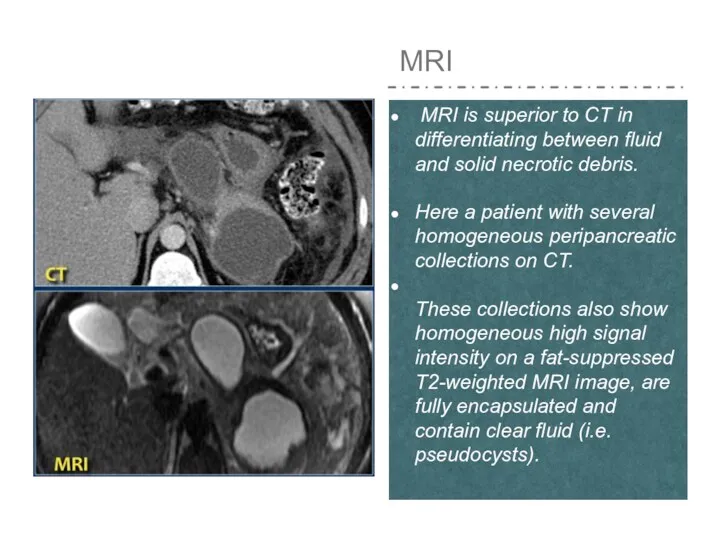
- 17. MRI MRI is superior to CT in differentiating between fluid and solid necrotic debris. Here a
- 18. This patient had an acute necrotizing pancreatitis with onset 2 months earlier. The CT-image shows a
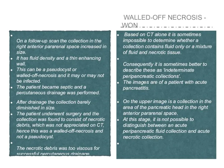
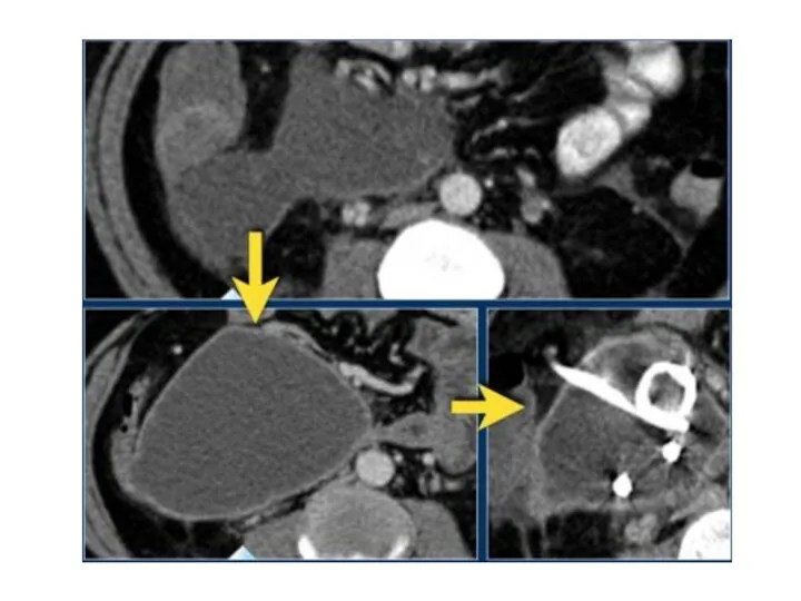
- 19. WALLED-OFF NECROSIS - WON Based on CT alone it is sometimes impossible to determine whether a
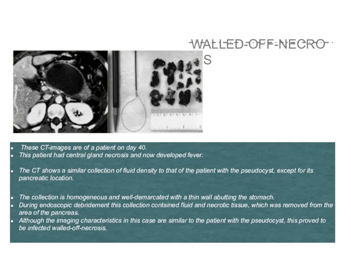
- 21. WALLED-OFF-NECROSIS These CT-images are of a patient on day 40. This patient had central gland necrosis
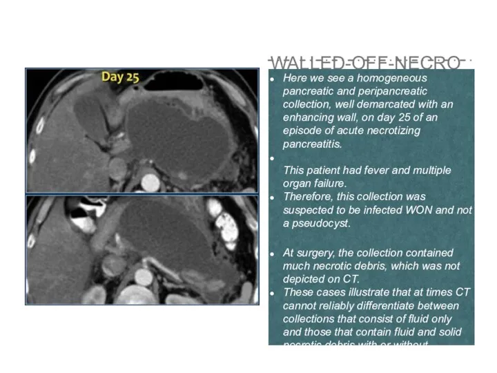
- 22. WALLED-OFF-NECROSIS Here we see a homogeneous pancreatic and peripancreatic collection, well demarcated with an enhancing wall,
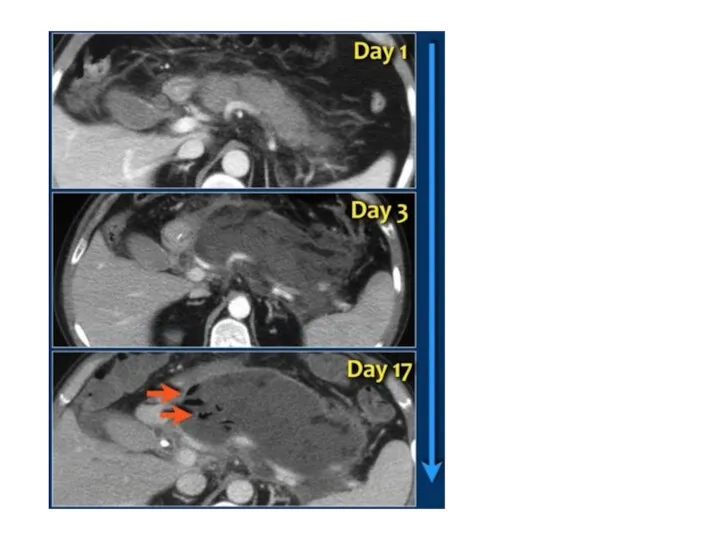
- 23. INFECTED NECROSIS Infected necrosis is: • Infection of necrotic pancreatic parenchyma or extrapancreatic fatty tissue -
- 26. Скачать презентацию
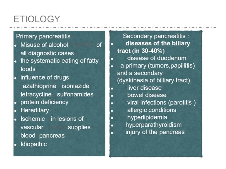
ETIOLOGY
Primary pancreatitis :
Misuse of alcohol (70-80% of all diagnostic
ETIOLOGY
Primary pancreatitis :
Misuse of alcohol (70-80% of all diagnostic
THE PATHOGENESIS OF CHRONIC PANCREATITIS
The main pathogenetic mechanism of
THE PATHOGENESIS OF CHRONIC PANCREATITIS
The main pathogenetic mechanism of
DURATION OF CHRONIC PANCREATITIS IS DIVIDED INTO 3 PHASES :
initial
DURATION OF CHRONIC PANCREATITIS IS DIVIDED INTO 3 PHASES :
initial
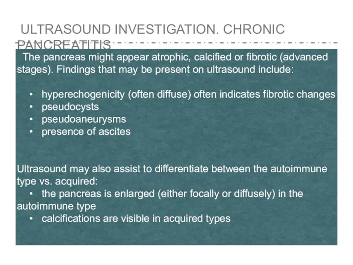
ULTRASOUND INVESTIGATION. CHRONIC PANCREATITIS
The pancreas might appear atrophic, calcified
ULTRASOUND INVESTIGATION. CHRONIC PANCREATITIS
The pancreas might appear atrophic, calcified
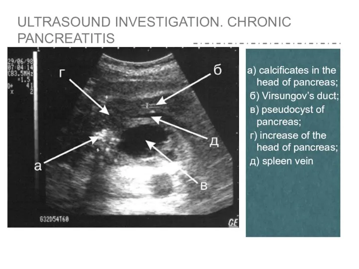
а) calcificates in the head of pancreas;
б) Virsungov’s duct;
б) Virsungov’s duct;
ULTRASOUND INVESTIGATION. CHRONIC CALCIFIED PANCREATITIS А) VIRSUNGOLITHIASIS Б) DILATED VIRSUNGOV’S
ULTRASOUND INVESTIGATION. CHRONIC CALCIFIED PANCREATITIS А) VIRSUNGOLITHIASIS Б) DILATED VIRSUNGOV’S
ENDOSCOPIC ULTRASOUND
has a vital diagnostic role because it
ENDOSCOPIC ULTRASOUND
has a vital diagnostic role because it
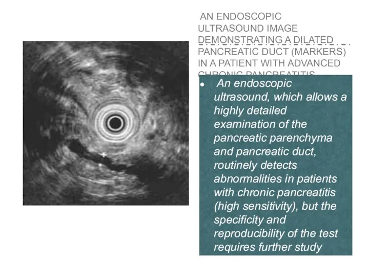
AN ENDOSCOPIC ULTRASOUND IMAGE DEMONSTRATING A DILATED PANCREATIC DUCT (MARKERS)
AN ENDOSCOPIC ULTRASOUND IMAGE DEMONSTRATING A DILATED PANCREATIC DUCT (MARKERS)
COMPUTER TOMOGRAMPHY
The diagnostic information similar to ultrasound, is indicated for suspected
COMPUTER TOMOGRAMPHY
The diagnostic information similar to ultrasound, is indicated for suspected
CT SCAN WITH CENTRAL PSEUDOCYST
Endoscopic ultrasound overcomes some
CT SCAN WITH CENTRAL PSEUDOCYST
Endoscopic ultrasound overcomes some

ENDOSCOPIC RETROGRADE CHOLANGYIOPANKREATO GRAPHY
reveals impaired patency of the main
ENDOSCOPIC RETROGRADE CHOLANGYIOPANKREATO GRAPHY
reveals impaired patency of the main
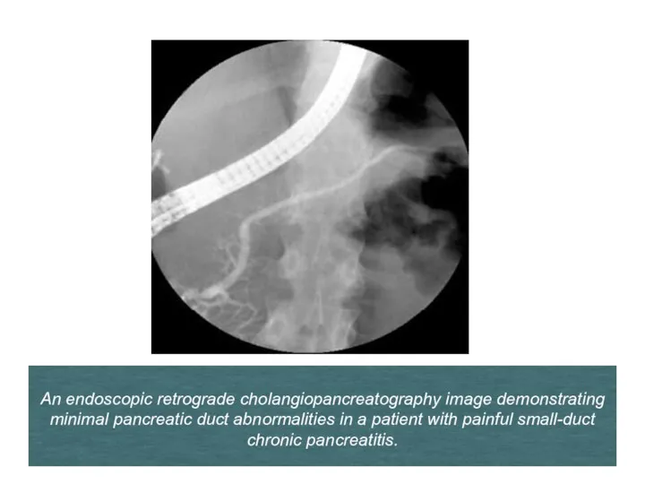
An endoscopic retrograde cholangiopancreatography image demonstrating minimal pancreatic duct abnormalities
An endoscopic retrograde cholangiopancreatography image demonstrating minimal pancreatic duct abnormalities
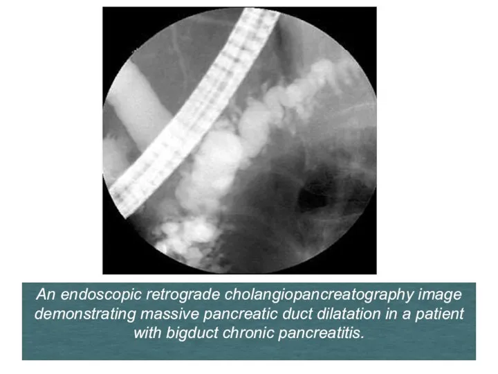
An endoscopic retrograde cholangiopancreatography image demonstrating massive pancreatic duct dilatation in
An endoscopic retrograde cholangiopancreatography image demonstrating massive pancreatic duct dilatation in
PANCREONECROSIS
Necrotizing Pancreatitis
Necrosis of pancreatic parenchyma or peripancreatic tissues occurs in
PANCREONECROSIS
Necrotizing Pancreatitis
Necrosis of pancreatic parenchyma or peripancreatic tissues occurs in
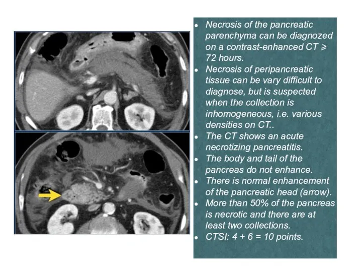
Necrosis of the pancreatic parenchyma can be diagnozed on a contrast-enhanced
Necrosis of the pancreatic parenchyma can be diagnozed on a contrast-enhanced
MRI
MRI is superior to CT in differentiating between fluid
MRI
MRI is superior to CT in differentiating between fluid
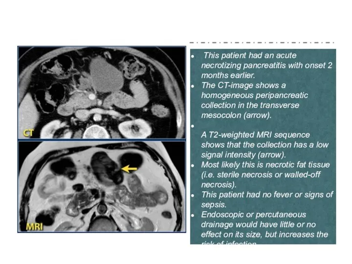
This patient had an acute necrotizing pancreatitis with onset 2
This patient had an acute necrotizing pancreatitis with onset 2
WALLED-OFF NECROSIS - WON
Based on CT alone it is
WALLED-OFF NECROSIS - WON
Based on CT alone it is
WALLED-OFF-NECROSIS
These CT-images are of a patient on day
WALLED-OFF-NECROSIS
These CT-images are of a patient on day
WALLED-OFF-NECROSIS
Here we see a homogeneous pancreatic and peripancreatic collection, well
WALLED-OFF-NECROSIS
Here we see a homogeneous pancreatic and peripancreatic collection, well
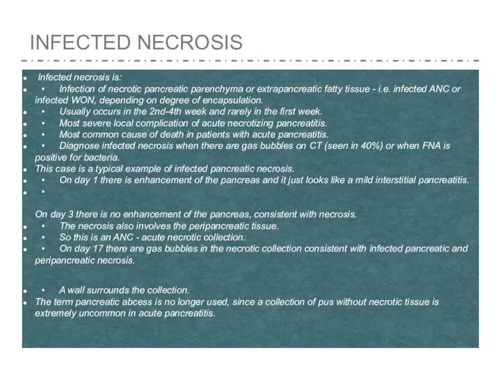
INFECTED NECROSIS
Infected necrosis is:
• Infection of necrotic pancreatic parenchyma or
INFECTED NECROSIS
Infected necrosis is:
• Infection of necrotic pancreatic parenchyma or
 Кандидоз полости рта у детей
 Коклюш
 Осторожно: клещи
 Гнойная инфекция кисти (панариций, флегмона)
 Рентгенологические методы исследования
 Проблемы питания и здорового образа жизни жителей современного города
 Черепные нервы
 Синтетические противомикробные средства. Лекция № 5
 Легионеллез
 Здоровые зубы
 Дієтичне харчування. Профілактика захворювань незбалансованого харчування. Харчові добавки
 Современные синтетические материалы в хирургии
 Хронический пылевой бронхит
 Мясоеды и вегетарианцы как две социальные группы
 Иммунный ответ. Определение, виды, особенности врожденного и адаптивного иммунитета
 Пищевые токсикоинфекции
 Устройство стоматологического кабинета
 Патологии органов зрения человека
 Гинекологические аспекты заболеваний молочных желёз
 Нарушение обмена веществ. Дистрофии
 Острое повреждение почек, влияние на исходы и возможности ранней коррекции
 Основы имплантации в стоматологии
 Тірегі имплант болғанда тіс протездерін қалыптастыру ерекшеліктері
 Правила обработки рук в соответствии с СанПиН. Методы и средства
 ЛФК при переломе позвоночника
 Трансплантация костного мозга
 Тұрғын үй қоғамдық ғимараттардың бөлмелерінде және тұрғын үй курылысын аймағында инфрадыбыс денгейін
 Невралгия тройничного нерва