- Эндоскопический подход в хирургии гнойных отитов

Содержание
- 2. VS
- 4. ТЕОРИТИЧЕСКИЕ ПЛЮСЫ И МИНУСЫ МИКРОСКОП ДВЕ РАБОЧИЕ РУКИ ОГРАНИЧЕННАЯ ВИДИМОСТЬ ЭНДОСКОП ОДНА РАБОЧАЯ РУКА ЯТРОГЕННЫЕ ТРАВМЫ
- 5. Джентельменский набор Figure 1. Instruments used. A: Traditional material for otological surgery added to curettes and
- 6. Figure 2. Ear endoscopic dissection. A: Endoscopic view (0 degrees and 4mm) of the middle ear,
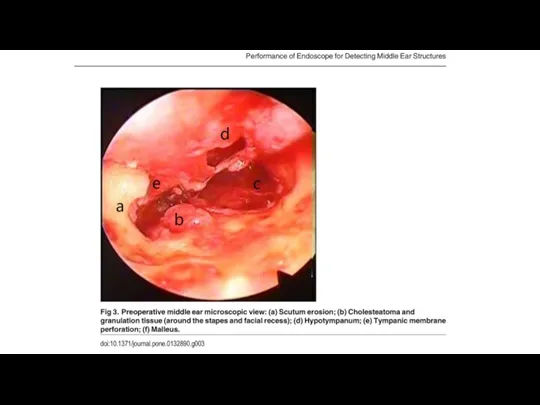
- 7. Figure 3. Ear endoscopic dissection. A: Endoscopic view (45 degrees and 4mm) of the middle ear,
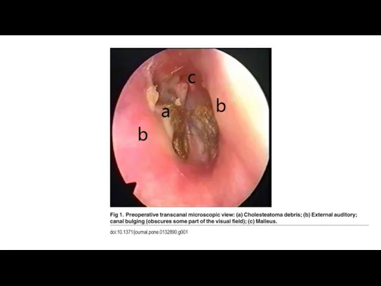
- 8. Figure 4. Ear endoscopic dissection. A: Endoscopic view (45 degrees and 4mm) of the middle ear.
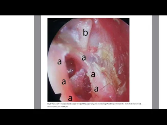
- 9. Figure 5. Ear endoscopic dissection. A: Endoscopic view (45 degrees and 4mm) of the middle ear
- 10. Figure 6. Ear endoscopic dissection. A: Endoscopic view (45 degrees and 4mm) of the middle ear
- 13. ИССЛЕДОВАНИЕ
- 14. Объекты исследования 58 пациентов Средний возраст 37 лет (15-63) 35 женщин, 23 мужчины Common complaints :
- 15. Манипуляции Общая анестезия, заушный разрез Lifted flap !Examination! Операция !Examination! =-=-=-=-
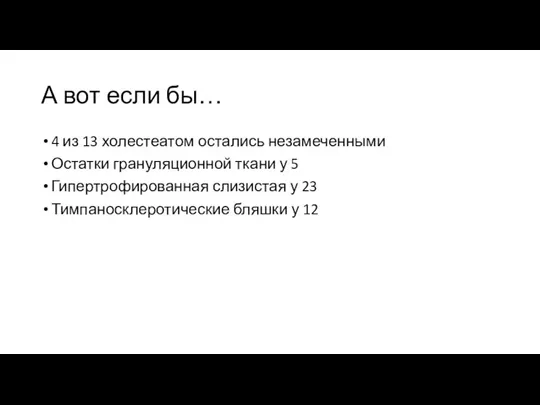
- 23. А вот если бы… 4 из 13 холестеатом остались незамеченными Остатки грануляционной ткани у 5 Гипертрофированная
- 25. Скачать презентацию
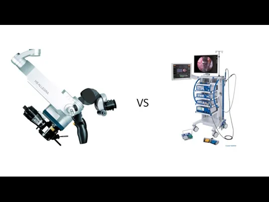
VS
VS

ТЕОРИТИЧЕСКИЕ ПЛЮСЫ И МИНУСЫ
МИКРОСКОП
ДВЕ РАБОЧИЕ РУКИ
ОГРАНИЧЕННАЯ ВИДИМОСТЬ
ЭНДОСКОП
ОДНА РАБОЧАЯ РУКА
ЯТРОГЕННЫЕ ТРАВМЫ
ИНДУЦИРОВАННАЯ
ТЕОРИТИЧЕСКИЕ ПЛЮСЫ И МИНУСЫ
МИКРОСКОП
ДВЕ РАБОЧИЕ РУКИ
ОГРАНИЧЕННАЯ ВИДИМОСТЬ
ЭНДОСКОП
ОДНА РАБОЧАЯ РУКА
ЯТРОГЕННЫЕ ТРАВМЫ
ИНДУЦИРОВАННАЯ
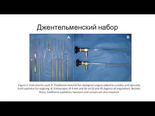
Джентельменский набор
Figure 1. Instruments used. A: Traditional material for otological
Джентельменский набор
Figure 1. Instruments used. A: Traditional material for otological
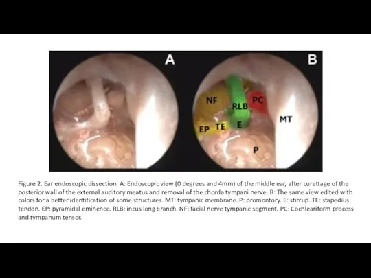
Figure 2. Ear endoscopic dissection. A: Endoscopic view (0 degrees and
Figure 2. Ear endoscopic dissection. A: Endoscopic view (0 degrees and
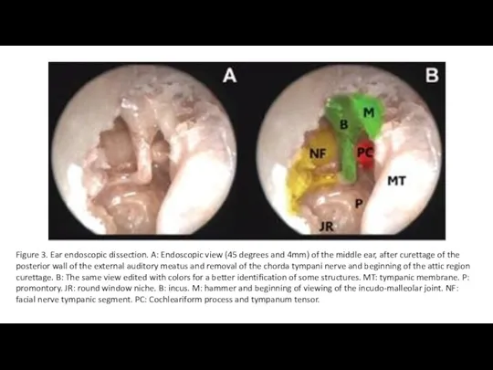
Figure 3. Ear endoscopic dissection. A: Endoscopic view (45 degrees and
Figure 3. Ear endoscopic dissection. A: Endoscopic view (45 degrees and

Figure 4. Ear endoscopic dissection. A: Endoscopic view (45 degrees and
Figure 4. Ear endoscopic dissection. A: Endoscopic view (45 degrees and

Figure 5. Ear endoscopic dissection. A: Endoscopic view (45 degrees and
Figure 5. Ear endoscopic dissection. A: Endoscopic view (45 degrees and
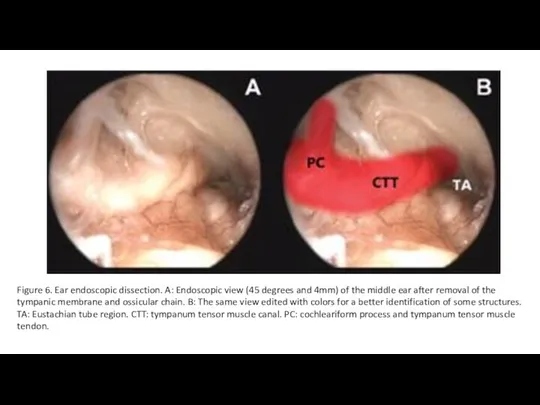
Figure 6. Ear endoscopic dissection. A: Endoscopic view (45 degrees and
Figure 6. Ear endoscopic dissection. A: Endoscopic view (45 degrees and

ИССЛЕДОВАНИЕ
ИССЛЕДОВАНИЕ
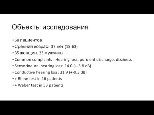
Объекты исследования
58 пациентов
Средний возраст 37 лет (15-63)
35 женщин, 23 мужчины
Common complaints
Объекты исследования
58 пациентов
Средний возраст 37 лет (15-63)
35 женщин, 23 мужчины
Common complaints
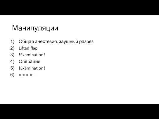
Манипуляции
Общая анестезия, заушный разрез
Lifted flap
!Examination!
Операция
!Examination!
=-=-=-=-
Манипуляции
Общая анестезия, заушный разрез
Lifted flap
!Examination!
Операция
!Examination!
=-=-=-=-


А вот если бы…
4 из 13 холестеатом остались незамеченными
Остатки грануляционной ткани
А вот если бы…
4 из 13 холестеатом остались незамеченными
Остатки грануляционной ткани
 Общая пропедевтика мочевыделительной системы
 Почки и надпочечники
 Penicillin is an antibiotic
 Бронхиальная астма
 Постхолецистэктомический синдром
 Владимир Петрович Филатов 1875 – 1956
 Косметика и гигиена
 Хронический гнойный средний отит
 Влияние алкоголя на женский организм
 Тубулопатия. Определение. Этиология
 Қан
Қан Психологическая профилактика алкогольной зависимости у медработников в условиях медицинского учреждения
 Принципы диагностики синдрома мальарбсорбции у детей
 Реконструкция проксимального отдела бедра после неудач в лечении травм
 Клинический разбор
 Создание новой модели медицинской организации, оказывающей первичную медико-санитарную помощь
 Бронхиальная астма, что нового?
 Санитарно-эпидемиологическая характеристика отходов
 Съемные ортодонтические аппараты различного типа действия
 Современные методы, позволяющие повысить эффективность имплантационных технологий
 Предмет и задачи патологии. Нозология
 Синдром слабости синусового узла
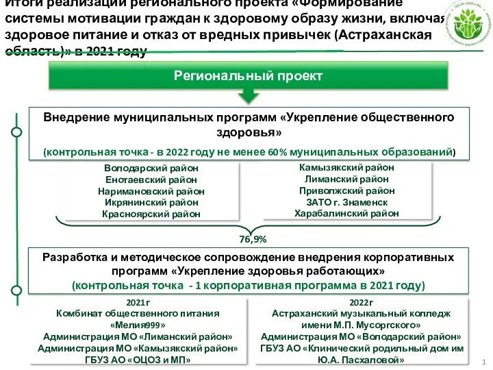 Реализация регионального проекта Укрепление общественного здоровья
 Нерв тіні
 Патофизиология ожоговой болезни. Интенсивная терапия ожоговой болезни и ожогового шока у детей
 Патогенез воспалительных заболеваний пародонта
 Сурфактантная терапия
 Фармакология как наука и учебная дисциплина