- ppt

Содержание
- 2. ECHINOCOCCUS GRANULOSUS Echinococcus granulosus, also called the hydatid worm, hyper tape-worm or dog tapeworm, is a
- 3. THE LIFECYCLE OF E. GRANULOSUS INVOLVES DOGS AND WILD CARNIVORES AS A DEFINITIVE HOST FOR THE
- 4. ECHINOCOCCUS GRANULOSUS WAS FIRST DOCUMENTED IN ALASKA BUT IS DISTRIBUTED WORLDWIDE. IT IS ESPECIALLY PREVALENT IN
- 5. TRANSMISSION E. granulosus requires two host types, a definitive host and an intermediate host. The definitive
- 6. ECHINOCOCCUS GRANULOSUS IS TRANSMITTED FROM THE INTERMEDIATE HOST (SHEEP) TO THE DEFINITIVE HOST (DOGS) BY FREQUENT
- 7. THE LIFE EXPECTANCY OF THE PARASITE, COUPLED WITH THE FREQUENCY OF ANTHELMINTHIC TREATMENTS, WILL ALSO PLAY
- 9. DIAGNOSIS Diagnosis in the definitive host, the dog, may be done by post mortem examination of
- 10. TREATMENT If a human becomes infected there are a variety of methods for treatment. The most
- 11. PREVENTION In order to prevent transmission to dogs from intermediate hosts, dogs can be given anthelminthic
- 12. ROPER DISPOSAL OF CARCASSES AND OFFAL AFTER HOME SLAUGHTER IS DIFFICULT IN POOR AND REMOTE COMMUNITIES
- 13. GEOGRAPHICAL DISTRIBUTION Echinococcus granulosus sensu lato occurs practically worldwide, and more frequently in rural, grazing areas
- 14. E. MULTILOCULARIS OCCURS IN THE NORTHERN HEMISPHERE, INCLUDING CENTRAL AND NORTHERN EUROPE, CENTRAL ASIA, NORTHERN RUSSIA,
- 15. CLINICAL PRESENTATION Echinococcus granulosus infections often remain asymptomatic for years before the cysts grow large enough
- 16. ECHINOCOCCUS MULTILOCULARIS AFFECTS THE LIVER AS A SLOW GROWING, DESTRUCTIVE TUMOR, OFTEN WITH ABDOMINAL PAIN AND
- 17. LIFE CYCLE The adult Echinococcus granulosus (sensu lato) (2—7 mm long) image resides in the small
- 18. DISEASE CAUSED – HYATID DISEASE SYMPTOMS: The time from ingestion of the eggs to developing symptoms
- 19. PREVENTIVE MEASURES Wash fruits and raw vegetables before eating. Wash hands before eating or smoking, after
- 20. MORPHOLOGY Echinococcus granulosus [this species causes hydatid disease in mammals, including humans] granulosus worms are small
- 21. SCIENTIFIC NAME- ECHINOCOCCUS GRANULOSUS HIGHER CLASSIFICATION- ECHINOCOCCUS ORDER- CYCLOPHYLLIDEA PHYLUM- PLATYHELMINTHIES FAMILY- TAENIIDAE CLASS- CESTODA
- 23. Скачать презентацию
ECHINOCOCCUS GRANULOSUS
Echinococcus granulosus, also called the hydatid worm, hyper tape-worm or
ECHINOCOCCUS GRANULOSUS
Echinococcus granulosus, also called the hydatid worm, hyper tape-worm or
THE LIFECYCLE OF E. GRANULOSUS INVOLVES DOGS AND WILD CARNIVORES AS
THE LIFECYCLE OF E. GRANULOSUS INVOLVES DOGS AND WILD CARNIVORES AS
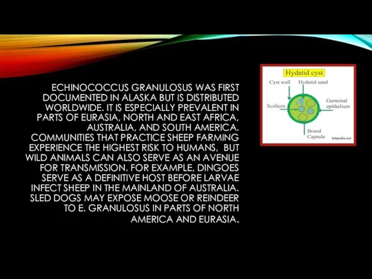
ECHINOCOCCUS GRANULOSUS WAS FIRST DOCUMENTED IN ALASKA BUT IS DISTRIBUTED WORLDWIDE.
ECHINOCOCCUS GRANULOSUS WAS FIRST DOCUMENTED IN ALASKA BUT IS DISTRIBUTED WORLDWIDE.
TRANSMISSION
E. granulosus requires two host types, a definitive host and an
TRANSMISSION
E. granulosus requires two host types, a definitive host and an
ECHINOCOCCUS GRANULOSUS IS TRANSMITTED FROM THE INTERMEDIATE HOST (SHEEP) TO THE
ECHINOCOCCUS GRANULOSUS IS TRANSMITTED FROM THE INTERMEDIATE HOST (SHEEP) TO THE
THE LIFE EXPECTANCY OF THE PARASITE, COUPLED WITH THE FREQUENCY OF
THE LIFE EXPECTANCY OF THE PARASITE, COUPLED WITH THE FREQUENCY OF
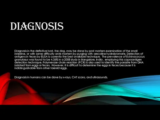
DIAGNOSIS
Diagnosis in the definitive host, the dog, may be done by
DIAGNOSIS
Diagnosis in the definitive host, the dog, may be done by
TREATMENT
If a human becomes infected there are a variety of methods
TREATMENT
If a human becomes infected there are a variety of methods
PREVENTION
In order to prevent transmission to dogs from intermediate hosts, dogs
PREVENTION
In order to prevent transmission to dogs from intermediate hosts, dogs
ROPER DISPOSAL OF CARCASSES AND OFFAL AFTER HOME SLAUGHTER IS DIFFICULT
ROPER DISPOSAL OF CARCASSES AND OFFAL AFTER HOME SLAUGHTER IS DIFFICULT
GEOGRAPHICAL DISTRIBUTION
Echinococcus granulosus sensu lato occurs practically worldwide, and more frequently
GEOGRAPHICAL DISTRIBUTION
Echinococcus granulosus sensu lato occurs practically worldwide, and more frequently
E. MULTILOCULARIS OCCURS IN THE NORTHERN HEMISPHERE, INCLUDING CENTRAL AND NORTHERN
E. MULTILOCULARIS OCCURS IN THE NORTHERN HEMISPHERE, INCLUDING CENTRAL AND NORTHERN
CLINICAL PRESENTATION
Echinococcus granulosus infections often remain asymptomatic for years before the
CLINICAL PRESENTATION
Echinococcus granulosus infections often remain asymptomatic for years before the
ECHINOCOCCUS MULTILOCULARIS AFFECTS THE LIVER AS A SLOW GROWING, DESTRUCTIVE TUMOR,
ECHINOCOCCUS MULTILOCULARIS AFFECTS THE LIVER AS A SLOW GROWING, DESTRUCTIVE TUMOR,
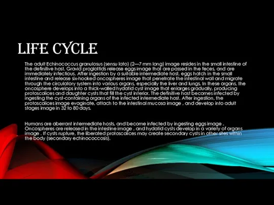
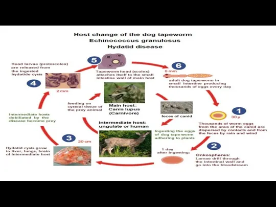
LIFE CYCLE
The adult Echinococcus granulosus (sensu lato) (2—7 mm long) image
LIFE CYCLE
The adult Echinococcus granulosus (sensu lato) (2—7 mm long) image
DISEASE CAUSED – HYATID DISEASE
SYMPTOMS:
The time from ingestion of the eggs
DISEASE CAUSED – HYATID DISEASE
SYMPTOMS:
The time from ingestion of the eggs
PREVENTIVE MEASURES
Wash fruits and raw vegetables before eating.
Wash hands before eating
PREVENTIVE MEASURES
Wash fruits and raw vegetables before eating.
Wash hands before eating
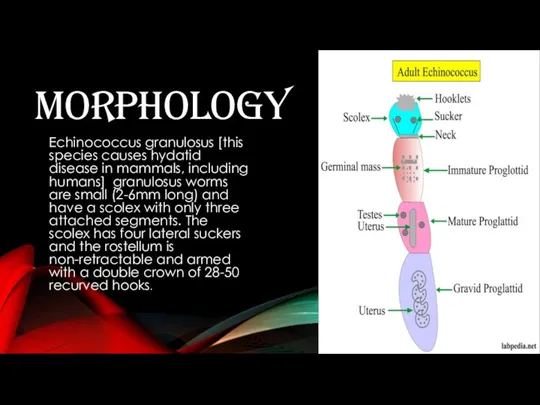
MORPHOLOGY
Echinococcus granulosus [this species causes hydatid disease in mammals, including humans]
MORPHOLOGY
Echinococcus granulosus [this species causes hydatid disease in mammals, including humans]
SCIENTIFIC NAME- ECHINOCOCCUS GRANULOSUS
HIGHER CLASSIFICATION- ECHINOCOCCUS
ORDER- CYCLOPHYLLIDEA
PHYLUM- PLATYHELMINTHIES
FAMILY- TAENIIDAE
CLASS- CESTODA
SCIENTIFIC NAME- ECHINOCOCCUS GRANULOSUS
HIGHER CLASSIFICATION- ECHINOCOCCUS
ORDER- CYCLOPHYLLIDEA
PHYLUM- PLATYHELMINTHIES
FAMILY- TAENIIDAE
CLASS- CESTODA
 ICPC-2 випадки тренування
 Ішек инфекциялары ауруларының асқынуы
 Аборты. Классификация. Способы производства. Ближайшие и отдаленные осложнения
 Астмалық статус
 Гипотиреоз
 Медико-генетическое консультирование (МГК)
 Хронобиология и хрономедицина. Временная организация клеток органов ротовой полости
 Алгоритм оказания первой помощи при ОНМК
 Синдромы при патологии сердечно-сосудистой системы
 Патологическая стираемость твердых тканей зубов
 Нарушения липидного обмена. Гиперлипидемия
 Ожоги и обморожения. Тепловой и солнечный удар. Укусы. Бешенство
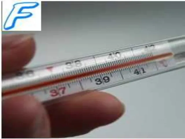 Лихорадка. Понятие о терморегуляции. Механизмы теплопродукции, теплоотдачи. Деятельность центра терморегуляции
 Клинические рекомендации по оказанию скорой медицинской помощи при перегревании и тепловом ударе
 Копрологические исследования
 Жүкті әйелдерге арналған стоматологиялық аурулардың алдын алу бағдарламасы
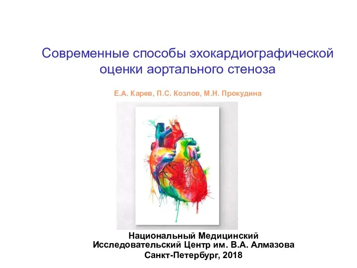 Современные способы эхокардиографической оценки аортального стеноза
 Қызықты клиникалық жағдай
 Системные заболевания соединительной ткани
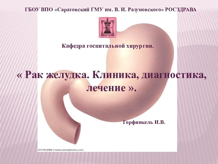 Рак желудка. Клиника, диагностика, лечение
 Жетілдірілген доға аппараттар
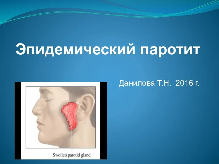 Эпидемический паротит
 Основы лучевой диагностики заболеваний легких
 Серологический метод исследования
 Первая помощь при травмах и ранениях
 остеомиелит новый
 Рубцовые поражения кожи. Косметологические методы коррекции
 Ведение беременности и родов при тазовом предлежании плода. Многоводие и маловодие