- Apoptosis and tumor suppressor proteins

Содержание
- 2. What is apoptosis? Apoptosis is a regulated cellular suicide mechanism characterized by nuclear condensation, cell shrinkage,
- 3. Importance of Apoptosis 1) Crucial for embryonic development -Errors in Apoptosis can lead to Birth Defects
- 4. Morphology Cell shrinkage (condensation of cytoplasm) Breakdown of mitochondria; release of cytochrome C Nuclear condensation Nuclear
- 5. How Apoptosis Differs from Necrosis? Apoptosis is intrinsically controlled, necrosis is not Apoptosis is more rapid
- 6. Mechanism I. Four stages of apoptosis have been defined: i. Committment to death by extracellular or
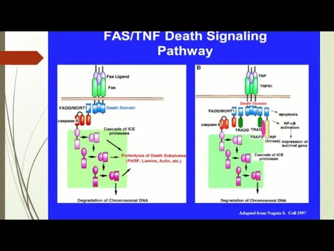
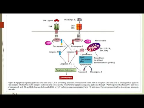
- 7. Death Factors Definition: cytokines that activate an apoptosis program by binding to their specific receptor. Typical
- 8. III. Activation of Caspase cascade i. Various stimuli described above eventually activate the executioner (caspase) cascade
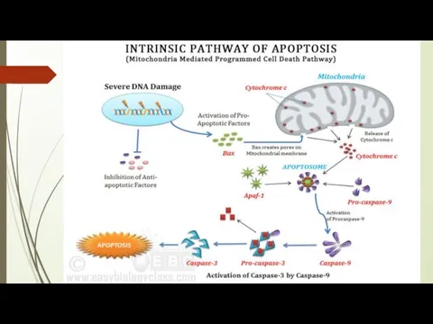
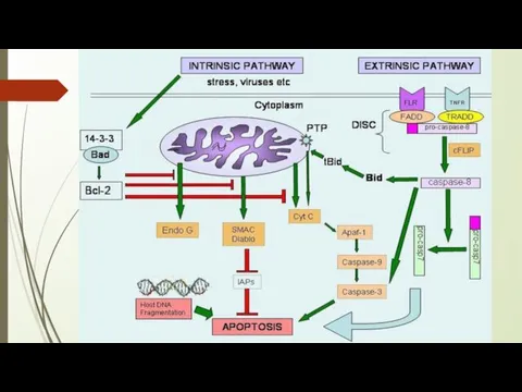
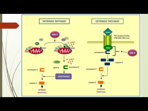
- 9. The intrinsic (mitochondrial) pathway of apoptosis. Death agonists cause changes in the inner mitochondrial membrane, resulting
- 10. Caspases are central initiators and executioners of apoptosis The term caspases is derived from cysteine-dependent aspartate-specific
- 17. Resistance to Fas signalling in cancer
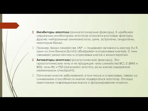
- 18. Ингибиторы апоптоза (антиапоптические факторы). К наиболее серьезным ингибиторам апоптоза относятся ростовые факторы. Другие: нейтральные аминокислоты, цинк,
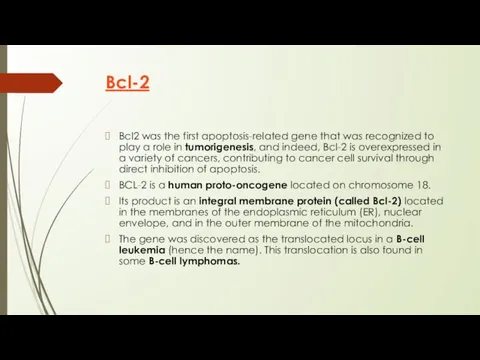
- 19. Bcl-2 Bcl2 was the first apoptosis-related gene that was recognized to play a role in tumorigenesis,
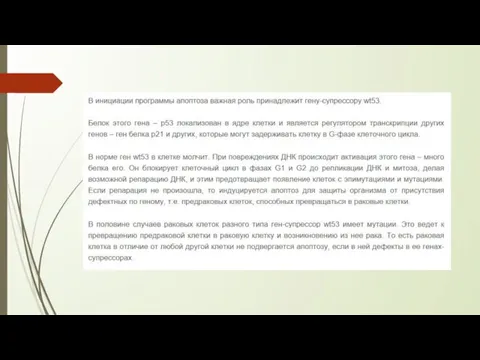
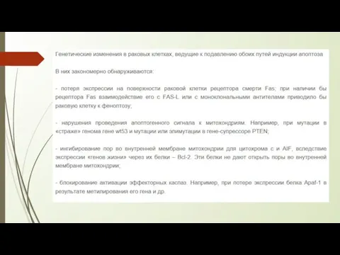
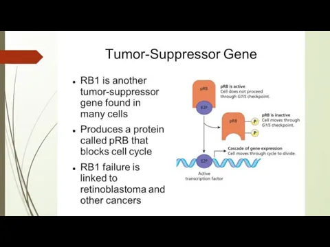
- 20. Белки супрессоры 1. Обнаружение повреждения в структуре ДНК. Этот факт - стимул для активации генов-супрессоров. 2.
- 21. Биологическая роль генов-супрессоров: они не пропускают в митоз клетку с поврежденной ДНК. Дефект гена-супрессора ведет к
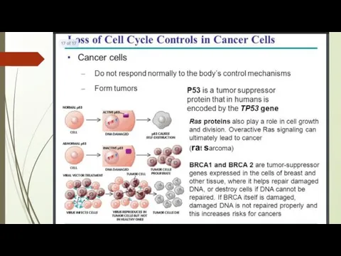
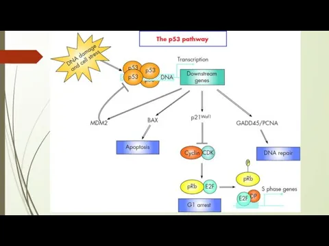
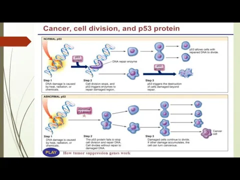
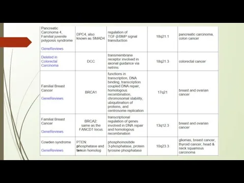
- 23. P53 protein Acts as a tumor suppressor gene 2 Main Functions: halts growth and division in
- 24. Сигнальный путь № 1 (связан с повреждением ДНК): 1. Повреждение ДНК 2. Активация гена р53 и
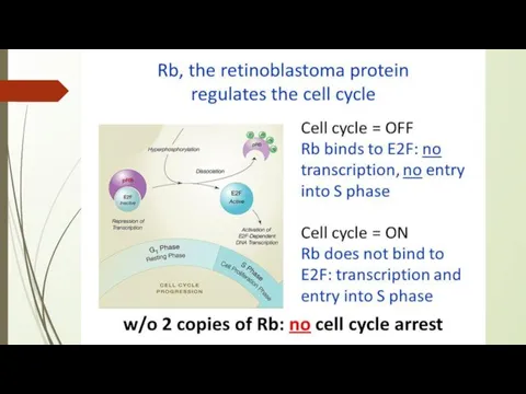
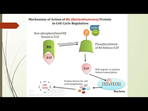
- 25. The p53 gene like the Rb gene, is a tumor suppressor gene, i.e., its activity stops
- 35. https://www.youtube.com/watch?v=8kbAQq_Pp8gb - Intrinsic Pathway https://www.youtube.com/watch?v=Aqf-n3pHv1I – Induction of apoptosis https://www.youtube.com/watch?v=1_s7KS2rit4 – Role of Mitochondria on apoptosis
- 36. Literature: https://www.researchgate.net/publication/221742318_Targeting_the_Fas-FasL_signaling_pathway_in_cancer_therapy https://www.cambridge.org/core/books/molecular-oncology/induction-of-apoptosis/0E2E934B7A64CCF86992F04BF081D8C2#fndtn-information https://www.cellsignal.com/contents/science-cst-pathways-apoptosis/regulation-of-apoptosis-interactive-pathway/pathways-apoptosis-regulation https://www.nature.com/articles/7290060 https://www.ncbi.nlm.nih.gov/books/NBK22268/ p53 https://themedicalbiochemistrypage.org/tumor-suppressors.php
- 37. http://humbio.ru/humbio/cytology/00118f2b.htm p53
- 39. Скачать презентацию
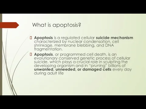
What is apoptosis?
Apoptosis is a regulated cellular suicide mechanism characterized by
What is apoptosis?
Apoptosis is a regulated cellular suicide mechanism characterized by
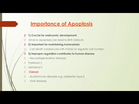
Importance of Apoptosis
1) Crucial for embryonic development
-Errors in Apoptosis can lead
Importance of Apoptosis
1) Crucial for embryonic development
-Errors in Apoptosis can lead
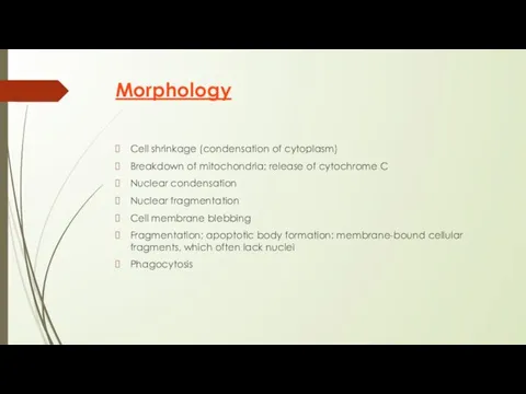
Morphology
Cell shrinkage (condensation of cytoplasm)
Breakdown of mitochondria; release of cytochrome C
Nuclear
Morphology
Cell shrinkage (condensation of cytoplasm)
Breakdown of mitochondria; release of cytochrome C
Nuclear
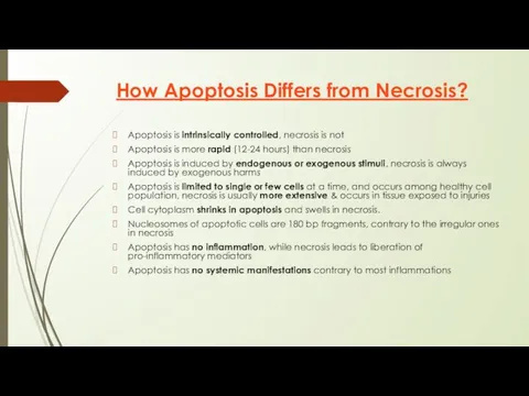
How Apoptosis Differs from Necrosis?
Apoptosis is intrinsically controlled, necrosis is not
Apoptosis
How Apoptosis Differs from Necrosis?
Apoptosis is intrinsically controlled, necrosis is not
Apoptosis
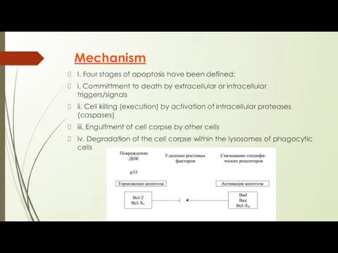
Mechanism
I. Four stages of apoptosis have been defined:
i. Committment to
Mechanism
I. Four stages of apoptosis have been defined:
i. Committment to
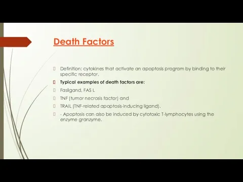
Death Factors
Definition: cytokines that activate an apoptosis program by binding to
Death Factors
Definition: cytokines that activate an apoptosis program by binding to
III. Activation of Caspase cascade
i. Various stimuli described above eventually
III. Activation of Caspase cascade
i. Various stimuli described above eventually
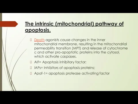
The intrinsic (mitochondrial) pathway of apoptosis.
Death agonists cause changes in the
The intrinsic (mitochondrial) pathway of apoptosis.
Death agonists cause changes in the
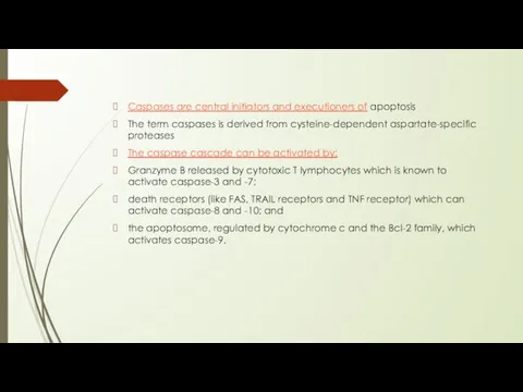
Caspases are central initiators and executioners of apoptosis
The term caspases is
Caspases are central initiators and executioners of apoptosis
The term caspases is
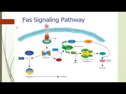
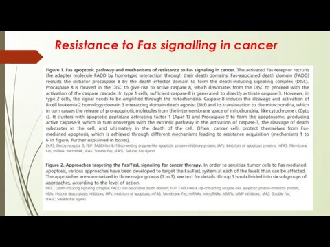
Resistance to Fas signalling in cancer
Resistance to Fas signalling in cancer
Ингибиторы апоптоза (антиапоптические факторы). К наиболее серьезным ингибиторам апоптоза относятся ростовые
Ингибиторы апоптоза (антиапоптические факторы). К наиболее серьезным ингибиторам апоптоза относятся ростовые
Bcl-2
Bcl2 was the first apoptosis-related gene that was recognized to play
Bcl-2
Bcl2 was the first apoptosis-related gene that was recognized to play
Белки супрессоры
1. Обнаружение повреждения в структуре ДНК. Этот факт -
Белки супрессоры
1. Обнаружение повреждения в структуре ДНК. Этот факт -
Биологическая роль генов-супрессоров: они не пропускают в митоз клетку с поврежденной
Биологическая роль генов-супрессоров: они не пропускают в митоз клетку с поврежденной
P53 protein
Acts as a tumor suppressor gene
2 Main Functions:
halts growth
P53 protein
Acts as a tumor suppressor gene
2 Main Functions:
halts growth
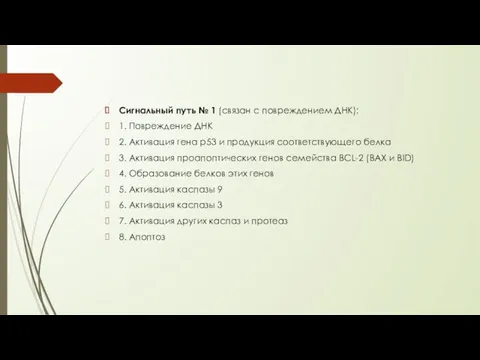
Сигнальный путь № 1 (связан с повреждением ДНК):
1. Повреждение ДНК
2. Активация
Сигнальный путь № 1 (связан с повреждением ДНК):
1. Повреждение ДНК
2. Активация
The p53 gene like the Rb gene, is a tumor suppressor
The p53 gene like the Rb gene, is a tumor suppressor
https://www.youtube.com/watch?v=8kbAQq_Pp8gb - Intrinsic Pathway
https://www.youtube.com/watch?v=Aqf-n3pHv1I – Induction of apoptosis
https://www.youtube.com/watch?v=1_s7KS2rit4 – Role of
https://www.youtube.com/watch?v=8kbAQq_Pp8gb - Intrinsic Pathway
https://www.youtube.com/watch?v=Aqf-n3pHv1I – Induction of apoptosis
https://www.youtube.com/watch?v=1_s7KS2rit4 – Role of
Literature:
https://www.researchgate.net/publication/221742318_Targeting_the_Fas-FasL_signaling_pathway_in_cancer_therapy
https://www.cambridge.org/core/books/molecular-oncology/induction-of-apoptosis/0E2E934B7A64CCF86992F04BF081D8C2#fndtn-information
https://www.cellsignal.com/contents/science-cst-pathways-apoptosis/regulation-of-apoptosis-interactive-pathway/pathways-apoptosis-regulation
https://www.nature.com/articles/7290060
https://www.ncbi.nlm.nih.gov/books/NBK22268/ p53
https://themedicalbiochemistrypage.org/tumor-suppressors.php
http://humbio.ru/humbio/cytology/00118f2b.htm p53
http://humbio.ru/humbio/cytology/00118f2b.htm p53
 Классный часСвятая мать добром спасет
 Презентация Развитие способностей и творческого потенциала всех детей в условиях реализации ФГОС ДО
 Клещевой энцифалит
 Этимология химических элементов
 20240130_17._delenie
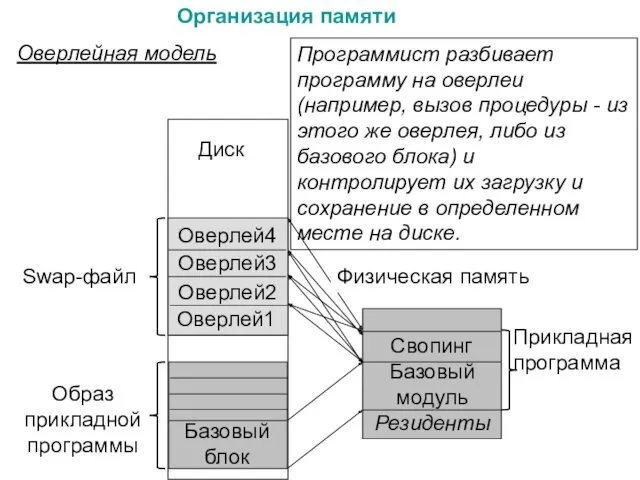 Организация памяти. (Лекция 5, 6)
 Особенности обучения взрослых и рекомендации по проведению тренинга
 Европейский иррационализм
 Роль религии в жизни общества
 Пространственная жесткость одноэтажных промышленных зданий
 Бази даних. Поняття звіту. Автоматичне створення звіту
 презентация Автоматизация звука С
 20231008_maslenitsa1
 Детские праздники и дни рождения Лазертаг
 ВКР: Реконструкции системы электроснабжения ОАО Балашихинский литейно-механический завод
 Урок по природе и экологии Красноярского края – 5 класс: Природные системы Красноярского края.
 Проект Безопасность детей через ознакомление с правилами дорожного движения
 Рельеф Земли. Равнины суши. Горы суши
 Мой любимый поэт Александр Сергеевич Пушкин
 Дисперсті жүйелердің оптикалық әдістері
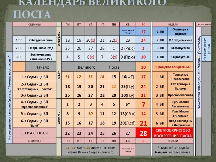 Календарь Великикого Поста
 Сочинение-описание картины Е. Н. Широкова Друзья
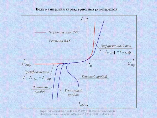 Вольт-амперная характеристика p-n-перехода
 Презентация День защиты детей
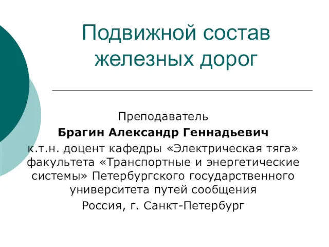 Подвижной состав железных дорог
 Коэффициент полезного действия механизма
 Тізе буыны туберкулезінің салыстырмалы диагностикасы
 Технология выращивания посадочного материала. Организация питомника. Лекция-1