Слайд 2

The tooth consists of: crown is the visible part of the
tooth, above the gums; root is the part of the tooth under the gums and inside the alveolar bone that keeps the tooth in place; gum margin(neck) is the area between the tooth crown and the root.
Enamel is the hardest and most highly mineralized substance of the body. It is one of the four major tissues which make up the tooth, along with dentin, cementum, and dental pulp. It is normally visible and must be supported by underlying dentin.
Слайд 3

96% of enamel consists of mineral, with water and organic material
comprising the rest. The normal color of enamel varies from light yellow to grayish white. At the edges of teeth where there is no dentin underlying the enamel, the color sometimes has a slightly blue tone. Since enamel is semitranslucent, the color of dentin and any restorative dental material underneath the enamel strongly affects the appearance of a tooth. Enamel varies in thickness over the surface of the tooth and is often thickest at the cusp, up to 2.5mm, and thinnest at its border. Enamel's primary mineral is hydroxylapatite, which is a crystalline calcium phosphate. The large amount of minerals in enamel accounts not only for its strength but also for its brittleness.
Слайд 4

Dentin is the substance between enamel or cementum and the pulp
chamber. It is secreted by the odontoblasts of the dental pulp. The formation of dentin is known as dentinogenesis. The porous, yellow-hued material is made up of 70% inorganic materials, 20% organic materials, and 10% water by weight.
Cementum is a specialized bone like substance covering the root of a tooth. It is approximately 45% inorganic material (mainly hydroxyapatite), 33% organic material (mainly collagen) and 22% water. Cementum is excreted by cementoblasts within the root of the tooth and is thickest at the root apex.
Слайд 5

The dental pulp is the central part of the tooth filled
with soft connective tissue. This tissue contains blood vessels and nerves that enter the tooth from a hole at the apex of the root. Along the border between the dentin and the pulp are odontoblasts, which initiate the formation of dentin.Other cells in the pulp include fibroblasts, preodontoblasts, macrophages and T lymphocytes. The pulp is commonly called "the nerve" of the tooth.
Слайд 6

Слайд 7


 Genito-Urinary Examination
 Test Execution
 Virus which gave birth to racism
 Aleksei Brusilov distinguished commander of the First World War
 Verb. General characteristics. Person and number
 The Influence of Topics on Listening Strategy Use for English for Academic Purposes
 Verb to be. Present simple
 Стереотипні поетичні образи в британській та американській поезії
 Monuments to cultural workers in Russia
 Reported speech
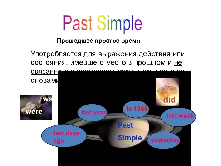 Past Simple. Прошедшее простое время
 My favorite holiday
 Английский для начинающих
 Family. Types of families
 Weekends. Leisure
 Usain Bolt
 Environmental pollution
 Hobby for pupils
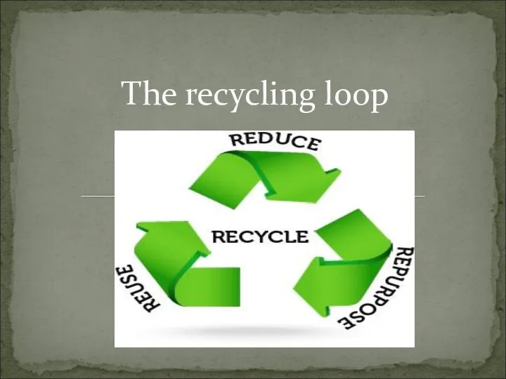 The recycling loop
 Preservation of cultural heritage of Kazakhstan
 Неопределенный артикль, 5 класс
 Start speaking. Английский устный, вариант 14
 Who knows the united kingdom perfectly?
 Household chores
 Random questions
 Colors
 Structure of English Words
 The judicial system of Great Britain