- Anatomy Of The Skin. Lecture 1

Содержание
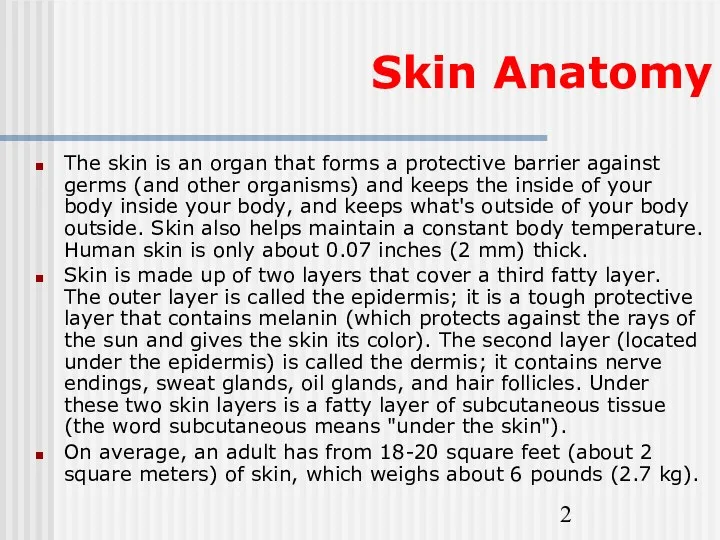
- 2. Skin Anatomy The skin is an organ that forms a protective barrier against germs (and other
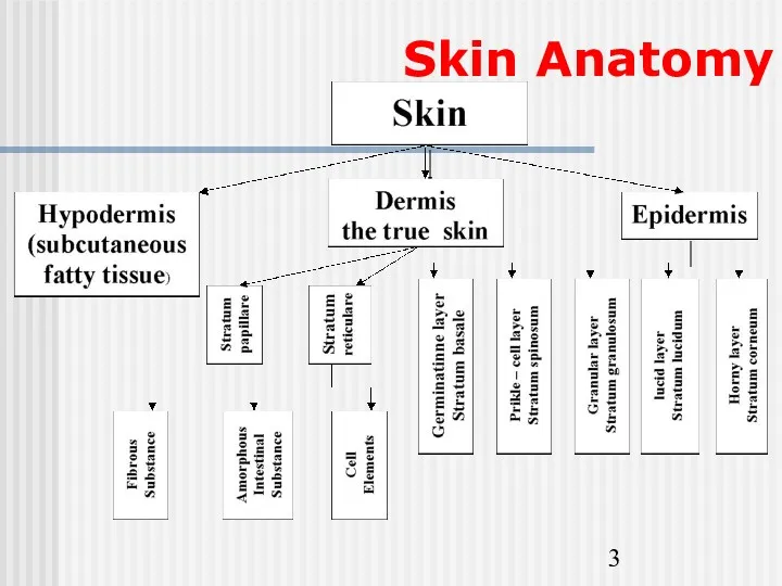
- 3. Skin Anatomy
- 4. Epidermis and it’s layers The epidermis is the most superficial layer of the skin and provides
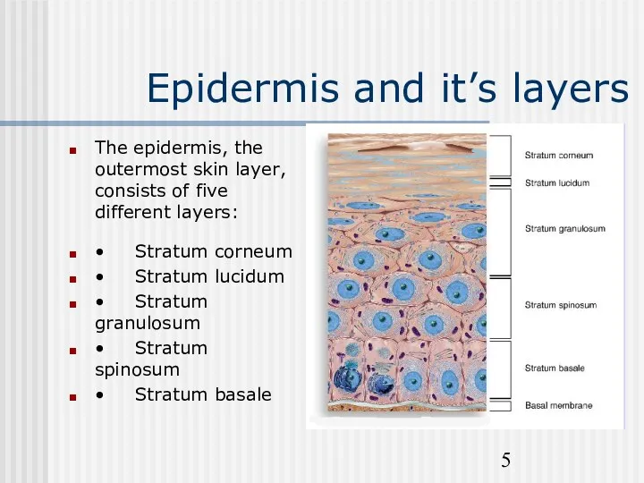
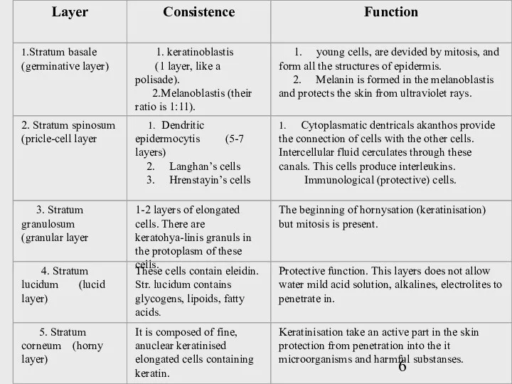
- 5. Epidermis and it’s layers The epidermis, the outermost skin layer, consists of five different layers: •
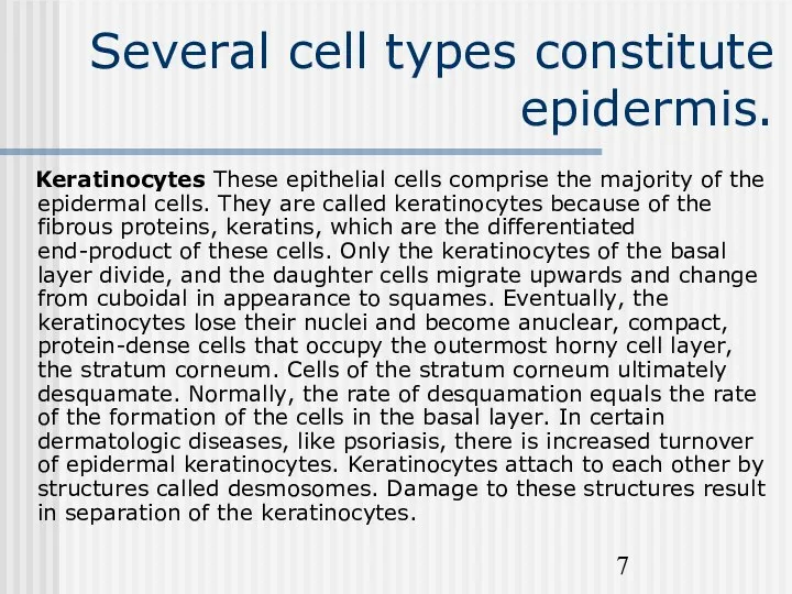
- 7. Several cell types constitute epidermis. Keratinocytes These epithelial cells comprise the majority of the epidermal cells.
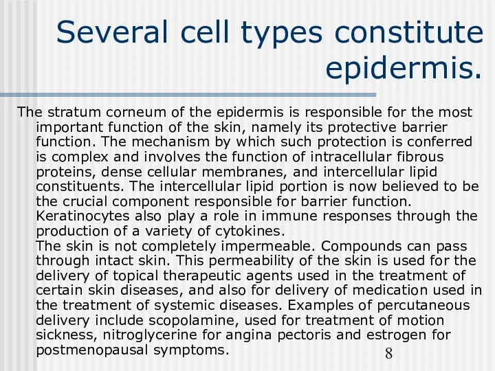
- 8. Several cell types constitute epidermis. The stratum corneum of the epidermis is responsible for the most
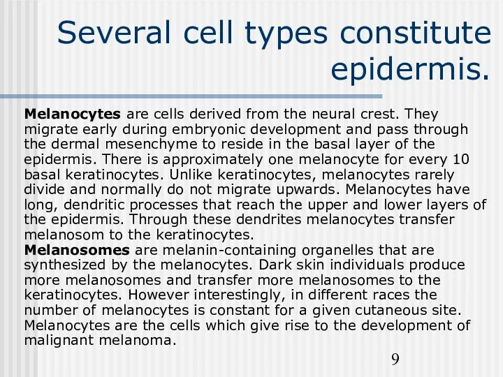
- 9. Several cell types constitute epidermis. Melanocytes are cells derived from the neural crest. They migrate early
- 10. Several cell types constitute the stratum bazale. The function of melanin is to provide protection against
- 11. Stratum germinativum (bazale) The stratum germinatum (SG) provides the germinal cells necessary for the regeneration of
- 12. Stratum spinosum The cells that divide in the statum germinativum soon begin to accumulate many desmosomes
- 13. Stratum granulosum The progressive maturation of a keratinocyte is charcterized by the accumulation of keratin, called
- 14. Stratum Lucidum Epidermis varies in thickness throughout the body depending mainly on frictional forces and is
- 15. Stratum corneum As a cell accumulates keratinohyalin granules, it is thought that rupture of lysosomal membranes
- 16. The dermal-epidermal basement membrane Between the epidermis and the dermis there is a basement membrane, composed
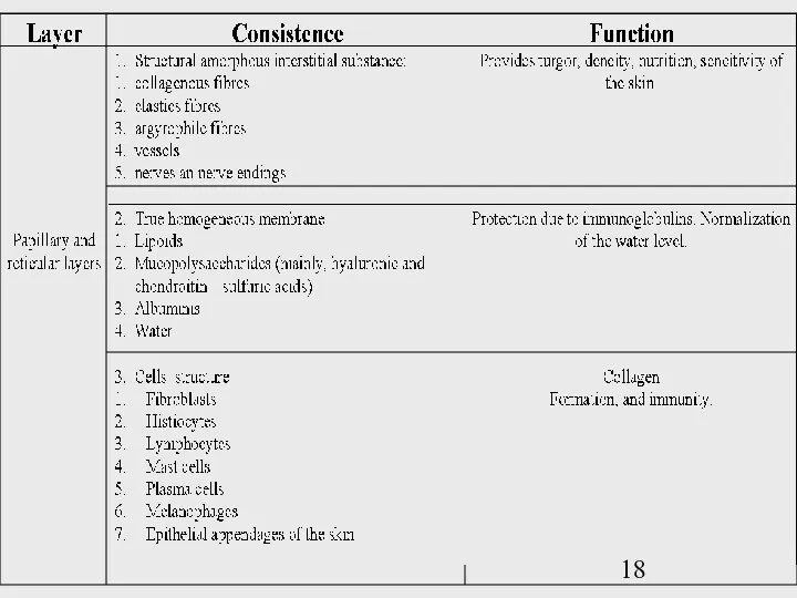
- 17. Dermis The dermis (D) assumes the important functions of thermoregulation and supports the vasular network to
- 19. Papillary dermis The papillary dermis (PD) contains vascular networks that have two important functions. The first
- 20. Reticular dermis The reticular layer of the dermis (RD) consists of dense irregular connective tissue, which
- 21. Dermis The dermis is the supporting layer of the epidermis. It consists of the fibrous components
- 22. Dermis 1. Fibrous Components and Ground Substance Collagens comprise 98% of the dermal fibrous component. They
- 23. Dermis 2. Blood Vessels - The skin is richly vascularized. The cutaneous vasculature is required for
- 24. Dermis 3. Nerves - Unmyelinated and myelinated sensory nerves are present in the dermis. Free nerve
- 25. Dermis 4. Epidermal appendages during fetal development, specialized epithelial derived structures develop from the epidermis, towards
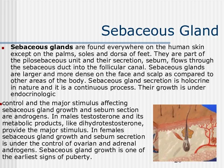
- 26. Sebaceous Gland Sebaceous glands are found everywhere on the human skin except on the palms, soles
- 27. Sweat Glands There are two types of sweat glands: eccrine and apocrine. Eccrine glands are found
- 28. Sweat Glands Thermal sweating occurs over most of the body integument. Emotional stress can induce eccrine
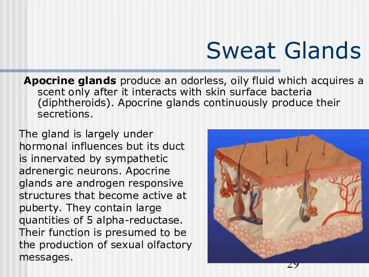
- 29. Sweat Glands Apocrine glands produce an odorless, oily fluid which acquires a scent only after it
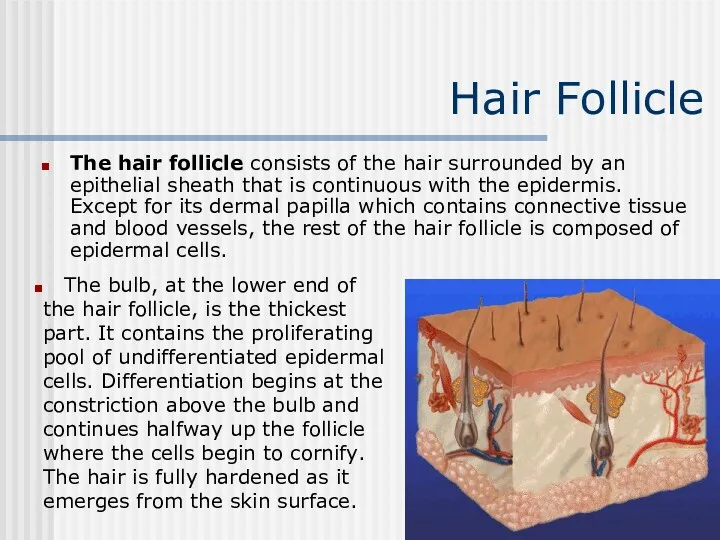
- 30. Hair Follicle The hair follicle consists of the hair surrounded by an epithelial sheath that is
- 31. Hair Follicle Human hair grows in cycles. The longer the hair growth phase of an individual,
- 32. Hair Follicle Conversely, telogen hairs are easily dislodged and account for the normal loss, defluvium, that
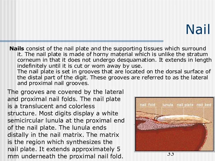
- 33. Nail Nails consist of the nail plate and the supporting tissues which surround it. The nail
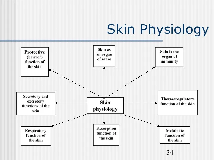
- 34. Skin Physiology
- 35. Skin Function A. Sensation (largest sensory organ in the body) B. Protection 1. Prevents dehydration 2.
- 36. Protective function
- 37. Epidermis Cell Layers (cells mature from inner to outer) A. Stratum Corneum (Cornified Layer) 1. Outermost
- 38. Sensory Apparatus of the Skin The skin is innervated with around one million afferent nerve fibers.
- 39. Sensory Apparatus of the Skin Sensory endings are of two main kinds: corpuscular, which embrace non-nervous
- 40. Sensory Apparatus of the Skin The Pacinian corpuscle is one of the encapsulated receptors. It is
- 41. Sensory Apparatus of the Skin Ruffini endings in the human digits have several expanded endings branching
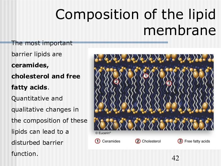
- 42. Composition of the lipid membrane The most important barrier lipids are ceramides, cholesterol and free fatty
- 43. Other functions of the skin The skin is structured to prevent loss of essential body fluids,
- 44. Protective functions of the skin
- 45. Natural Moisturising Factors
- 46. Skin surface lipids
- 47. The skin‘s immune system They also play a role in immunosurveillance against viral infections. Langerhans cells
- 48. SALT
- 49. Other functions of the skin Melanin pigment of the skin protects the nuclear structures against damage
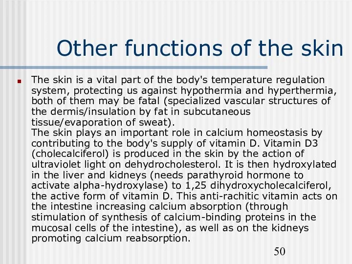
- 50. Other functions of the skin The skin is a vital part of the body's temperature regulation
- 52. Скачать презентацию
Skin Anatomy
The skin is an organ that forms a protective
Skin Anatomy
The skin is an organ that forms a protective
Skin Anatomy
Skin Anatomy
Epidermis and it’s layers
The epidermis is the most superficial layer of
Epidermis and it’s layers
The epidermis is the most superficial layer of
Epidermis and it’s layers
The epidermis, the outermost skin layer,
consists
Epidermis and it’s layers
The epidermis, the outermost skin layer, consists
Several cell types constitute epidermis.
Keratinocytes These epithelial cells comprise
Several cell types constitute epidermis.
Keratinocytes These epithelial cells comprise
Several cell types constitute epidermis.
The stratum corneum of the epidermis is
Several cell types constitute epidermis.
The stratum corneum of the epidermis is
Several cell types constitute epidermis.
Melanocytes are cells derived from the
Several cell types constitute epidermis.
Melanocytes are cells derived from the
Several cell types constitute the stratum bazale.
The function of melanin
Several cell types constitute the stratum bazale.
The function of melanin
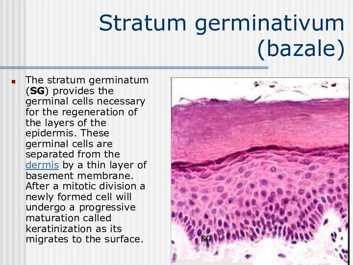
Stratum germinativum (bazale)
The stratum germinatum (SG) provides the germinal cells necessary
Stratum germinativum (bazale)
The stratum germinatum (SG) provides the germinal cells necessary
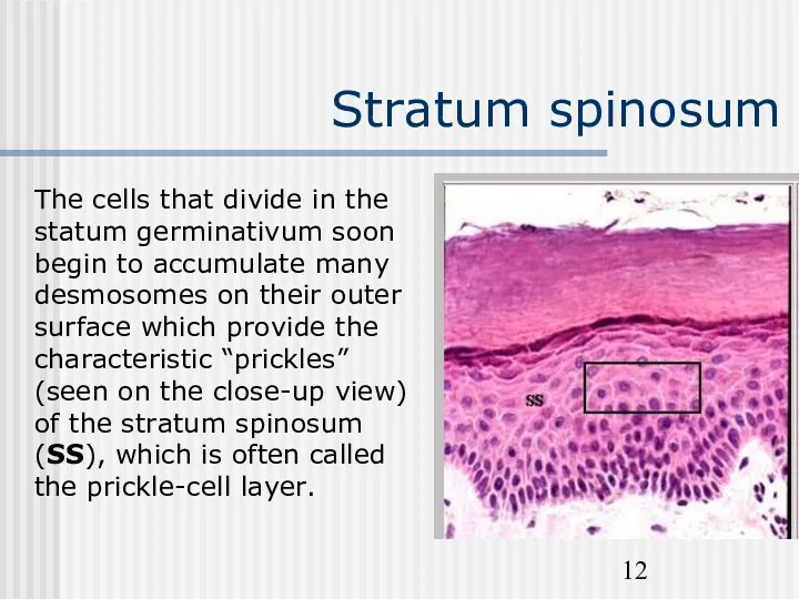
Stratum spinosum
The cells that divide in the statum germinativum soon
Stratum spinosum
The cells that divide in the statum germinativum soon
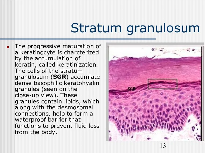
Stratum granulosum
The progressive maturation of a keratinocyte is charcterized by
Stratum granulosum
The progressive maturation of a keratinocyte is charcterized by
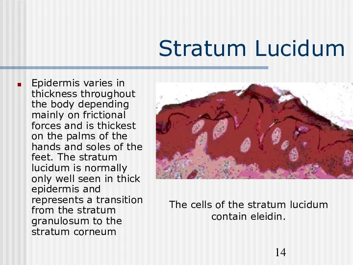
Stratum Lucidum
Epidermis varies in thickness throughout the body depending mainly
Stratum Lucidum
Epidermis varies in thickness throughout the body depending mainly
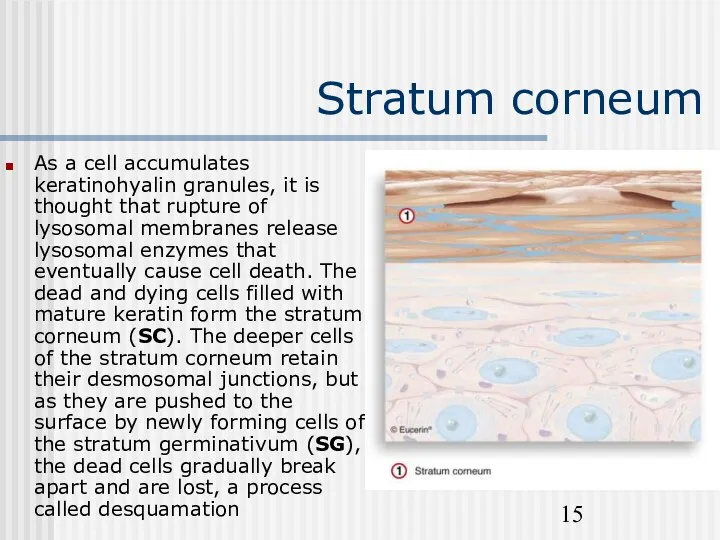
Stratum corneum
As a cell accumulates keratinohyalin granules, it is thought
Stratum corneum
As a cell accumulates keratinohyalin granules, it is thought
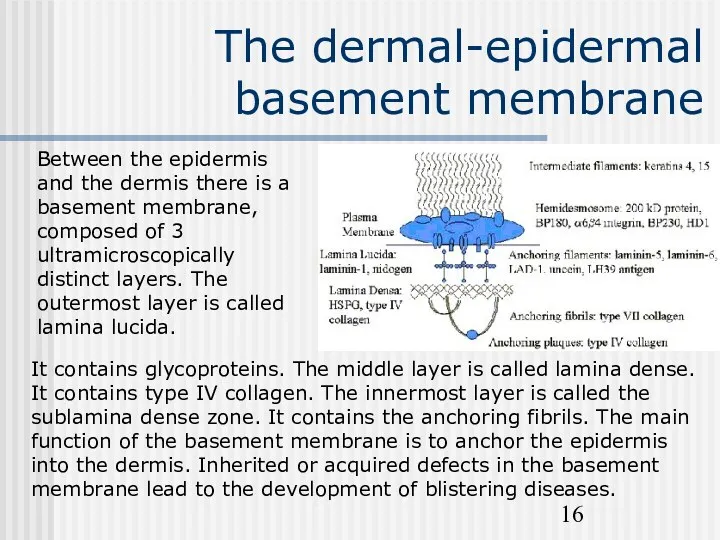
The dermal-epidermal basement membrane
Between the epidermis and the dermis there is
The dermal-epidermal basement membrane
Between the epidermis and the dermis there is
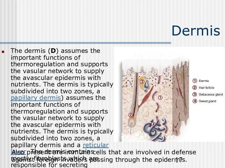
Dermis
The dermis (D) assumes the important functions of thermoregulation and
Dermis
The dermis (D) assumes the important functions of thermoregulation and

Papillary dermis
The papillary dermis (PD) contains vascular networks that have
Papillary dermis
The papillary dermis (PD) contains vascular networks that have
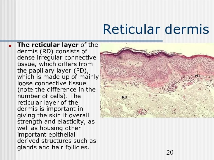
Reticular dermis
The reticular layer of the dermis (RD) consists of
Reticular dermis
The reticular layer of the dermis (RD) consists of
Dermis
The dermis is the supporting layer of the epidermis. It consists
Dermis
The dermis is the supporting layer of the epidermis. It consists
Dermis
1. Fibrous Components and Ground Substance Collagens comprise 98% of the
Dermis
1. Fibrous Components and Ground Substance Collagens comprise 98% of the
Dermis
2. Blood Vessels - The skin is richly vascularized. The
Dermis
2. Blood Vessels - The skin is richly vascularized. The
Dermis
3. Nerves - Unmyelinated and myelinated sensory nerves are present
Dermis
3. Nerves - Unmyelinated and myelinated sensory nerves are present
Dermis
4. Epidermal appendages during fetal development, specialized epithelial derived structures
Dermis
4. Epidermal appendages during fetal development, specialized epithelial derived structures
Sebaceous Gland
Sebaceous glands are found everywhere on the human skin
Sebaceous Gland
Sebaceous glands are found everywhere on the human skin
Sweat Glands
There are two types of sweat glands: eccrine and
Sweat Glands
There are two types of sweat glands: eccrine and
Sweat Glands
Thermal sweating occurs over most of the body integument.
Sweat Glands
Thermal sweating occurs over most of the body integument.
Sweat Glands
Apocrine glands produce an odorless, oily fluid which acquires
Sweat Glands
Apocrine glands produce an odorless, oily fluid which acquires
Hair Follicle
The hair follicle consists of the hair surrounded by
Hair Follicle
The hair follicle consists of the hair surrounded by
Hair Follicle
Human hair grows in cycles. The longer the hair
Hair Follicle
Human hair grows in cycles. The longer the hair
Hair Follicle
Conversely, telogen hairs are easily dislodged and account for
Hair Follicle
Conversely, telogen hairs are easily dislodged and account for
Nail
Nails consist of the nail plate and the supporting tissues
Nail
Nails consist of the nail plate and the supporting tissues
Skin Physiology
Skin Physiology
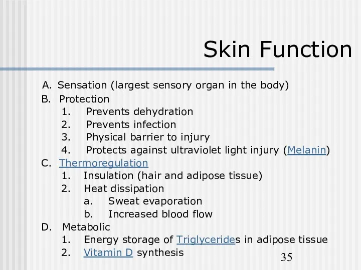
Skin Function
A. Sensation (largest sensory organ in the body)
B. Protection
Skin Function
A. Sensation (largest sensory organ in the body)
B. Protection
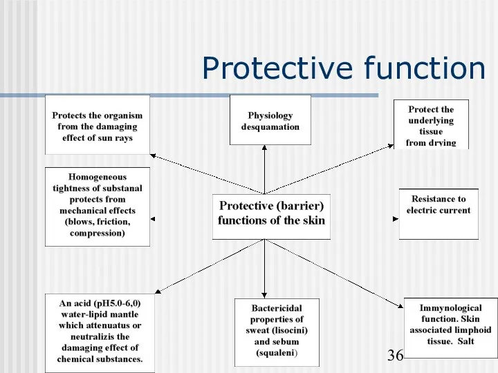
Protective function
Protective function
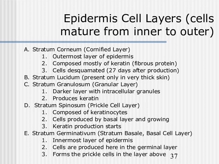
Epidermis Cell Layers (cells mature from inner to outer)
A. Stratum
Epidermis Cell Layers (cells mature from inner to outer)
A. Stratum
Sensory Apparatus of the Skin
The skin is innervated with around
Sensory Apparatus of the Skin
The skin is innervated with around
Sensory Apparatus of the Skin
Sensory endings are of two main
Sensory Apparatus of the Skin
Sensory endings are of two main
Sensory Apparatus of the Skin
The Pacinian corpuscle is one of
Sensory Apparatus of the Skin
The Pacinian corpuscle is one of
Sensory Apparatus of the Skin
Ruffini endings in the human
Sensory Apparatus of the Skin
Ruffini endings in the human
Composition of the lipid membrane
The most important barrier lipids are ceramides,
Composition of the lipid membrane
The most important barrier lipids are ceramides,
Other functions of the skin
The skin is structured to prevent loss
Other functions of the skin
The skin is structured to prevent loss
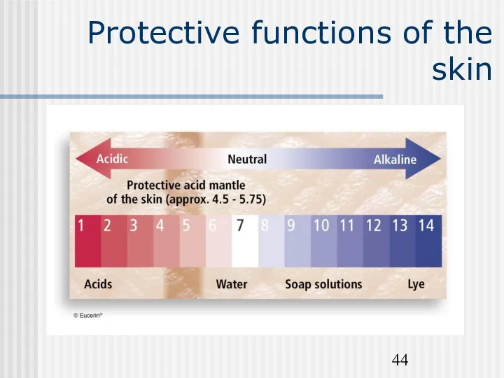
Protective functions of the skin
Protective functions of the skin
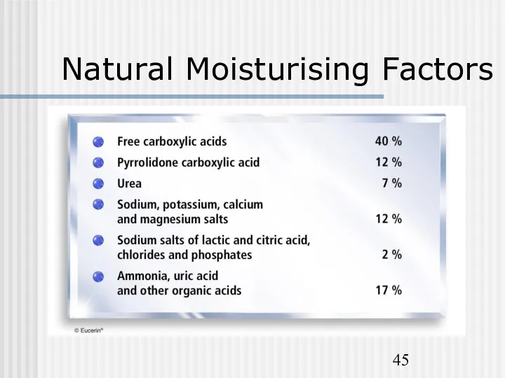
Natural Moisturising Factors
Natural Moisturising Factors
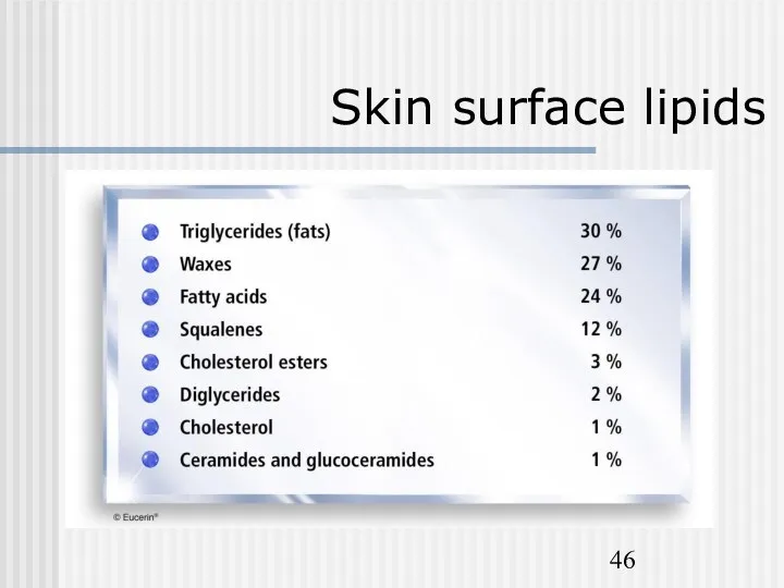
Skin surface lipids
Skin surface lipids
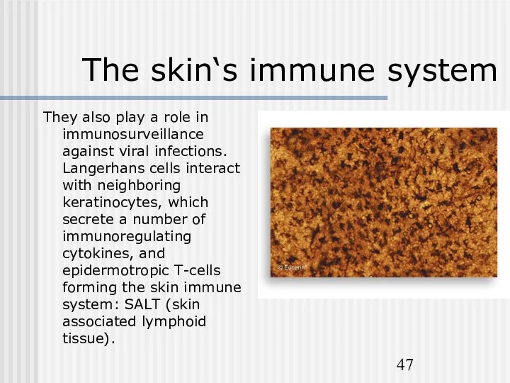
The skin‘s immune system
They also play a role in immunosurveillance against
The skin‘s immune system
They also play a role in immunosurveillance against
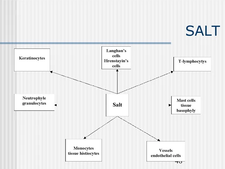
SALT
SALT

Other functions of the skin
Melanin pigment of the skin protects
Other functions of the skin
Melanin pigment of the skin protects
Other functions of the skin
The skin is a vital part of
Other functions of the skin
The skin is a vital part of
 Вода – Н2О. Значение воды для здоровья человека. Источники загрязнения питьевой воды
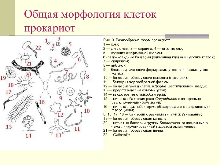 Общая морфология клеток прокариот
 Полевые культуры как экологическая система. Понятие биологического потенциала продуктивности растений
 Синапсқа жалпы шолу
 Мутагенез. Причины мутационного процесса
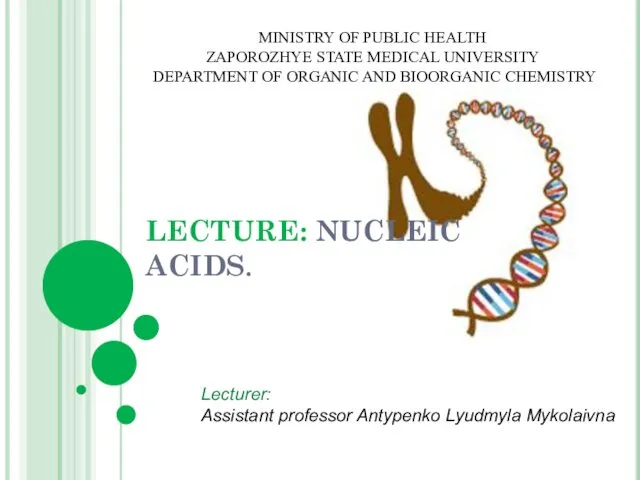 Nucleic acids
 Микроскоп
 Летают ли черепахи. Красноухая пресноводная черепаха
 Анатомия и физиология больших пищеварительных желез
 Волокнистые соединительные ткани. Происхож-дение, морфология и функции клеток рыхлой волокнистой соединительной ткани
 Общая характеристика нарушений голоса. История изучения
 Lancelet - its importance in evolution
 Российские учёные-биологи. Их вклад в науку.
 Урок-презентация по биологии Голосеменные.
 Презентация: Борьба организма с инфекцией. Иммунитет
 Строение легких. Газообмен в легких и тканях
 Цитология - наука о клетке
 Отдел голосеменные
 Пищеварительная система
 Презентация к уроку Систематические группы птиц
 Роль животных на Земле
 Презентация урока 6 класс ФГОС.
 Биологическая эволюция человека
 Презентация к уроку Класс Земноводные
 Қызанақтың адам организміне пайдасы
 Получение молочной кислоты. Продуценты. Практическое использование
 Генетика - теоретическая основа селекции
 Les étapes de l'évolution chimique. Эволюция ферментных систем