- Animal Reproduction

Содержание
- 2. Overview: Pairing Up for Sexual Reproduction Each earthworm produces sperm and eggs; in a few weeks,
- 3. How can each of these earthworms be both male and female?
- 4. Both asexual and sexual reproduction occur in the animal kingdom Sexual reproduction is the creation of
- 5. Asexual reproduction of a sea anemone
- 6. Budding = new individuals arise from outgrowths of existing ones. Fragmentation = breaking of the body
- 7. Sexual Reproduction: An Evolutionary Enigma Sexual females have half as many daughters as asexual females; this
- 8. The “reproductive handicap” of sex: Sexual females have half as many daughters as asexual females. Asexual
- 9. Sexual reproduction results in genetic recombination, which provides potential advantages: An increase in variation in offspring,
- 10. Reproductive Cycles and Patterns Ovulation is the release of mature eggs at the midpoint of a
- 11. Sexual reproduction is a special problem for organisms that seldom encounter a mate. One solution is
- 12. Individuals of some species undergo sex reversals. Some species exhibit male to female reversal (for example,
- 13. Fertilization depends on mechanisms that bring together sperm and eggs of the same species The mechanisms
- 14. External fertilization Eggs
- 15. In internal fertilization, sperm are deposited in or near the female reproductive tract, and fertilization occurs
- 16. Ensuring the Survival of Offspring All species produce more offspring than the environment can handle, and
- 17. Species with internal fertilization provide greater protection of the embryos and more parental care. The embryos
- 18. Parental care in an invertebrate
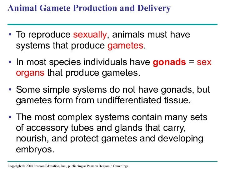
- 19. Animal Gamete Production and Delivery To reproduce sexually, animals must have systems that produce gametes. In
- 20. Most insects have separate sexes with complex reproductive systems. In many insects, the female has a
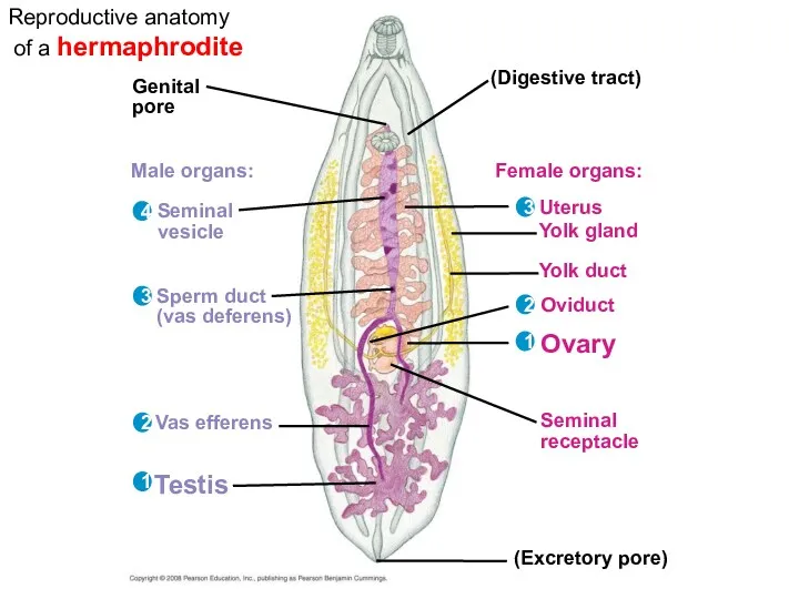
- 21. Genital pore (Digestive tract) Male organs: Seminal vesicle Sperm duct (vas deferens) Vas efferens Testis Female
- 22. A cloaca is a common opening between the external environment and the digestive, excretory, and reproductive
- 23. Reproductive organs produce and transport gametes The following section focuses on the human reproductive system.
- 24. Ovaries = Female Gonads The female gonads, the ovaries, lie in the abdominal cavity. Each ovary
- 25. Ovulation expels an egg cell from the follicle. The remaining follicular tissue grows within the ovary,
- 26. Oviducts and Uterus The egg cell travels from the ovary to the uterus via an oviduct,
- 27. Mammary Glands The mammary glands are not part of the reproductive system but are important to
- 28. Testes = Male Gonads The testes consist of highly coiled tubes surrounded by connective tissue. Sperm
- 29. Ducts From the seminiferous tubules of a testis, mature sperm pass into the coiled tubules of
- 30. Accessory Glands Semen is composed of sperm plus secretions from three sets of accessory glands. The
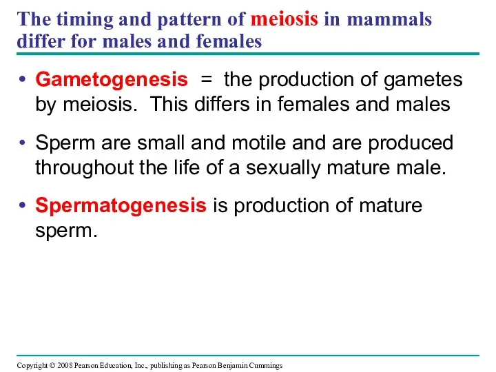
- 31. The timing and pattern of meiosis in mammals differ for males and females Gametogenesis = the
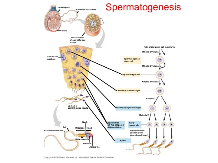
- 32. Spermatogenesis Epididymis Seminiferous tubule Testis Cross section of seminiferous tubule Sertoli cell nucleus Primordial germ cell
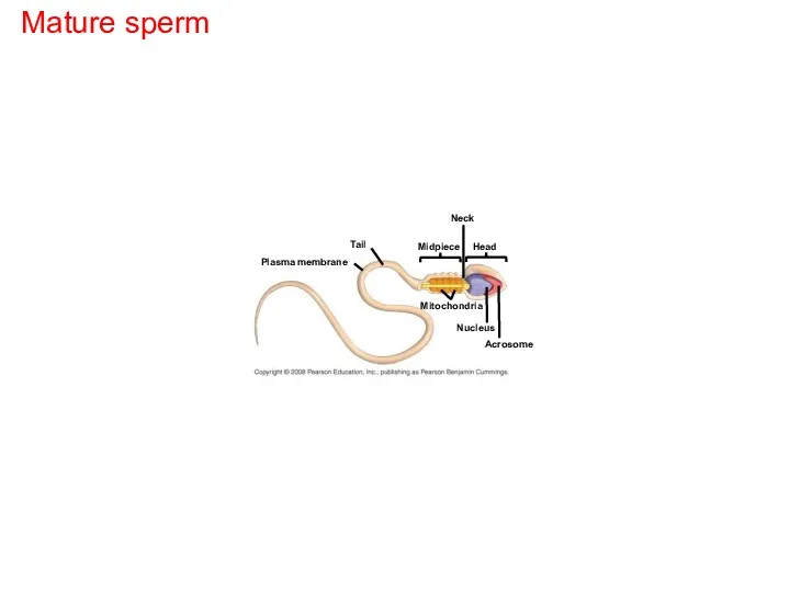
- 33. Mature sperm Plasma membrane Tail Neck Midpiece Head Mitochondria Nucleus Acrosome
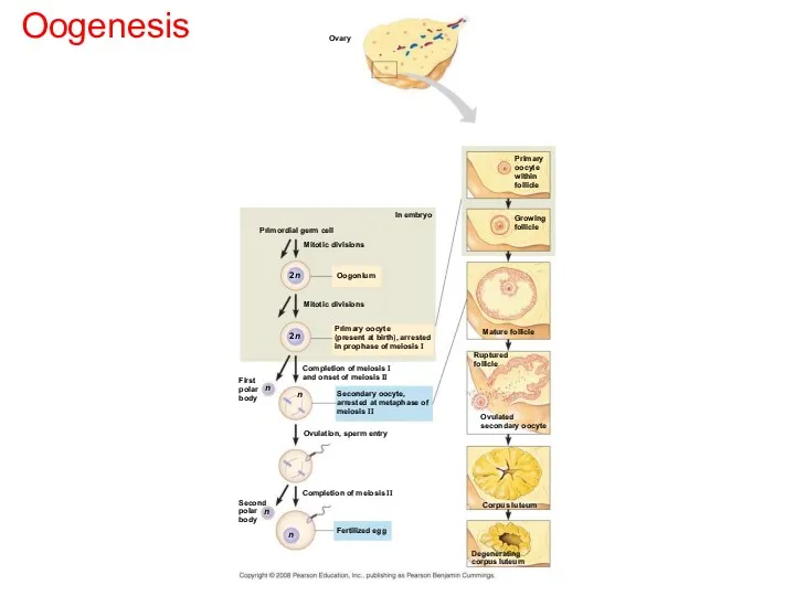
- 34. Eggs contain stored nutrients and are much larger. Oogenesis is development of mature oocytes (eggs) and
- 35. Oogenesis Ovary In embryo Primordial germ cell Mitotic divisions Oogonium Mitotic divisions Primary oocyte (present at
- 36. Spermatogenesis differs from oogenesis: In oogenesis, one egg forms from each cycle of meiosis; in spermatogenesis
- 37. The interplay of tropic and sex hormones regulates mammalian reproduction Human reproduction is coordinated by hormones
- 38. The sex hormones are androgens, estrogens, and progesterone. Sex hormones regulate: The development of primary sex
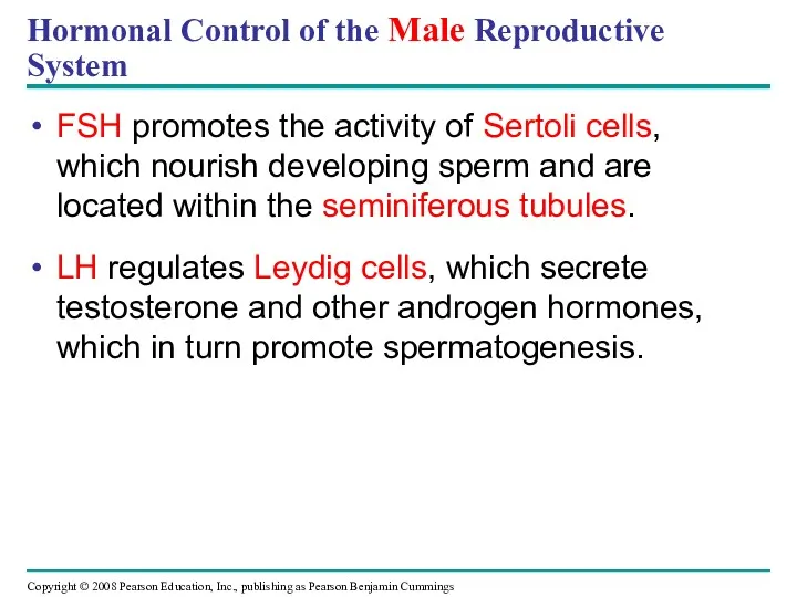
- 39. Hormonal Control of the Male Reproductive System FSH promotes the activity of Sertoli cells, which nourish
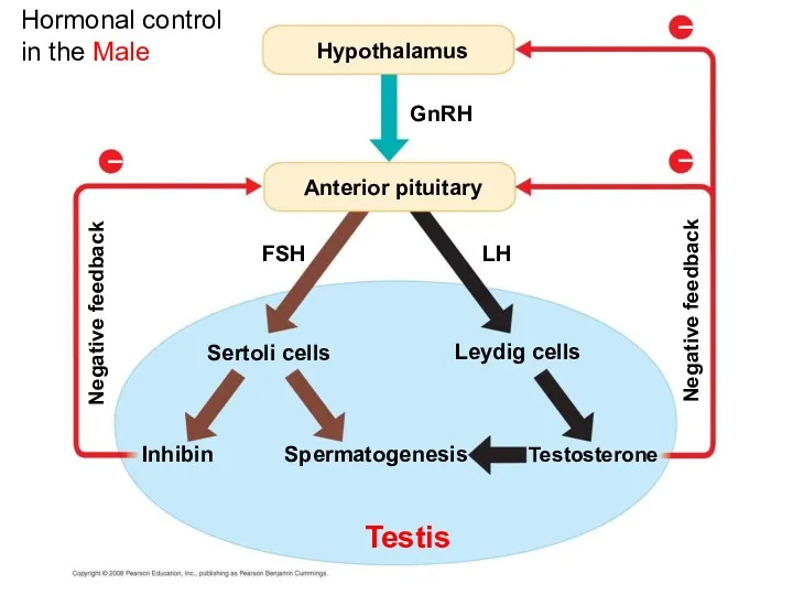
- 40. Hormonal control in the Male Hypothalamus GnRH FSH Anterior pituitary Sertoli cells Leydig cells Inhibin Spermatogenesis
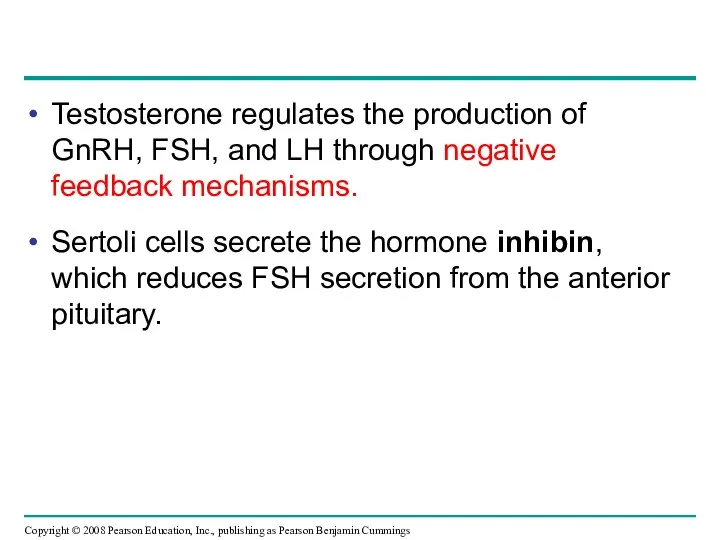
- 41. Testosterone regulates the production of GnRH, FSH, and LH through negative feedback mechanisms. Sertoli cells secrete
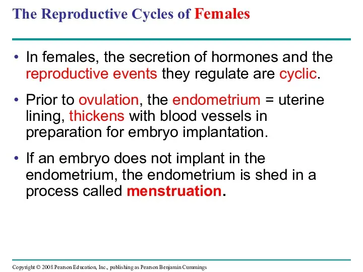
- 42. The Reproductive Cycles of Females In females, the secretion of hormones and the reproductive events they
- 43. Hormones closely link the two cycles of female reproduction: Changes in the uterus / uterine lining
- 44. The reproductive cycle of the human female (a) Control by hypothalamus Hypothalamus GnRH Anterior pituitary 1
- 45. Control by hypothalamus Inhibited by combination of estradiol and progesterone Stimulated by high levels of estradiol
- 46. Ovarian hormones in blood Peak causes LH surge Estradiol level very low Estradiol Progesterone Ovulation Progesterone
- 47. The Ovarian Cycle The sequential release of GnRH then FSH and LH stimulates follicle growth. Follicle
- 48. Following ovulation, the follicular tissue left behind transforms into the corpus luteum; this is the luteal
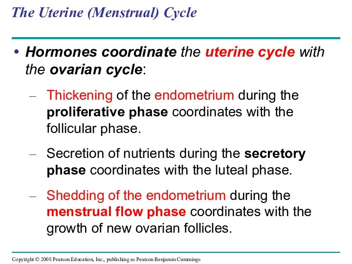
- 49. The Uterine (Menstrual) Cycle Hormones coordinate the uterine cycle with the ovarian cycle: Thickening of the
- 50. A new cycle begins if no embryo implants in the endometrium. Cells of the uterine lining
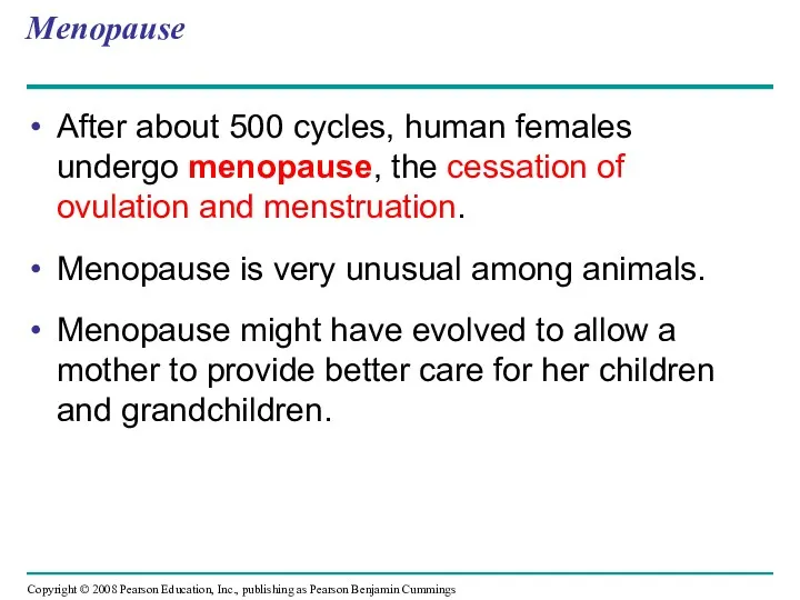
- 51. Menopause After about 500 cycles, human females undergo menopause, the cessation of ovulation and menstruation. Menopause
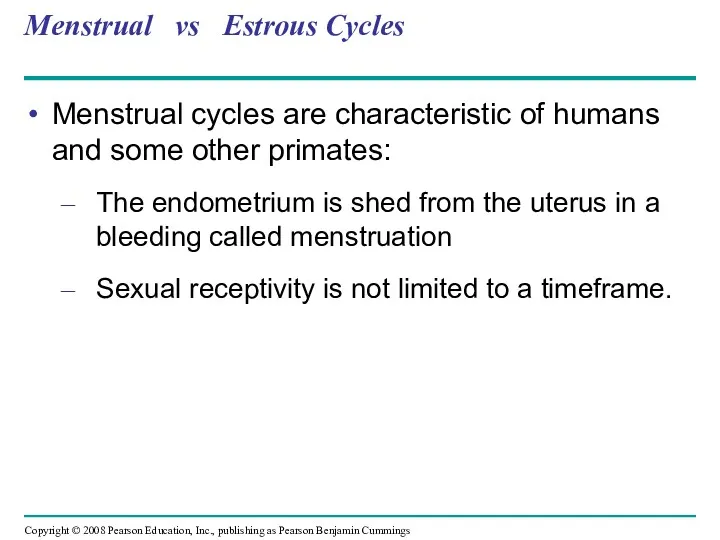
- 52. Menstrual vs Estrous Cycles Menstrual cycles are characteristic of humans and some other primates: The endometrium
- 53. Estrous cycles are characteristic of most mammals: The endometrium is reabsorbed by the uterus Sexual receptivity
- 54. In placental mammals, an embryo develops fully within the mother’s uterus An egg develops into an
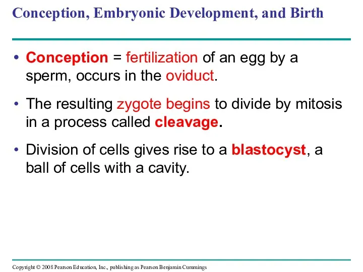
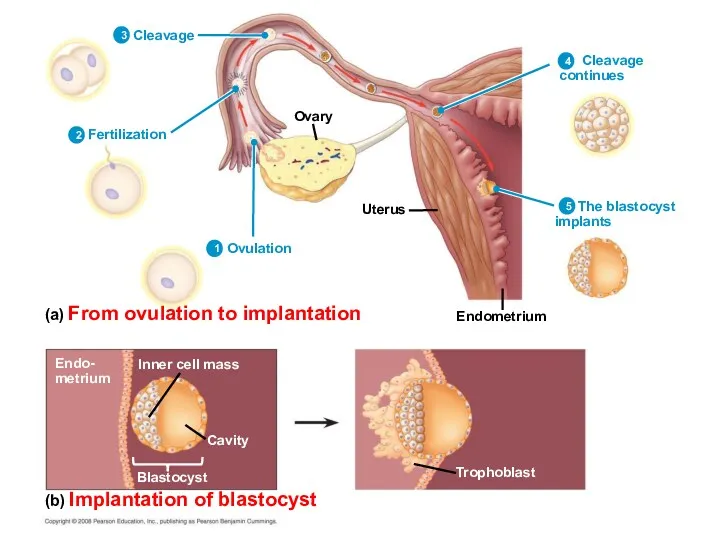
- 55. Conception, Embryonic Development, and Birth Conception = fertilization of an egg by a sperm, occurs in
- 56. Ovary Uterus Endometrium (a) From ovulation to implantation (b) Implantation of blastocyst Cleavage Fertilization Ovulation Cleavage
- 57. After blastocyst formation, the embryo implants into the endometrium. The embryo releases human chorionic gonadotropin (hCG),
- 58. Pregnancies can terminate spontaneously due to chromosomal or developmental abnormalities. An ectopic pregnancy occurs when a
- 59. First Trimester Human gestation can be divided into three trimesters of about three months each. The
- 60. During its first 2 to 4 weeks, the embryo obtains nutrients directly from the endometrium. Meanwhile,
- 61. Placental circulation Placenta Uterus Umbilical cord Chorionic villus, containing fetal capillaries Maternal blood pools Maternal arteries
- 62. Splitting of the embryo during the first month of development results in genetically identical twins. Release
- 63. Changes occur in the mother: Growth of the placenta Cessation of ovulation and the menstrual cycle
- 64. Human fetal development (a) 5 weeks (b) 14 weeks (c) 20 weeks
- 65. (a) 5 weeks
- 66. (b) 14 weeks
- 67. (c) 20 weeks
- 68. Second Trimester During the second trimester: The fetus grows and is very active The mother may
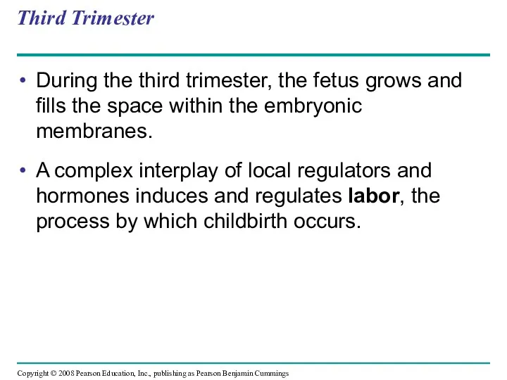
- 69. Third Trimester During the third trimester, the fetus grows and fills the space within the embryonic
- 70. Labor Estradiol Oxytocin from ovaries Induces oxytocin receptors on uterus from fetus and mother’s posterior pituitary
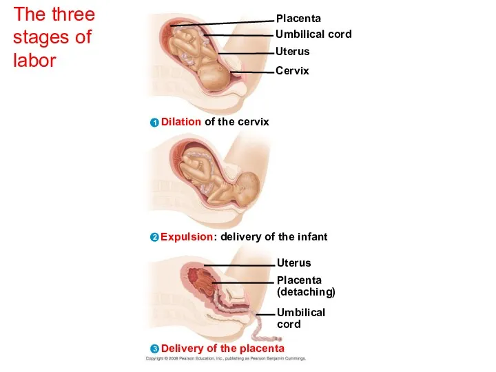
- 71. The three stages of labor Placenta Umbilical cord Uterus Cervix Dilation of the cervix 1
- 72. Expulsion: delivery of the infant 2
- 73. Delivery of the placenta Uterus Placenta (detaching) Umbilical cord 3
- 74. The three stages of labor 3 2 1 Dilation of the cervix Placenta Umbilical cord Uterus
- 75. Birth, or parturition, is brought about by a series of strong, rhythmic uterine contractions. First the
- 76. Maternal Immune Tolerance of the Embryo and Fetus A woman’s acceptance of her “foreign” offspring is
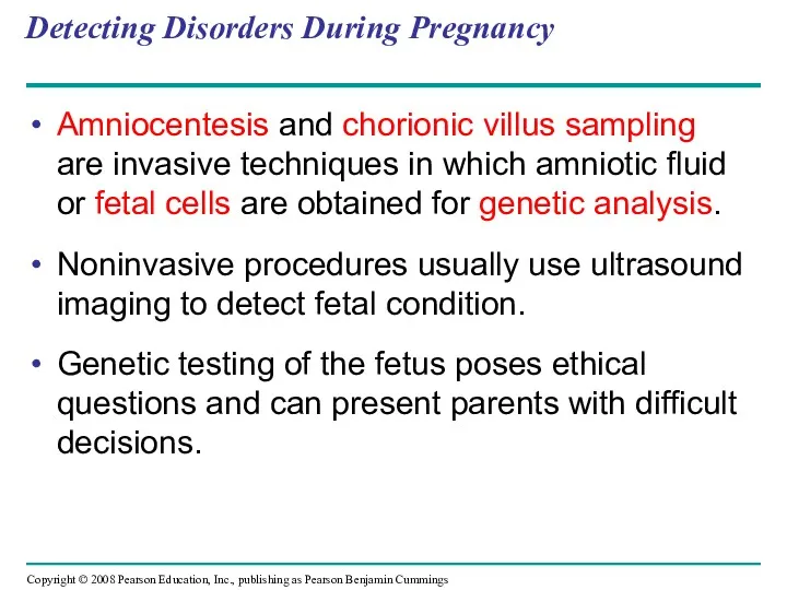
- 77. Detecting Disorders During Pregnancy Amniocentesis and chorionic villus sampling are invasive techniques in which amniotic fluid
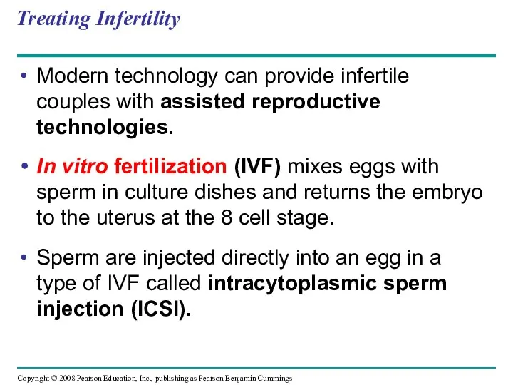
- 78. Treating Infertility Modern technology can provide infertile couples with assisted reproductive technologies. In vitro fertilization (IVF)
- 79. Gametogenesis Spermatogenesis Oogenesis Primary spermatocyte Primary oocyte Polar body Secondary spermatocytes Secondary oocyte Spermatids Sperm Polar
- 80. You should now be able to: Distinguish between asexual and sexual reproduction. Explain how hermaphroditism may
- 82. Скачать презентацию
Overview: Pairing Up for Sexual Reproduction
Each earthworm produces sperm and eggs;
Overview: Pairing Up for Sexual Reproduction
Each earthworm produces sperm and eggs;
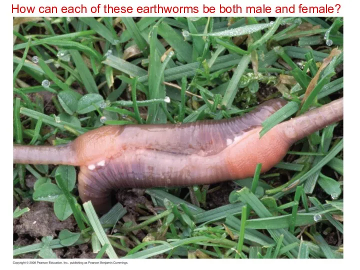
How can each of these earthworms be both male and
How can each of these earthworms be both male and
Both asexual and sexual reproduction occur in the animal kingdom
Sexual reproduction
Both asexual and sexual reproduction occur in the animal kingdom
Sexual reproduction
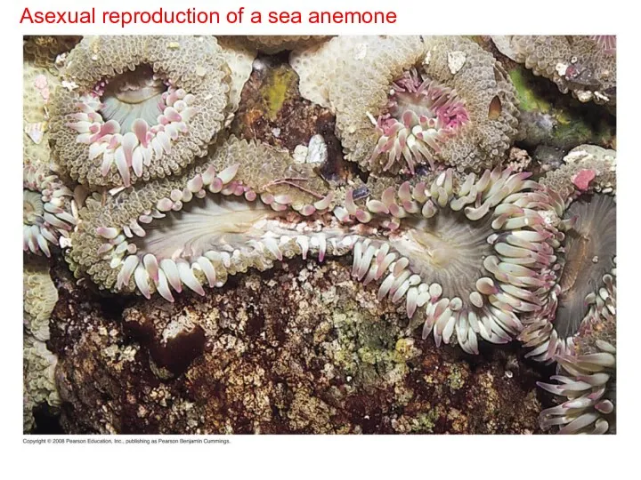
Asexual reproduction of a sea anemone
Asexual reproduction of a sea anemone
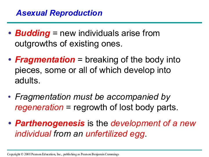
Budding = new individuals arise from outgrowths of existing ones.
Fragmentation =
Budding = new individuals arise from outgrowths of existing ones.
Fragmentation =
Sexual Reproduction: An Evolutionary Enigma
Sexual females have half as many daughters
Sexual Reproduction: An Evolutionary Enigma
Sexual females have half as many daughters
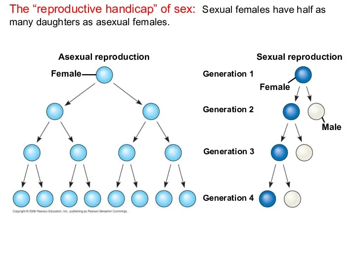
The “reproductive handicap” of sex: Sexual females have half as many
The “reproductive handicap” of sex: Sexual females have half as many
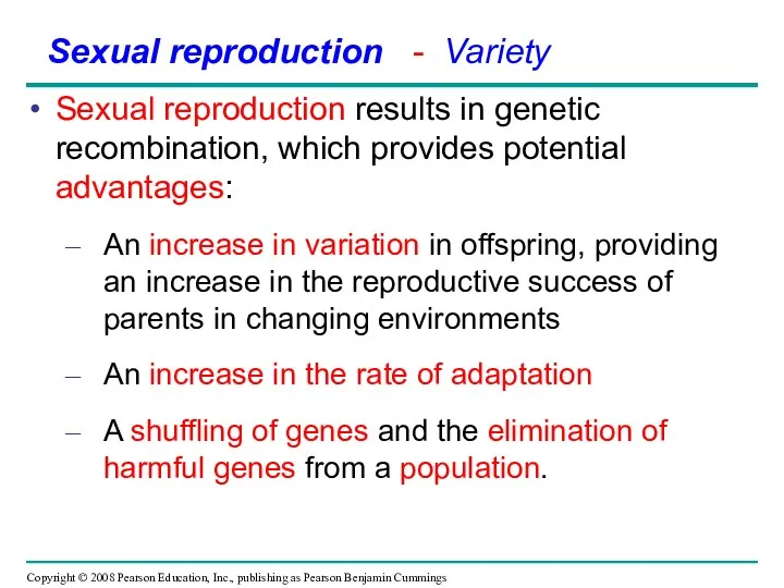
Sexual reproduction results in genetic recombination, which provides potential advantages:
An increase
Sexual reproduction results in genetic recombination, which provides potential advantages:
An increase
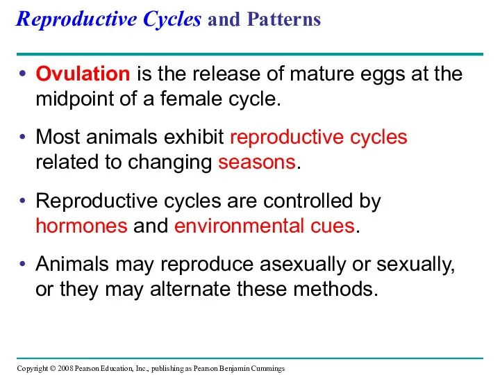
Reproductive Cycles and Patterns
Ovulation is the release of mature eggs at
Reproductive Cycles and Patterns
Ovulation is the release of mature eggs at
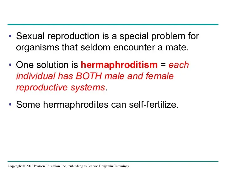
Sexual reproduction is a special problem for organisms that seldom encounter
Sexual reproduction is a special problem for organisms that seldom encounter
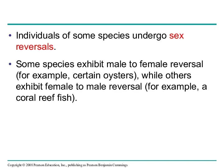
Individuals of some species undergo sex reversals.
Some species exhibit male to
Individuals of some species undergo sex reversals.
Some species exhibit male to
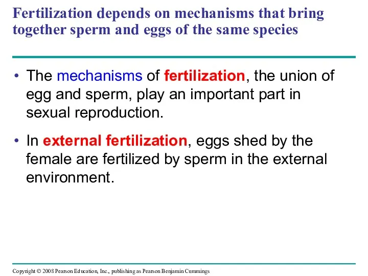
Fertilization depends on mechanisms that bring together sperm and eggs of
Fertilization depends on mechanisms that bring together sperm and eggs of
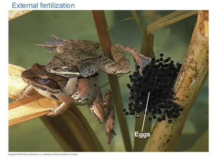
External fertilization
Eggs
External fertilization
Eggs
In internal fertilization, sperm are deposited in or near the female
In internal fertilization, sperm are deposited in or near the female
Ensuring the Survival of Offspring
All species produce more offspring than the
Ensuring the Survival of Offspring
All species produce more offspring than the
Species with internal fertilization provide greater protection of the embryos and
Species with internal fertilization provide greater protection of the embryos and

Parental care
in an invertebrate
Parental care
in an invertebrate
Animal Gamete Production and Delivery
To reproduce sexually, animals must have systems
Animal Gamete Production and Delivery
To reproduce sexually, animals must have systems
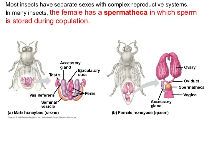
Most insects have separate sexes with complex reproductive systems.
In many insects,
Most insects have separate sexes with complex reproductive systems.
In many insects,
Genital
pore
(Digestive tract)
Male organs:
Seminal
vesicle
Sperm duct
(vas deferens)
Vas efferens
Testis
Female organs:
Uterus
Yolk gland
Yolk duct
Oviduct
Ovary
Seminal
receptacle
(Excretory pore)
4
3
2
1
3
2
1
Reproductive anatomy
Genital
pore
(Digestive tract)
Male organs:
Seminal
vesicle
Sperm duct
(vas deferens)
Vas efferens
Testis
Female organs:
Uterus
Yolk gland
Yolk duct
Oviduct
Ovary
Seminal
receptacle
(Excretory pore)
4
3
2
1
3
2
1
Reproductive anatomy
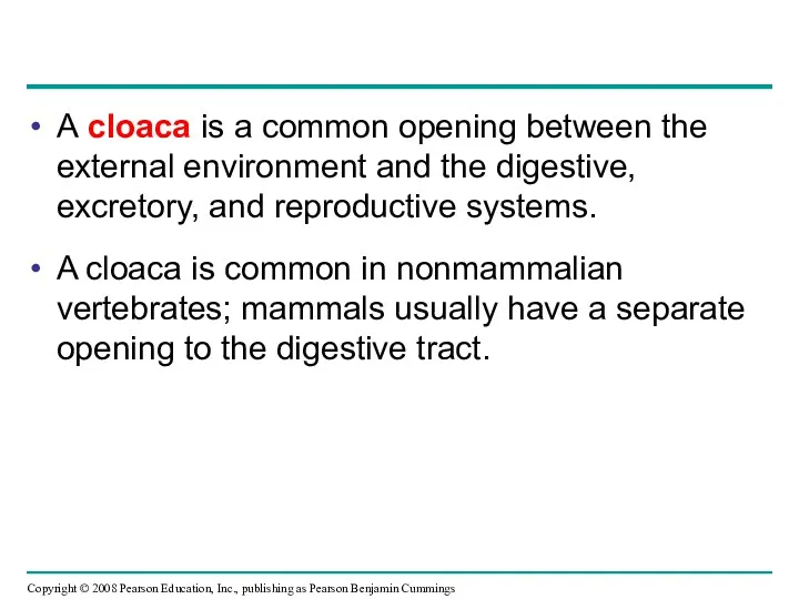
A cloaca is a common opening between the external environment and
A cloaca is a common opening between the external environment and
Reproductive organs produce and transport gametes
The following section focuses on the
Reproductive organs produce and transport gametes
The following section focuses on the
Ovaries = Female Gonads
The female gonads, the ovaries, lie in the
Ovaries = Female Gonads
The female gonads, the ovaries, lie in the
Ovulation expels an egg cell from the follicle.
The remaining follicular tissue
Ovulation expels an egg cell from the follicle.
The remaining follicular tissue
Oviducts and Uterus
The egg cell travels from the ovary to the
Oviducts and Uterus
The egg cell travels from the ovary to the
Mammary Glands
The mammary glands are not part of the reproductive system
Mammary Glands
The mammary glands are not part of the reproductive system
Testes = Male Gonads
The testes consist of highly coiled tubes surrounded
Testes = Male Gonads
The testes consist of highly coiled tubes surrounded
Ducts
From the seminiferous tubules of a testis, mature sperm pass into
Ducts
From the seminiferous tubules of a testis, mature sperm pass into
Accessory Glands
Semen is composed of sperm plus secretions from three sets
Accessory Glands
Semen is composed of sperm plus secretions from three sets
The timing and pattern of meiosis in mammals differ for males
The timing and pattern of meiosis in mammals differ for males
Spermatogenesis
Epididymis
Seminiferous tubule
Testis
Cross section
of seminiferous
tubule
Sertoli cell
nucleus
Primordial germ cell in embryo
Mitotic divisions
Spermatogonial
stem cell
Mitotic
Spermatogenesis
Epididymis
Seminiferous tubule
Testis
Cross section
of seminiferous
tubule
Sertoli cell
nucleus
Primordial germ cell in embryo
Mitotic divisions
Spermatogonial
stem cell
Mitotic
Mature sperm
Plasma membrane
Tail
Neck
Midpiece
Head
Mitochondria
Nucleus
Acrosome
Mature sperm
Plasma membrane
Tail
Neck
Midpiece
Head
Mitochondria
Nucleus
Acrosome
Eggs contain stored nutrients and are much larger.
Oogenesis is development of
Eggs contain stored nutrients and are much larger.
Oogenesis is development of
Oogenesis
Ovary
In embryo
Primordial germ cell
Mitotic divisions
Oogonium
Mitotic divisions
Primary oocyte
(present at birth), arrested
in prophase
Oogenesis
Ovary
In embryo
Primordial germ cell
Mitotic divisions
Oogonium
Mitotic divisions
Primary oocyte (present at birth), arrested in prophase
Spermatogenesis differs from oogenesis:
In oogenesis, one egg forms from each cycle
Spermatogenesis differs from oogenesis:
In oogenesis, one egg forms from each cycle
The interplay of tropic and sex hormones regulates mammalian reproduction
Human reproduction
The interplay of tropic and sex hormones regulates mammalian reproduction
Human reproduction
The sex hormones are androgens, estrogens, and progesterone.
Sex hormones regulate:
The development
The sex hormones are androgens, estrogens, and progesterone.
Sex hormones regulate:
The development
Hormonal Control of the Male Reproductive System
FSH promotes the activity of
Hormonal Control of the Male Reproductive System
FSH promotes the activity of
Hormonal control in the Male
Hypothalamus
GnRH
FSH
Anterior pituitary
Sertoli cells
Leydig cells
Inhibin
Spermatogenesis
Testosterone
Testis
LH
Negative feedback
Negative feedback
–
–
–
Hormonal control in the Male
Hypothalamus
GnRH
FSH
Anterior pituitary
Sertoli cells
Leydig cells
Inhibin
Spermatogenesis
Testosterone
Testis
LH
Negative feedback
Negative feedback
–
–
–
Testosterone regulates the production of GnRH, FSH, and LH through negative
Testosterone regulates the production of GnRH, FSH, and LH through negative
The Reproductive Cycles of Females
In females, the secretion of hormones and
The Reproductive Cycles of Females
In females, the secretion of hormones and
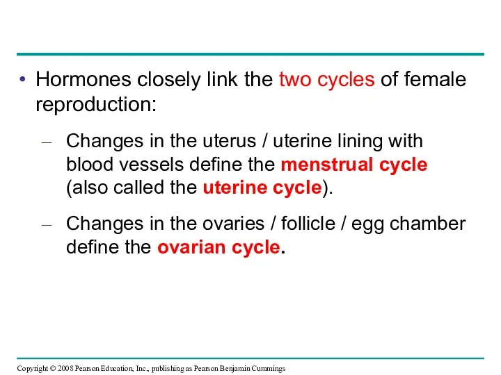
Hormones closely link the two cycles of female reproduction:
Changes in the
Hormones closely link the two cycles of female reproduction:
Changes in the
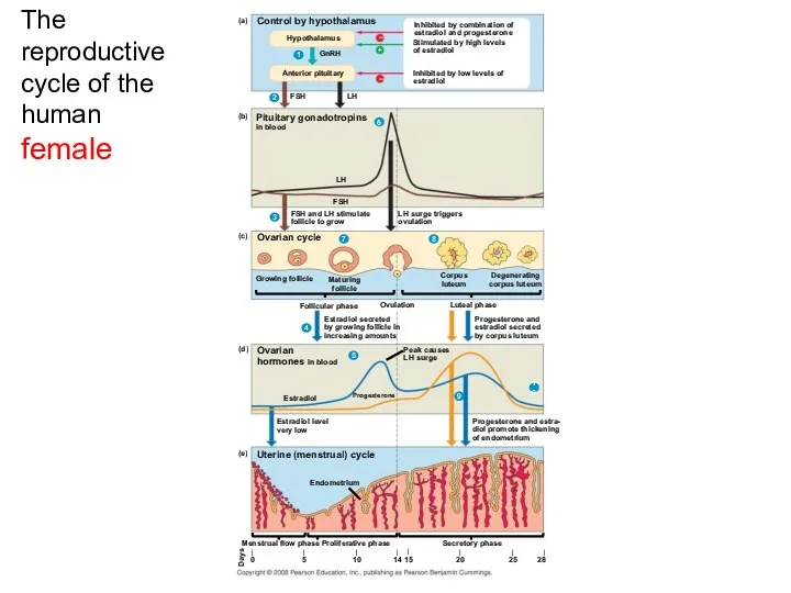
The reproductive cycle of the human female
(a)
Control by hypothalamus
Hypothalamus
GnRH
Anterior pituitary
1
Inhibited by
The reproductive cycle of the human female
(a)
Control by hypothalamus
Hypothalamus
GnRH
Anterior pituitary
1
Inhibited by
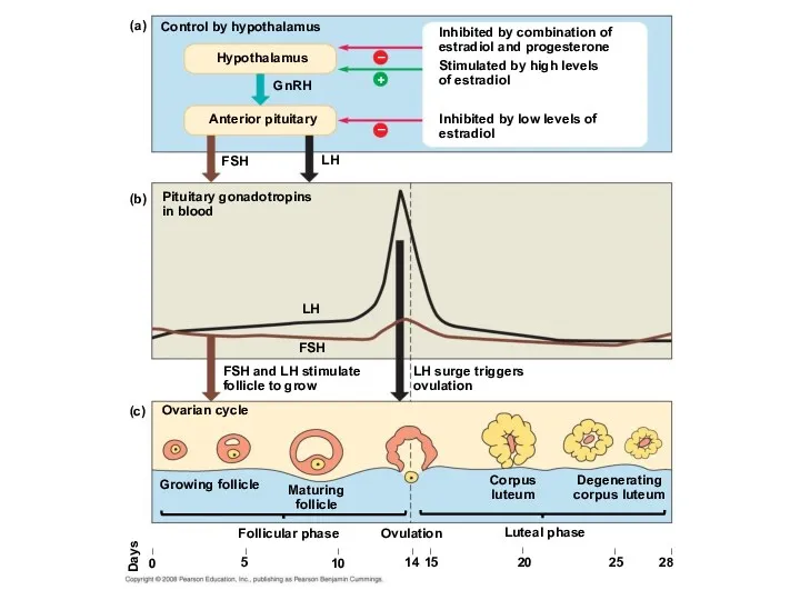
Control by hypothalamus
Inhibited by combination of estradiol and progesterone
Stimulated by high
Control by hypothalamus
Inhibited by combination of estradiol and progesterone
Stimulated by high

Ovarian hormones in blood
Peak causes
LH surge
Estradiol level very low
Estradiol
Progesterone
Ovulation
Progesterone and estra-
diol
Ovarian hormones in blood
Peak causes
LH surge
Estradiol level very low
Estradiol
Progesterone
Ovulation
Progesterone and estra- diol
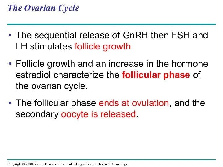
The Ovarian Cycle
The sequential release of GnRH then FSH and LH
The Ovarian Cycle
The sequential release of GnRH then FSH and LH
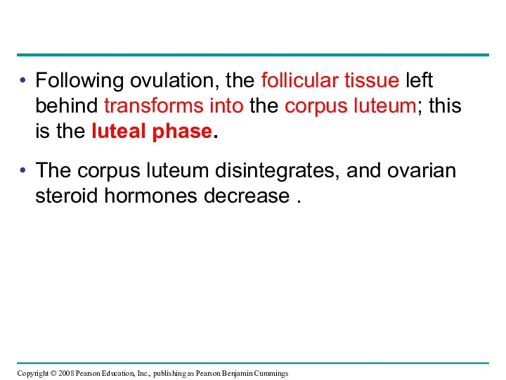
Following ovulation, the follicular tissue left behind transforms into the corpus
Following ovulation, the follicular tissue left behind transforms into the corpus
The Uterine (Menstrual) Cycle
Hormones coordinate the uterine cycle with the ovarian
The Uterine (Menstrual) Cycle
Hormones coordinate the uterine cycle with the ovarian
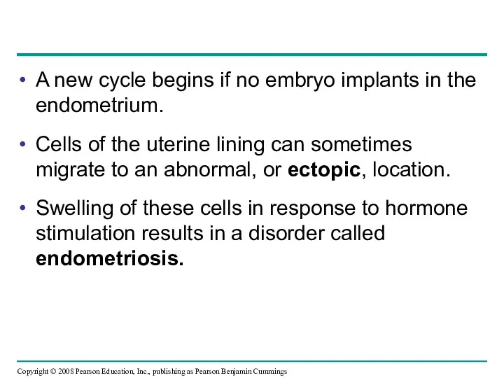
A new cycle begins if no embryo implants in the endometrium.
Cells
A new cycle begins if no embryo implants in the endometrium.
Cells
Menopause
After about 500 cycles, human females undergo menopause, the cessation of
Menopause
After about 500 cycles, human females undergo menopause, the cessation of
Menstrual vs Estrous Cycles
Menstrual cycles are characteristic of humans and some
Menstrual vs Estrous Cycles
Menstrual cycles are characteristic of humans and some
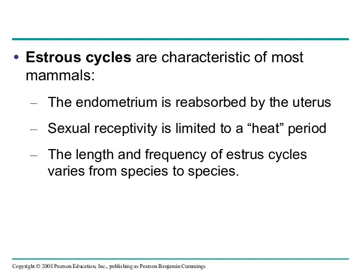
Estrous cycles are characteristic of most mammals:
The endometrium is reabsorbed by
Estrous cycles are characteristic of most mammals:
The endometrium is reabsorbed by
In placental mammals, an embryo develops fully within the mother’s uterus
An
In placental mammals, an embryo develops fully within the mother’s uterus
An
Conception, Embryonic Development, and Birth
Conception = fertilization of an egg by
Conception, Embryonic Development, and Birth
Conception = fertilization of an egg by
Ovary
Uterus
Endometrium
(a) From ovulation to implantation
(b) Implantation of blastocyst
Cleavage
Fertilization
Ovulation
Cleavage continues
The
Ovary
Uterus
Endometrium
(a) From ovulation to implantation
(b) Implantation of blastocyst
Cleavage
Fertilization
Ovulation
Cleavage continues
The
After blastocyst formation, the embryo implants into the endometrium.
The embryo releases
After blastocyst formation, the embryo implants into the endometrium.
The embryo releases
Pregnancies can terminate spontaneously due to chromosomal or developmental abnormalities.
An ectopic
Pregnancies can terminate spontaneously due to chromosomal or developmental abnormalities.
An ectopic
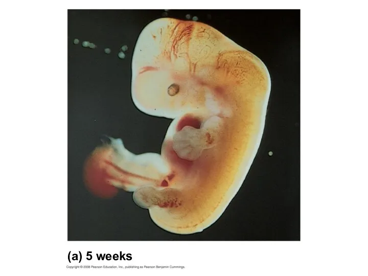
First Trimester
Human gestation can be divided into three trimesters of about
First Trimester
Human gestation can be divided into three trimesters of about
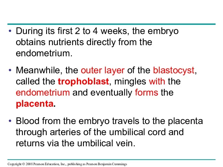
During its first 2 to 4 weeks, the embryo obtains nutrients
During its first 2 to 4 weeks, the embryo obtains nutrients
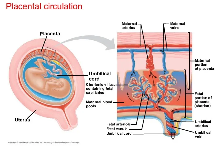
Placental circulation
Placenta
Uterus
Umbilical cord
Chorionic villus,
containing fetal
capillaries
Maternal blood
pools
Maternal
arteries
Maternal
veins
Maternal
portion
of placenta
Fetal arteriole
Fetal venule
Umbilical cord
Fetal
portion of
placenta
(chorion)
Umbilical
arteries
Umbilical
vein
Placental circulation
Placenta
Uterus
Umbilical cord
Chorionic villus,
containing fetal
capillaries
Maternal blood
pools
Maternal
arteries
Maternal
veins
Maternal
portion
of placenta
Fetal arteriole
Fetal venule
Umbilical cord
Fetal
portion of
placenta
(chorion)
Umbilical
arteries
Umbilical
vein
Splitting of the embryo during the first month of development results
Splitting of the embryo during the first month of development results

Changes occur in the mother:
Growth of the placenta
Cessation of ovulation and
Changes occur in the mother:
Growth of the placenta
Cessation of ovulation and
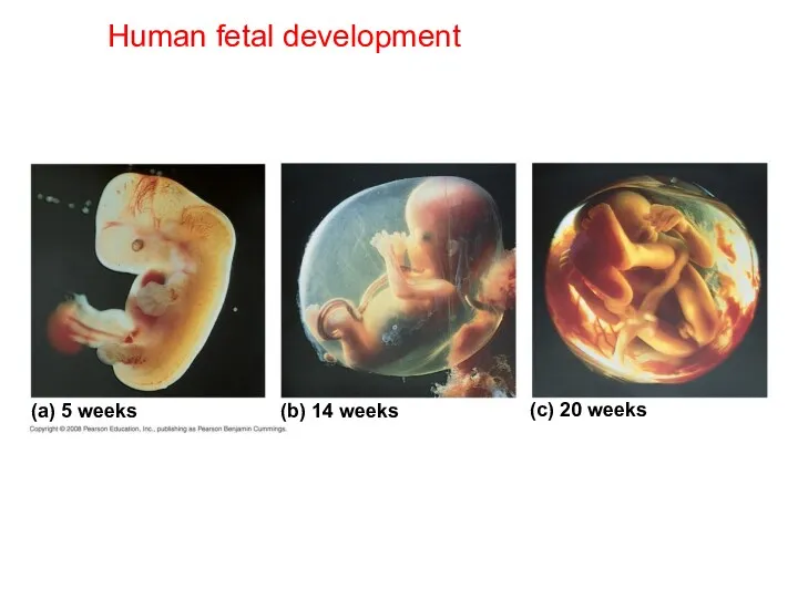
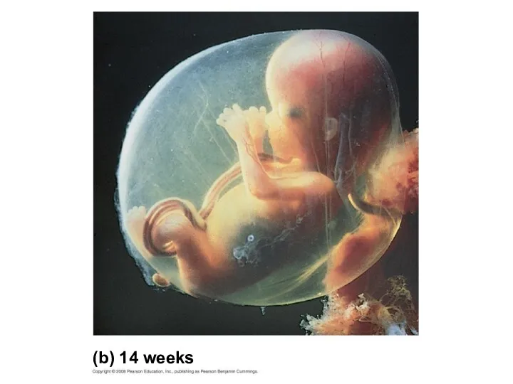
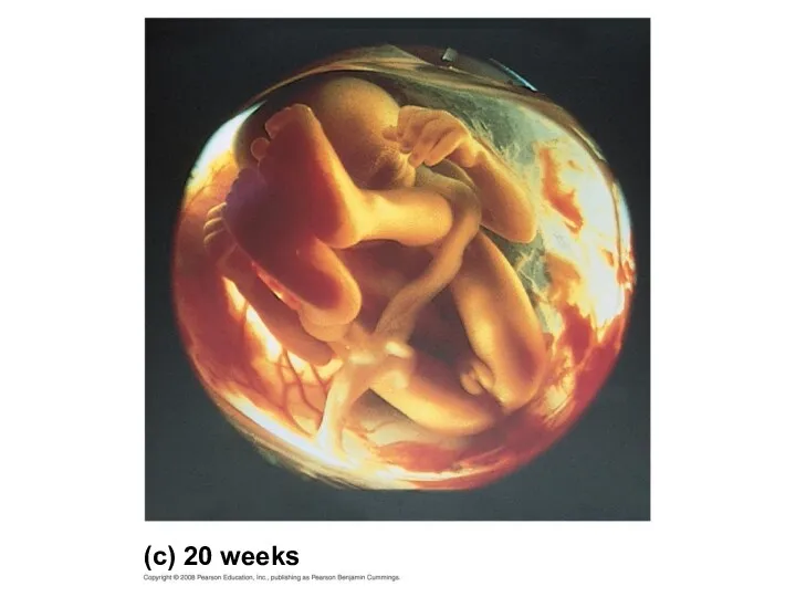
Human fetal development
(a) 5 weeks
(b) 14 weeks
(c) 20 weeks
Human fetal development
(a) 5 weeks
(b) 14 weeks
(c) 20 weeks
(a) 5 weeks
(a) 5 weeks
(b) 14 weeks
(b) 14 weeks
(c) 20 weeks
(c) 20 weeks
Second Trimester
During the second trimester:
The fetus grows and is very active
The
Second Trimester
During the second trimester:
The fetus grows and is very active
The
Third Trimester
During the third trimester, the fetus grows and fills the
Third Trimester
During the third trimester, the fetus grows and fills the
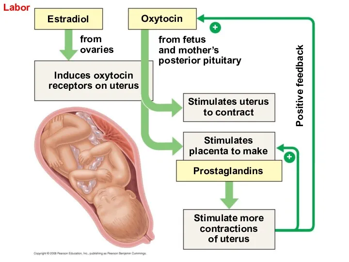
Labor
Estradiol
Oxytocin
from
ovaries
Induces oxytocin
receptors on uterus
from fetus
and mother’s
posterior pituitary
Stimulates uterus
to contract
Stimulates placenta to
Labor
Estradiol
Oxytocin
from
ovaries
Induces oxytocin
receptors on uterus
from fetus
and mother’s
posterior pituitary
Stimulates uterus
to contract
Stimulates placenta to
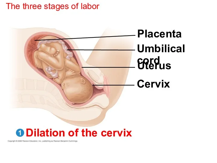
The three stages of labor
Placenta
Umbilical cord
Uterus
Cervix
Dilation of the cervix
1
The three stages of labor
Placenta
Umbilical cord
Uterus
Cervix
Dilation of the cervix
1
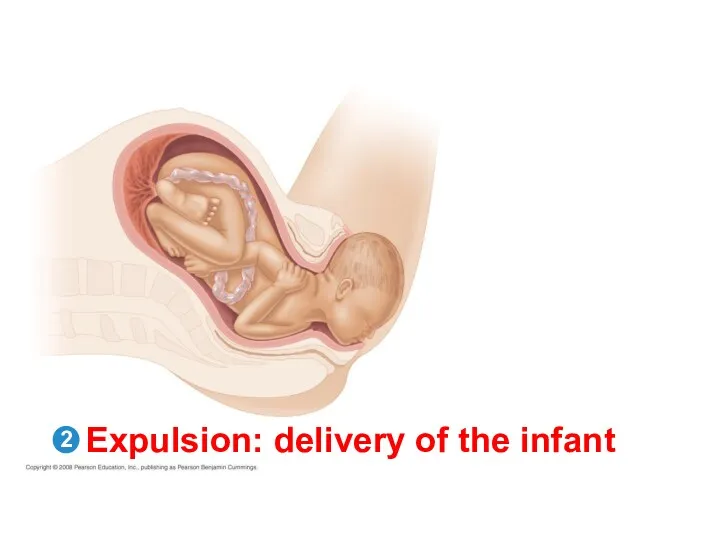
Expulsion: delivery of the infant
2
Expulsion: delivery of the infant
2
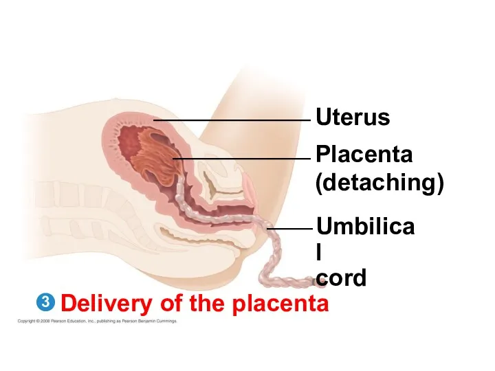
Delivery of the placenta
Uterus
Placenta
(detaching)
Umbilical
cord
3
Delivery of the placenta
Uterus
Placenta
(detaching)
Umbilical
cord
3
The three stages of labor
3
2
1
Dilation of the cervix
Placenta
Umbilical cord
Uterus
Cervix
Expulsion: delivery of
The three stages of labor
3
2
1
Dilation of the cervix
Placenta
Umbilical cord
Uterus
Cervix
Expulsion: delivery of
Birth, or parturition, is brought about by a series of strong,
Birth, or parturition, is brought about by a series of strong,
Maternal Immune Tolerance of the Embryo and Fetus
A woman’s acceptance of
Maternal Immune Tolerance of the Embryo and Fetus
A woman’s acceptance of
Detecting Disorders During Pregnancy
Amniocentesis and chorionic villus sampling are invasive techniques
Detecting Disorders During Pregnancy
Amniocentesis and chorionic villus sampling are invasive techniques
Treating Infertility
Modern technology can provide infertile couples with assisted reproductive technologies.
In
Treating Infertility
Modern technology can provide infertile couples with assisted reproductive technologies.
In
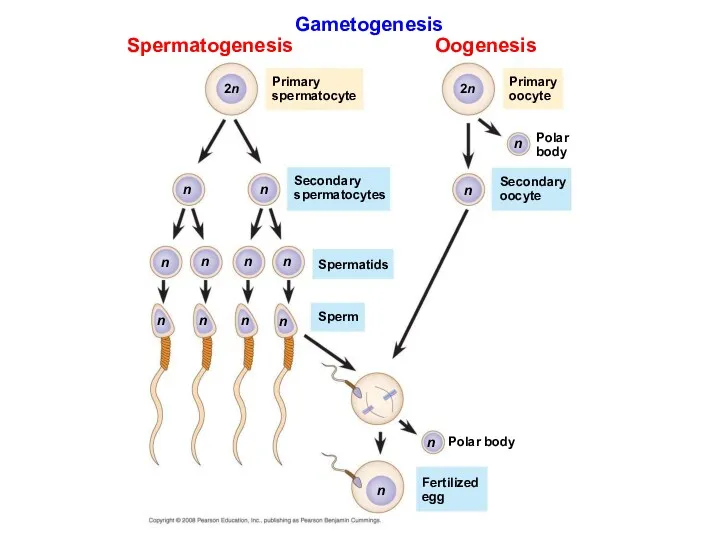
Gametogenesis
Spermatogenesis
Oogenesis
Primary
spermatocyte
Primary
oocyte
Polar
body
Secondary
spermatocytes
Secondary
oocyte
Spermatids
Sperm
Polar body
Fertilized
egg
n
2n
2n
n
n
n
n
n
n
n
n
n
n
n
n
n
Gametogenesis
Spermatogenesis
Oogenesis
Primary
spermatocyte
Primary
oocyte
Polar
body
Secondary
spermatocytes
Secondary
oocyte
Spermatids
Sperm
Polar body
Fertilized
egg
n
2n
2n
n
n
n
n
n
n
n
n
n
n
n
n
n
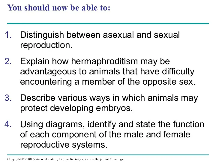
You should now be able to:
Distinguish between asexual and sexual reproduction.
Explain
You should now be able to:
Distinguish between asexual and sexual reproduction.
Explain
 Клеточное строение листа
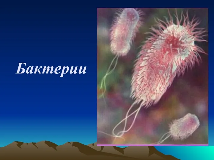 Бактерии. Распространение бактерий
 Чибис-птица года - 2010
 Свойства живых организмов
 Генетика людини
 Покровные и проводящие растительные ткани
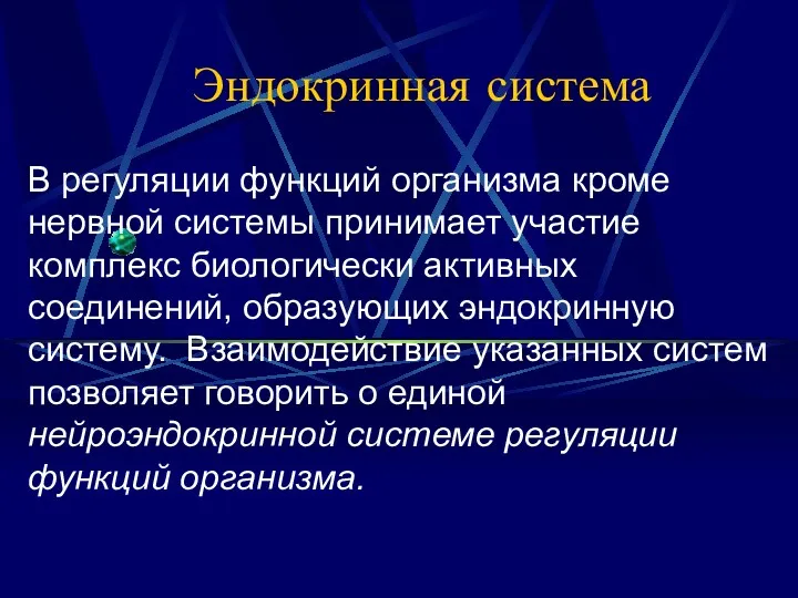 Эндокринная система
 Оплодотворение у цветковых растений
 ПРЕЗЕНТАЦИЯ ДЛЯ ИНТЕРАКТИВНОЙ ДОСКИ. ТЕСТ. МЕХАНИЗМЫ ЭВОЛЮЦИИ.
 Конкурс:Экология. Книга. Мы. Лучший библиотечный цветник
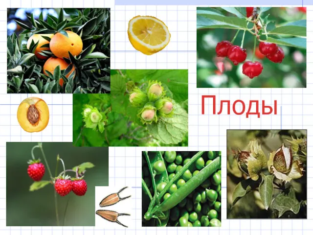 Плод. Строение плода
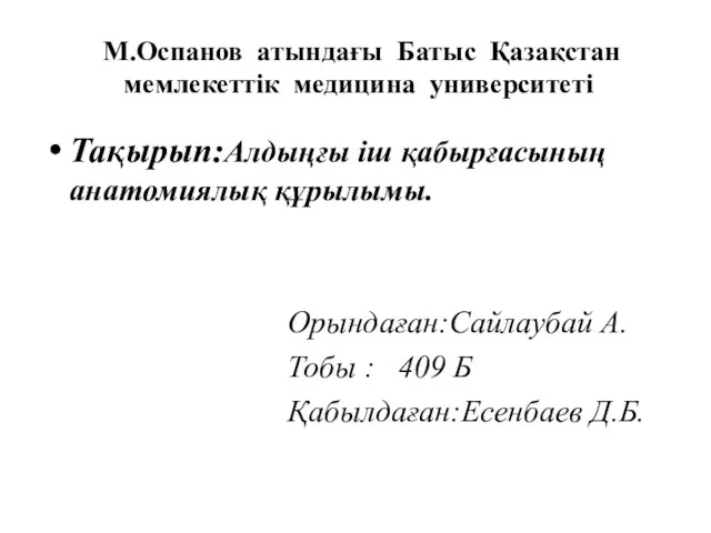 Алдыңғы іш қабырғасының анатомиялық құрылымы
 Обмен белков-3
 Биотические факторы среды. Отношения организмов
 Основы молекулярной биологии. Биосинтез белка. Транскрипция
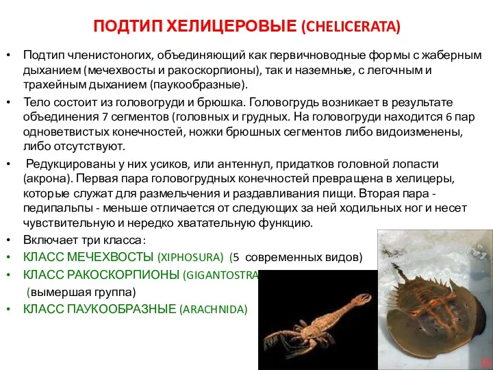 Подтип Хелицеровые (Сhelicerata)
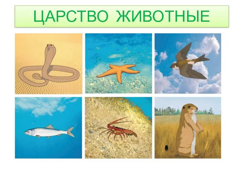 Царство Животные
 Живая и неживая природа
 История развития организмов по эрам
 Знатоки растений
 Загальна характеристика класу Кісткові риби
 Внешнее строение листа
 Отряд Черепахи
 Клеточная теория
 Нервная система животных. Рефлекс. Инстинкт
 Ядовитые растения Самарской области
 Анализаторы. Органы слуха и равновесия
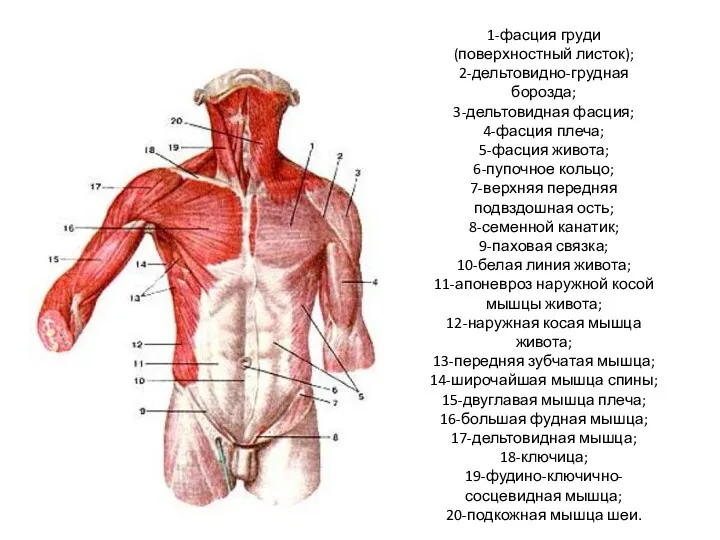 Мышцы туловища