- Bone Classification

Содержание
- 2. BONE CLASSIFICATION Bone Classification: Long Bones Short Bones Sesamoid Bones Flat Bones Irregular Bones Wormian Bones
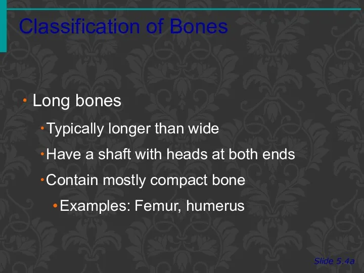
- 3. Classification of Bones Slide 5.4a Long bones Typically longer than wide Have a shaft with heads
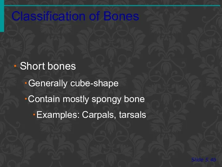
- 4. Classification of Bones Slide 5.4b Short bones Generally cube-shape Contain mostly spongy bone Examples: Carpals, tarsals
- 5. Classification of Bones on the Basis of Shape Slide 5.4c Figure 5.1
- 6. Classification of Bones Slide 5.5a Flat bones Thin and flattened Usually curved Thin layers of compact
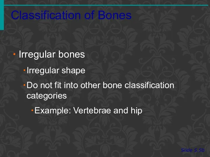
- 7. Classification of Bones Slide 5.5b Irregular bones Irregular shape Do not fit into other bone classification
- 8. Classification of Bones on the Basis of Shape Slide 5.5c Figure 5.1
- 9. PARTS OF A LONG BONE Epiphysis Distal Proximal Diaphysis Metaphysis Compact bone Spongy bone Articular cartilage
- 10. MICROSCOPIC STRUCTURE Bone cells are called osteocytes in a lacuna Osteocytes transport nutrients and wastes by
- 11. COMPACT BONE Osteon Haversian System Central canal Perforating canal Volkmann’s canal Osteocytes Lamellae Lacunae Bone matrix
- 12. Microscopic Anatomy of Bone Slide 5.10b Figure 5.3
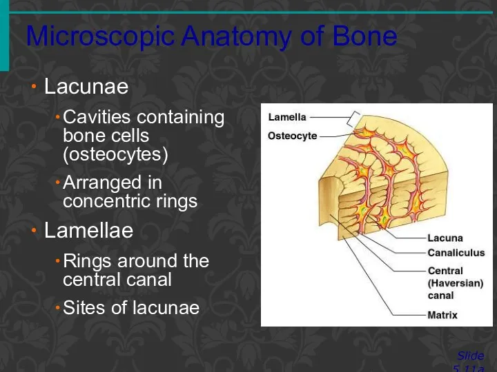
- 13. Microscopic Anatomy of Bone Slide 5.11a Lacunae Cavities containing bone cells (osteocytes) Arranged in concentric rings
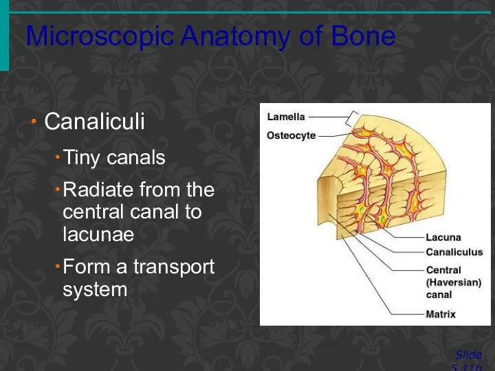
- 14. Microscopic Anatomy of Bone Slide 5.11b Canaliculi Tiny canals Radiate from the central canal to lacunae
- 15. BONE DEVELOPMENT AND GROWTH Parts of the skeletal system begin to develop during the first few
- 16. INTRAMEMBRANOUS BONES Intramembranous Bones These bones originate within sheetlike layers of connective tissues They are the
- 17. ENDOCHONDRAL BONES Endochondral Bones Bones begin as hyaline cartilage Form models for future bones These are
- 18. ENDOCHONDRAL OSSIFICATION Hyaline cartilage model Primary ossification center Secondary ossification centers Epiphyseal plate Osteoblasts vs. osteoclasts
- 19. BONE FUNCTION Bones shape, support, and protect body structures
- 20. SUPPORT, PROTECTION, AND MOVEMENT Support, Movement & Protection Gives shape to head, etc. Supports body’s weight
- 21. BLOOD CELL FORMATION Blood Cell Formation Also known as hematopoiesis Occurs in the red bone marrow
- 22. INORGANIC SALT STORAGE Inorganic Salt Storage Calcium Phosphate Magnesium Sodium Potassium
- 23. SKELETAL ORGANIZATION The actual number of bones in the human skeleton varies from person to person
- 24. DIVISIONS OF THE SKELETON Axial Skeleton Skull Spine Rib cage Appendicular Skeleton Upper limbs Lower limbs
- 25. SKULL Is composed of the cranium (brain case) and the facial bones
- 26. CRANIUM Sphenoid Bone (1) Base of cranium Sides of skull Floors and sides of orbits Sella
- 27. CRANIUM Ethmoid Bone (1) Roof and walls of nasal cavity Floor of cranium Wall of orbits
- 28. FACIAL SKELETON Maxillary Bones (2) Upper jaw Anterior roof of mouth Floors of orbits Sides of
- 29. FACIAL SKELETON Frontal sinus Ethmoidal sinuses Sphenoidal sinus Maxillary sinus
- 30. FACIAL SKELETON Palatine Bones (2) ‘L’ shaped bones located behind the maxillae Posterior section of hard
- 31. FACIAL SKELETON Zygomatic Bones (2) Prominences of cheeks Lateral walls of orbits Floors of orbits Temporal
- 32. FACIAL SKELETON Lacrimal Bones (2) Medial walls of orbits Groove from orbit to nasal cavity Nasal
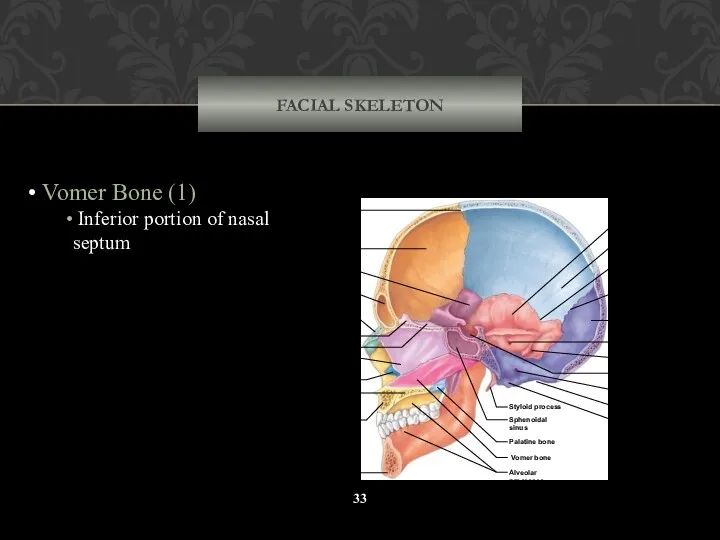
- 33. FACIAL SKELETON Vomer Bone (1) Inferior portion of nasal septum Coronal suture Frontal bone Nasal bone
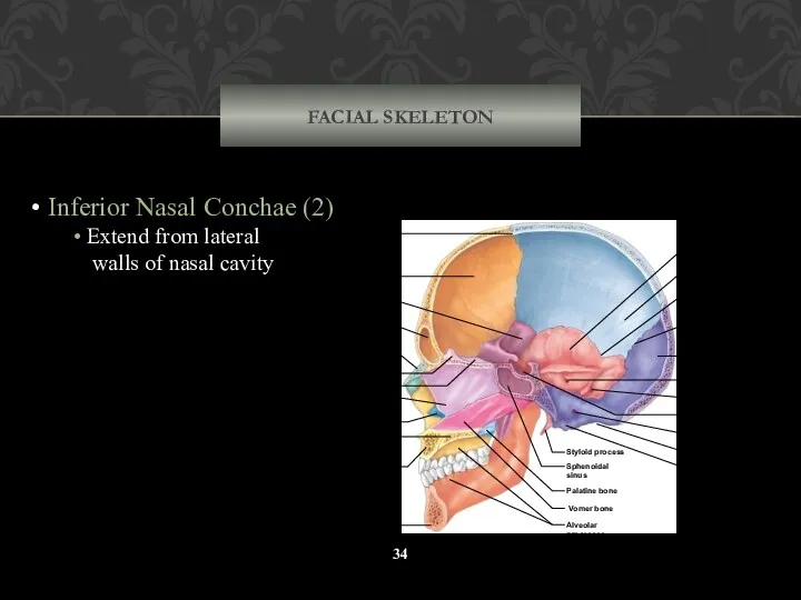
- 34. FACIAL SKELETON Inferior Nasal Conchae (2) Extend from lateral walls of nasal cavity Coronal suture Frontal
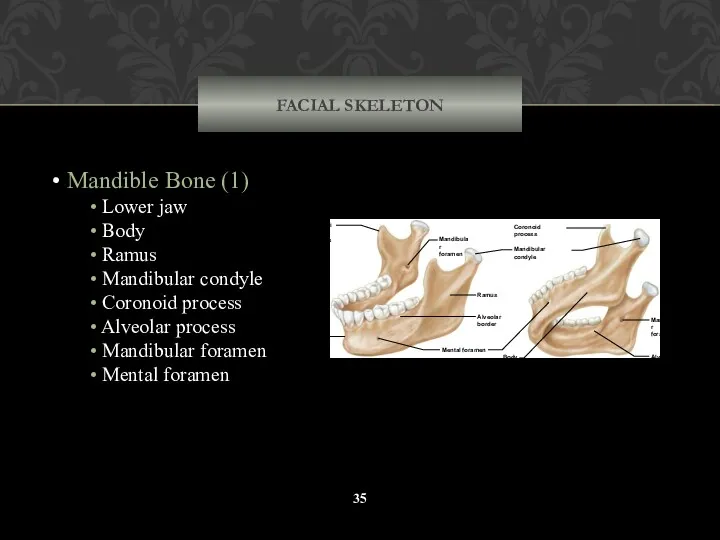
- 35. FACIAL SKELETON Mandible Bone (1) Lower jaw Body Ramus Mandibular condyle Coronoid process Alveolar process Mandibular
- 36. VERTEBRAL COLUMN The vertebral column, or spinal column, consists of many vertebrae separated by cartilaginous intervertebral
- 37. VERTEBRAL COLUMN Cervical vertebrae (7) Thoracic vertebrae (12) Lumbar vertebrae (5) Sacral (4-5 fused segments) Sacrum
- 38. VERTEBRAL COLUMN Cervical curvature Thoracic curvature Lumbar curvature Sacral curvature Rib facets Vertebral prominens Intervertebral discs
- 39. TYPICAL VERTEBRAE Includes the following parts: Vertebral body Pedicles Lamina Spinous process Transverse processes Vertebral foramen
- 40. CERVICAL VERTEBRAE Atlas – 1st; supports head Axis – 2nd; dens pivots to turn head Transverse
- 41. THORACIC VERTEBRAE Body Superior articular process Spinous process Transverse process Inferior articular process Intervertebral disc Anterior
- 42. LUMBAR VERTEBRAE Large bodies Thick, short spinous processes (c) Lumbar vertebra Lamina Pedicle Body Vertebral foramen
- 43. SACRUM 4-5 fused segments Median sacral crest Posterior sacral foramina Posterior wall of pelvic cavity Sacral
- 44. COCCYX 3-4 fused segments Sacral canal Tubercle of median sacral crest Auricular surface Posterior sacral foramen
- 45. THORACIC CAGE The thoracic cage includes the ribs, the thoracic vertebrae, the sternum, and the costal
- 46. THORACIC CAGE Ribs (12) Sternum Thoracic vertebrae (12) Costal cartilages Supports shoulder girdle and upper limbs
- 47. RIBS Humans have 12 pairs of ribs: True ribs (7) False ribs (5), of which: Floating
- 48. RIB STRUCTURE Shaft Head – posterior end; articulates with vertebrae Tubercle – articulates with vertebrae Costal
- 49. STERNUM Three (3) parts of the sternum: Manubrium Body Xiphoid process 1 2 3 4 5
- 50. PECTORAL GIRDLE Also known as the shoulder girdle Clavicles Scapulae Supports upper limbs True shoulder joint
- 51. CLAVICLES Articulate with manubrium Articulate with scapulae (acromion process) A-C joint Sternum Costal cartilage Rib Scapula
- 52. Acromion process Coracoid process Spine Glenoid cavity Suprascapular notch Superior border Supra- glenoid tubercle Infra- glenoid
- 53. UPPER LIMB Humerus Radius Ulna (Interosseous membrane) Carpals Metacarpals Phalanges Olecranon process Head of radius Neck
- 54. HUMERUS Head Greater tubercle Lesser tubercle Anatomical neck Surgical neck Deltoid tuberosity Capitulum Trochlea Coronoid fossa
- 55. RADIUS Lateral forearm bone Head Radial tuberosity Styloid process Styloid process Ulnar notch of radius Styloid
- 56. ULNA Medial forearm bone Trochlear notch Olecranon process Coronoid process Styloid process Styloid process Ulnar notch
- 57. WRIST AND HAND Carpal Bones (16 total bones) Scaphoid Lunate Triquetral Pisiform Hamate Capitate Trapezoid Trapezium
- 58. PELVIC GIRDLE Coxal Bones (2) Supports trunk of body Protects viscera Forms pelvic cavity Sacrum Sacral
- 59. HIP BONES Also known as the coxae: Acetabulum There are three (3) bones: 1. Ilium Iliac
- 60. GREATER AND LESSER PELVES Greater Pelvis Lumbar vertebrae posteriorly Iliac bones laterally Abdominal wall anteriorly Lesser
- 61. DIFFERENCES BETWEEN MALE FEMALE PELVES Female pelvis Iliac bones more flared Broader hips Pubic arch angle
- 62. LOWER LIMB Femur Patella Tibia Fibula Tarsals Metatarsals Phalanges Metatarsals Fibula Tibia T ibia Patella Femur
- 63. FEMUR Longest bone of body Head Fovea capitis Neck Greater trochanter Lesser trochanter Linea aspera Condyles
- 64. PATELLA Anterior surface of the knee joint Flat sesamoid bone located in the quadriceps tendon Metatarsals
- 65. TIBIA Medial to fibula Condyles Tibial tuberosity Makes the medial malleolus Tibia Fibula Medial malleolus Tibial
- 66. FIBULA Lateral to tibia Long, slender Head Makes the lateral malleolus Non-weight bearing Tibia Fibula Medial
- 67. FOOT Tarsal Bones (14) Calcaneus Talus Navicular Cuboid Lateral (3rd) cuneiform Intermediate (2nd) cuneiform Medial (1st)
- 68. FOOT Calcaneus Talus Navicular Cuboid Lateral cuneiform Intermediate cuneiform Medial cuneiform Proximal phalanx Middle phalanx Distal
- 69. LIFESPAN CHANGES Decrease in height at about age 30 Calcium levels fall Bones become brittle Osteoclasts
- 70. Joints Slide 5.43 Copyright © 2003 Pearson Education, Inc. publishing as Benjamin Cummings Articulations of bones
- 71. Functional Classification of Joints Slide 5.44 Synarthroses – immovable joints Amphiarthroses – slightly moveable joints Diarthroses
- 72. Structural Classification of Joints Slide 5.45 Copyright © 2003 Pearson Education, Inc. publishing as Benjamin Cummings
- 73. Fibrous Joints Bones united by fibrous tissue – synarthrosis or largely immovable.
- 74. Cartilaginous Joints – mostly amphiarthrosis Slide 5.47 Copyright © 2003 Pearson Education, Inc. publishing as Benjamin
- 75. Synovial Joints Slide 5.48 Copyright © 2003 Pearson Education, Inc. publishing as Benjamin Cummings Articulating bones
- 76. Features of Synovial Joints- Diarthroses Articular cartilage (hyaline cartilage) covers the ends of bones Joint surfaces
- 77. Structures Associated with the Synovial Joint Slide 5.50 Copyright © 2003 Pearson Education, Inc. publishing as
- 78. The Synovial Joint Slide 5.51 Copyright © 2003 Pearson Education, Inc. publishing as Benjamin Cummings Figure
- 79. Types of Synovial Joints Based on Shape Slide 5.52a Copyright © 2003 Pearson Education, Inc. publishing
- 81. Скачать презентацию

BONE CLASSIFICATION
Bone Classification:
Long Bones
Short Bones
Sesamoid Bones
Flat
BONE CLASSIFICATION
Bone Classification:
Long Bones
Short Bones
Sesamoid Bones
Flat
Classification of Bones
Slide 5.4a
Long bones
Typically longer than wide
Have a shaft with
Classification of Bones
Slide 5.4a
Long bones
Typically longer than wide
Have a shaft with
Classification of Bones
Slide 5.4b
Short bones
Generally cube-shape
Contain mostly spongy bone
Examples: Carpals, tarsals
Classification of Bones
Slide 5.4b
Short bones
Generally cube-shape
Contain mostly spongy bone
Examples: Carpals, tarsals
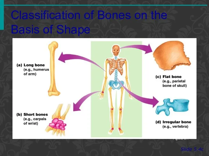
Classification of Bones on the Basis of Shape
Slide 5.4c
Figure 5.1
Classification of Bones on the Basis of Shape
Slide 5.4c
Figure 5.1

Classification of Bones
Slide 5.5a
Flat bones
Thin and flattened
Usually curved
Thin layers of compact
Classification of Bones
Slide 5.5a
Flat bones
Thin and flattened
Usually curved
Thin layers of compact
Classification of Bones
Slide 5.5b
Irregular bones
Irregular shape
Do not fit into other bone
Classification of Bones
Slide 5.5b
Irregular bones
Irregular shape
Do not fit into other bone

Classification of Bones on the Basis of Shape
Slide 5.5c
Figure 5.1
Classification of Bones on the Basis of Shape
Slide 5.5c
Figure 5.1
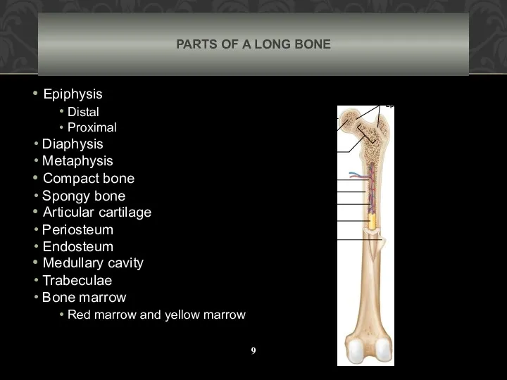
PARTS OF A LONG BONE
Epiphysis
Distal
Proximal
Diaphysis
Metaphysis
Compact
PARTS OF A LONG BONE
Epiphysis
Distal
Proximal
Diaphysis
Metaphysis
Compact
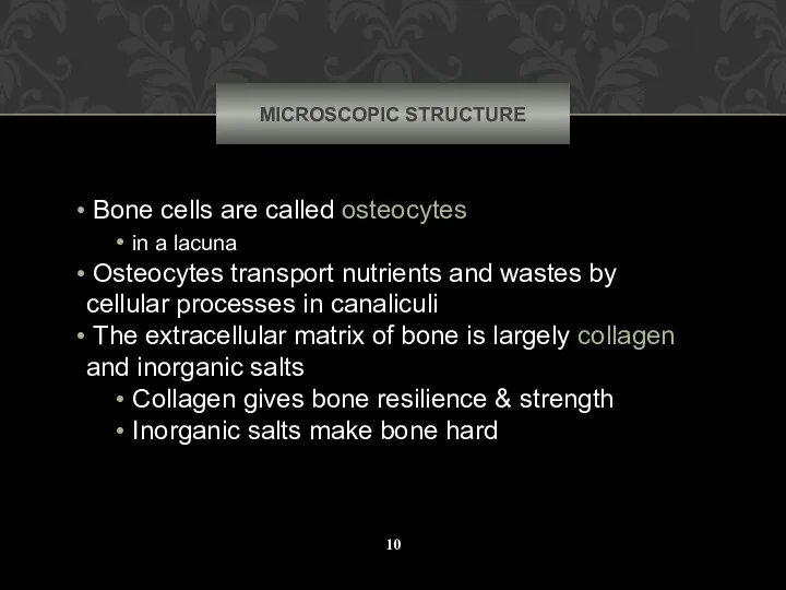
MICROSCOPIC STRUCTURE
Bone cells are called osteocytes
in a lacuna
Osteocytes
MICROSCOPIC STRUCTURE
Bone cells are called osteocytes
in a lacuna
Osteocytes
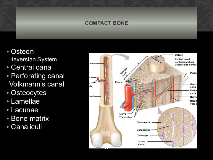
COMPACT BONE
Osteon
Haversian System
Central canal
Perforating canal Volkmann’s canal
Osteocytes
COMPACT BONE
Osteon
Haversian System
Central canal
Perforating canal Volkmann’s canal
Osteocytes
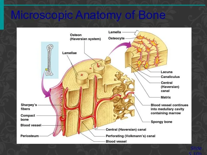
Microscopic Anatomy of Bone
Slide 5.10b
Figure 5.3
Microscopic Anatomy of Bone
Slide 5.10b
Figure 5.3
Microscopic Anatomy of Bone
Slide 5.11a
Lacunae
Cavities containing bone cells (osteocytes)
Arranged in concentric
Microscopic Anatomy of Bone
Slide 5.11a
Lacunae
Cavities containing bone cells (osteocytes)
Arranged in concentric
Microscopic Anatomy of Bone
Slide 5.11b
Canaliculi
Tiny canals
Radiate from the central canal
Microscopic Anatomy of Bone
Slide 5.11b
Canaliculi
Tiny canals
Radiate from the central canal
BONE DEVELOPMENT
AND GROWTH
Parts of the skeletal system begin to
BONE DEVELOPMENT
AND GROWTH
Parts of the skeletal system begin to
INTRAMEMBRANOUS BONES
Intramembranous Bones
These bones originate within sheetlike layers of
INTRAMEMBRANOUS BONES
Intramembranous Bones
These bones originate within sheetlike layers of
ENDOCHONDRAL BONES
Endochondral Bones
Bones begin as hyaline cartilage
Form models
ENDOCHONDRAL BONES
Endochondral Bones
Bones begin as hyaline cartilage
Form models
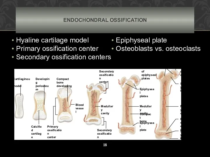
ENDOCHONDRAL OSSIFICATION
Hyaline cartilage model
Primary ossification center
Secondary ossification centers
ENDOCHONDRAL OSSIFICATION
Hyaline cartilage model
Primary ossification center
Secondary ossification centers
BONE FUNCTION
Bones shape, support, and protect body structures
BONE FUNCTION
Bones shape, support, and protect body structures
SUPPORT, PROTECTION,
AND MOVEMENT
Support, Movement & Protection
Gives shape to
SUPPORT, PROTECTION,
AND MOVEMENT
Support, Movement & Protection
Gives shape to
BLOOD CELL FORMATION
Blood Cell Formation
Also known as hematopoiesis
Occurs
BLOOD CELL FORMATION
Blood Cell Formation
Also known as hematopoiesis
Occurs
INORGANIC SALT STORAGE
Inorganic Salt Storage
Calcium
Phosphate
Magnesium
Sodium
INORGANIC SALT STORAGE
Inorganic Salt Storage
Calcium
Phosphate
Magnesium
Sodium
SKELETAL ORGANIZATION
The actual number of bones in the human skeleton
SKELETAL ORGANIZATION
The actual number of bones in the human skeleton
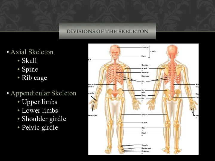
DIVISIONS OF THE SKELETON
Axial Skeleton
Skull
Spine
Rib
DIVISIONS OF THE SKELETON
Axial Skeleton
Skull
Spine
Rib
SKULL
Is composed of the cranium (brain case) and the facial
SKULL
Is composed of the cranium (brain case) and the facial
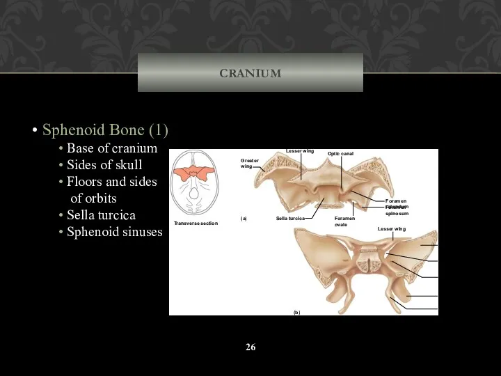
CRANIUM
Sphenoid Bone (1)
Base of cranium
Sides of skull
Floors
CRANIUM
Sphenoid Bone (1)
Base of cranium
Sides of skull
Floors
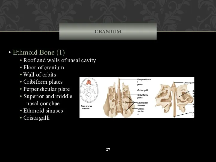
CRANIUM
Ethmoid Bone (1)
Roof and walls of nasal cavity
Floor
CRANIUM
Ethmoid Bone (1)
Roof and walls of nasal cavity
Floor
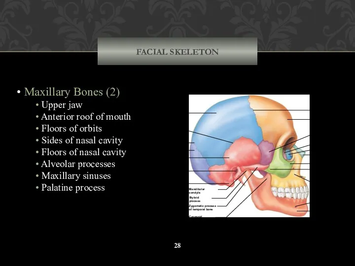
FACIAL SKELETON
Maxillary Bones (2)
Upper jaw
Anterior roof of mouth
FACIAL SKELETON
Maxillary Bones (2)
Upper jaw
Anterior roof of mouth
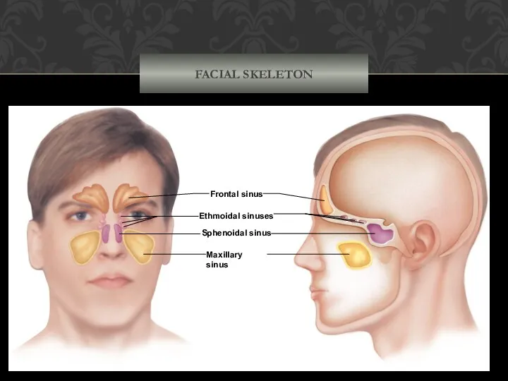
FACIAL SKELETON
Frontal sinus
Ethmoidal sinuses
Sphenoidal sinus
Maxillary sinus
FACIAL SKELETON
Frontal sinus
Ethmoidal sinuses
Sphenoidal sinus
Maxillary sinus
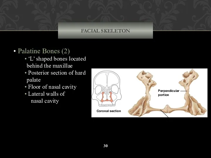
FACIAL SKELETON
Palatine Bones (2)
‘L’ shaped bones located behind the
FACIAL SKELETON
Palatine Bones (2)
‘L’ shaped bones located behind the
FACIAL SKELETON
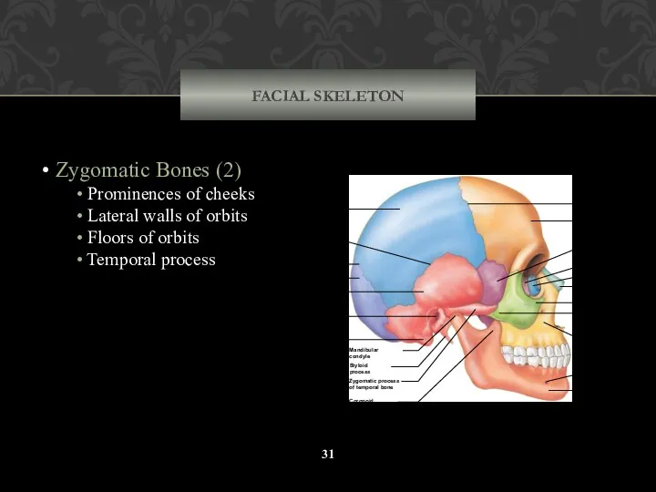
Zygomatic Bones (2)
Prominences of cheeks
Lateral walls of
FACIAL SKELETON
Zygomatic Bones (2)
Prominences of cheeks
Lateral walls of
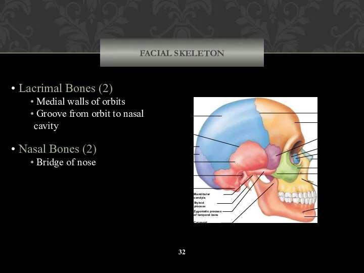
FACIAL SKELETON
Lacrimal Bones (2)
Medial walls of orbits
Groove from
FACIAL SKELETON
Lacrimal Bones (2)
Medial walls of orbits
Groove from
FACIAL SKELETON
Vomer Bone (1)
Inferior portion of nasal septum
Coronal suture
Frontal
FACIAL SKELETON
Vomer Bone (1)
Inferior portion of nasal septum
Coronal suture
Frontal
FACIAL SKELETON
Inferior Nasal Conchae (2)
Extend from lateral
walls of
FACIAL SKELETON
Inferior Nasal Conchae (2)
Extend from lateral
walls of
FACIAL SKELETON
Mandible Bone (1)
Lower jaw
Body
Ramus
Mandibular condyle
FACIAL SKELETON
Mandible Bone (1)
Lower jaw
Body
Ramus
Mandibular condyle
VERTEBRAL COLUMN
The vertebral column, or spinal column, consists of many
VERTEBRAL COLUMN
The vertebral column, or spinal column, consists of many
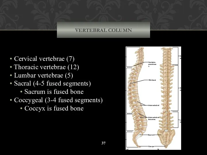
VERTEBRAL COLUMN
Cervical vertebrae (7)
Thoracic vertebrae (12)
Lumbar vertebrae (5)
VERTEBRAL COLUMN
Cervical vertebrae (7)
Thoracic vertebrae (12)
Lumbar vertebrae (5)
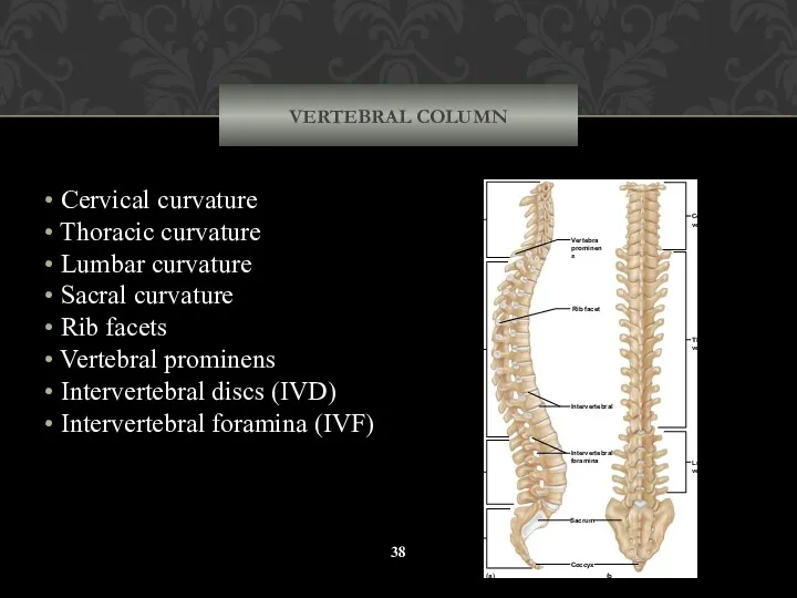
VERTEBRAL COLUMN
Cervical curvature
Thoracic curvature
Lumbar curvature
Sacral curvature
Rib
VERTEBRAL COLUMN
Cervical curvature
Thoracic curvature
Lumbar curvature
Sacral curvature
Rib
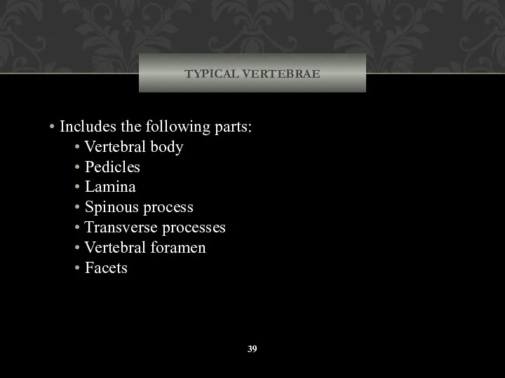
TYPICAL VERTEBRAE
Includes the following parts:
Vertebral body
Pedicles
Lamina
Spinous
TYPICAL VERTEBRAE
Includes the following parts:
Vertebral body
Pedicles
Lamina
Spinous
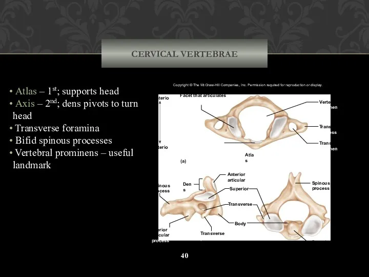
CERVICAL VERTEBRAE
Atlas – 1st; supports head
Axis – 2nd; dens
CERVICAL VERTEBRAE
Atlas – 1st; supports head
Axis – 2nd; dens
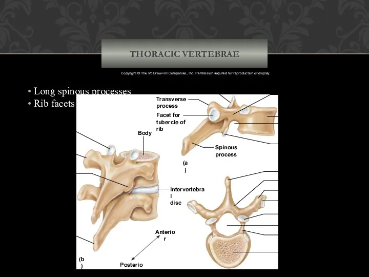
THORACIC VERTEBRAE
Body
Superior
articular
process
Spinous
process
Transverse
process
Inferior articular
process
Intervertebral
disc
Anterior
Posterior
Body
Pedicle
Vertebral foramen
Superior articular process
Facet for tubercle of rib
Transverse process
Lamina
Spinous
THORACIC VERTEBRAE
Body
Superior
articular
process
Spinous
process
Transverse
process
Inferior articular
process
Intervertebral
disc
Anterior
Posterior
Body
Pedicle
Vertebral foramen
Superior articular process
Facet for tubercle of rib
Transverse process
Lamina
Spinous
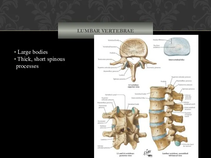
LUMBAR VERTEBRAE
Large bodies
Thick, short spinous processes
(c) Lumbar vertebra
Lamina
Pedicle
Body
Vertebral foramen
Spinous
LUMBAR VERTEBRAE
Large bodies
Thick, short spinous processes
(c) Lumbar vertebra
Lamina
Pedicle
Body
Vertebral foramen
Spinous
SACRUM
4-5 fused segments
Median sacral crest
Posterior sacral foramina
Posterior
SACRUM
4-5 fused segments
Median sacral crest
Posterior sacral foramina
Posterior
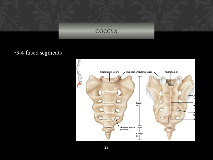
COCCYX
3-4 fused segments
Sacral canal
Tubercle
of median
sacral crest
Auricular
surface
Posterior sacral
foramen
Sacral hiatus
Coccyx
Sacrum
Superior articular process
Sacral promontory
Anterior
COCCYX
3-4 fused segments
Sacral canal
Tubercle
of median
sacral crest
Auricular
surface
Posterior sacral
foramen
Sacral hiatus
Coccyx
Sacrum
Superior articular process
Sacral promontory
Anterior
THORACIC CAGE
The thoracic cage includes the ribs, the thoracic vertebrae,
THORACIC CAGE
The thoracic cage includes the ribs, the thoracic vertebrae,
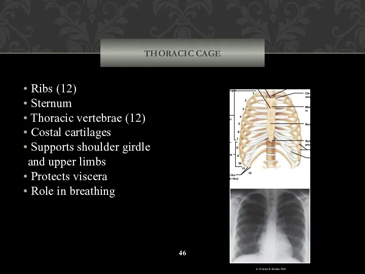
THORACIC CAGE
Ribs (12)
Sternum
Thoracic vertebrae (12)
Costal cartilages
Supports
THORACIC CAGE
Ribs (12)
Sternum
Thoracic vertebrae (12)
Costal cartilages
Supports
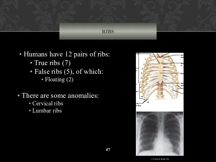
RIBS
Humans have 12 pairs of ribs:
True ribs (7)
False
RIBS
Humans have 12 pairs of ribs:
True ribs (7)
False
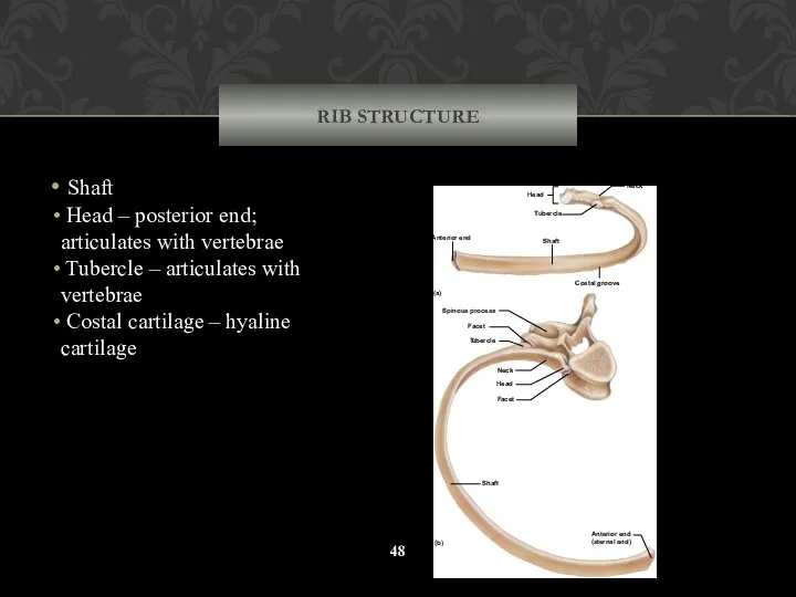
RIB STRUCTURE
Shaft
Head – posterior end; articulates with vertebrae
Tubercle
RIB STRUCTURE
Shaft
Head – posterior end; articulates with vertebrae
Tubercle
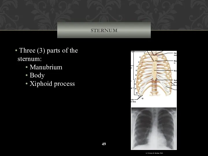
STERNUM
Three (3) parts of the sternum:
Manubrium
Body
Xiphoid process
1
2
3
4
5
6
7
8
9
10
1
1
12
True
STERNUM
Three (3) parts of the sternum:
Manubrium
Body
Xiphoid process
1
2
3
4
5
6
7
8
9
10
1
1
12
True
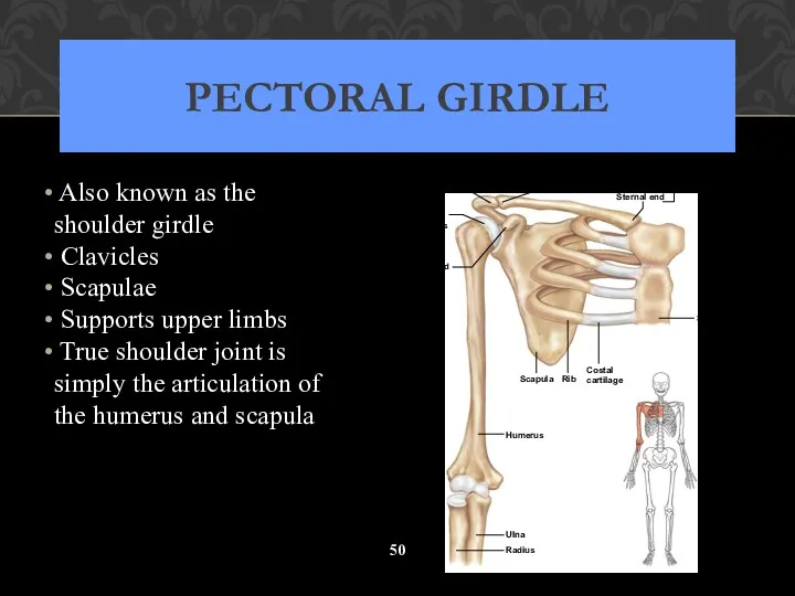
PECTORAL GIRDLE
Also known as the shoulder girdle
Clavicles
Scapulae
PECTORAL GIRDLE
Also known as the shoulder girdle
Clavicles
Scapulae
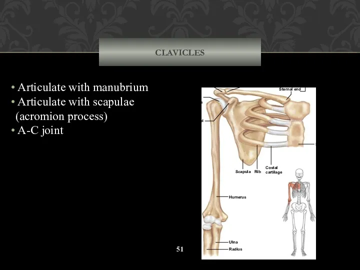
CLAVICLES
Articulate with manubrium
Articulate with scapulae (acromion process)
A-C joint
Sternum
Costal
cartilage
Rib
Scapula
Humerus
Ulna
Radius
Clavicle
(a)
Coracoid
process
Head
CLAVICLES
Articulate with manubrium
Articulate with scapulae (acromion process)
A-C joint
Sternum
Costal
cartilage
Rib
Scapula
Humerus
Ulna
Radius
Clavicle
(a)
Coracoid
process
Head
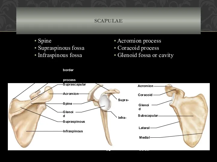
Acromion
process
Coracoid
process
Spine
Glenoid
cavity
Suprascapular
notch
Superior
border
Supra-
glenoid
tubercle
Infra-
glenoid
tubercle
Coracoid
process
Acromion
process
Supraspinous
fossa
Infraspinous
fossa
Glenoid
cavity
Lateral
(axillary) border
Subscapular
fossa
Medial
(vertebral)
border
(a)
(b)
(c)
SCAPULAE
Spine
Supraspinous fossa
Infraspinous fossa
Acromion process
Coracoid process
Acromion
process
Coracoid
process
Spine
Glenoid
cavity
Suprascapular
notch
Superior
border
Supra-
glenoid
tubercle
Infra-
glenoid
tubercle
Coracoid
process
Acromion
process
Supraspinous
fossa
Infraspinous
fossa
Glenoid
cavity
Lateral
(axillary) border
Subscapular
fossa
Medial
(vertebral)
border
(a)
(b)
(c)
SCAPULAE
Spine
Supraspinous fossa
Infraspinous fossa
Acromion process
Coracoid process
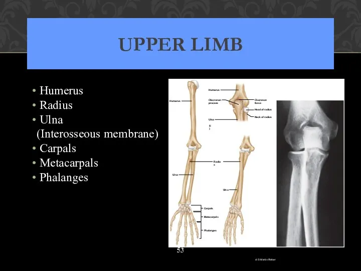
UPPER LIMB
Humerus
Radius
Ulna
(Interosseous membrane)
Carpals
Metacarpals
Phalanges
Olecranon
process
Head of radius
Neck
UPPER LIMB
Humerus
Radius
Ulna
(Interosseous membrane)
Carpals
Metacarpals
Phalanges
Olecranon
process
Head of radius
Neck
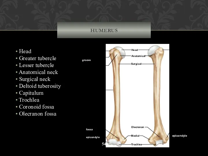
HUMERUS
Head
Greater tubercle
Lesser tubercle
Anatomical neck
Surgical neck
Deltoid
HUMERUS
Head
Greater tubercle
Lesser tubercle
Anatomical neck
Surgical neck
Deltoid
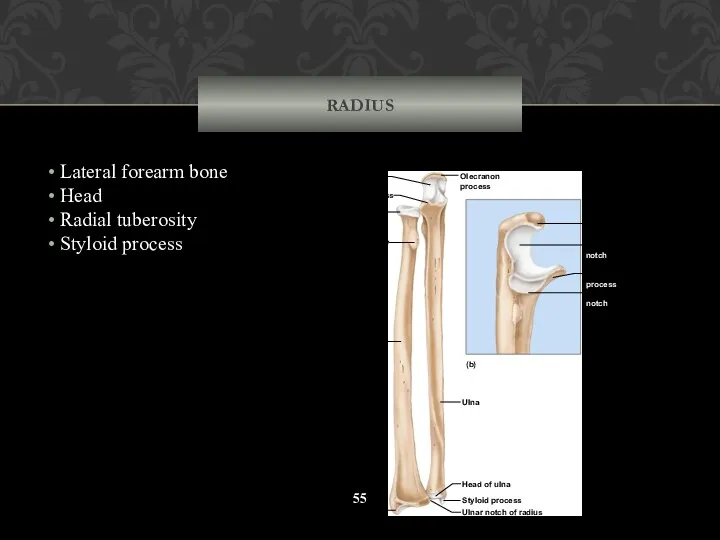
RADIUS
Lateral forearm bone
Head
Radial tuberosity
Styloid process
Styloid process
Ulnar notch
RADIUS
Lateral forearm bone
Head
Radial tuberosity
Styloid process
Styloid process
Ulnar notch
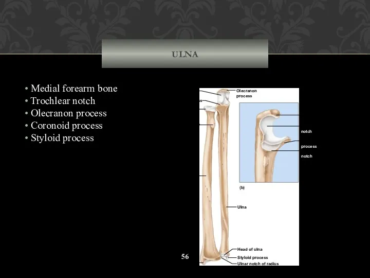
ULNA
Medial forearm bone
Trochlear notch
Olecranon process
Coronoid process
Styloid
ULNA
Medial forearm bone
Trochlear notch
Olecranon process
Coronoid process
Styloid
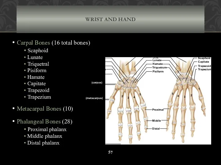
WRIST AND HAND
Carpal Bones (16 total bones)
Scaphoid
Lunate
Triquetral
WRIST AND HAND
Carpal Bones (16 total bones)
Scaphoid
Lunate
Triquetral
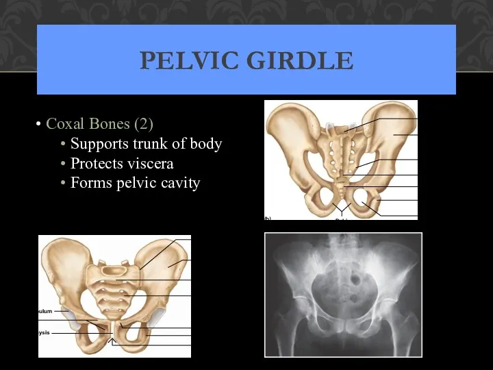
PELVIC GIRDLE
Coxal Bones (2)
Supports trunk of body
Protects viscera
PELVIC GIRDLE
Coxal Bones (2)
Supports trunk of body
Protects viscera
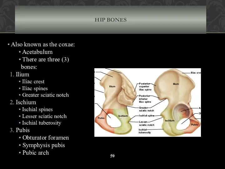
HIP BONES
Also known as the coxae:
Acetabulum
There are
HIP BONES
Also known as the coxae:
Acetabulum
There are
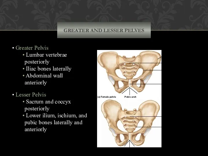
GREATER AND LESSER PELVES
Greater Pelvis
Lumbar vertebrae posteriorly
Iliac bones
GREATER AND LESSER PELVES
Greater Pelvis
Lumbar vertebrae posteriorly
Iliac bones
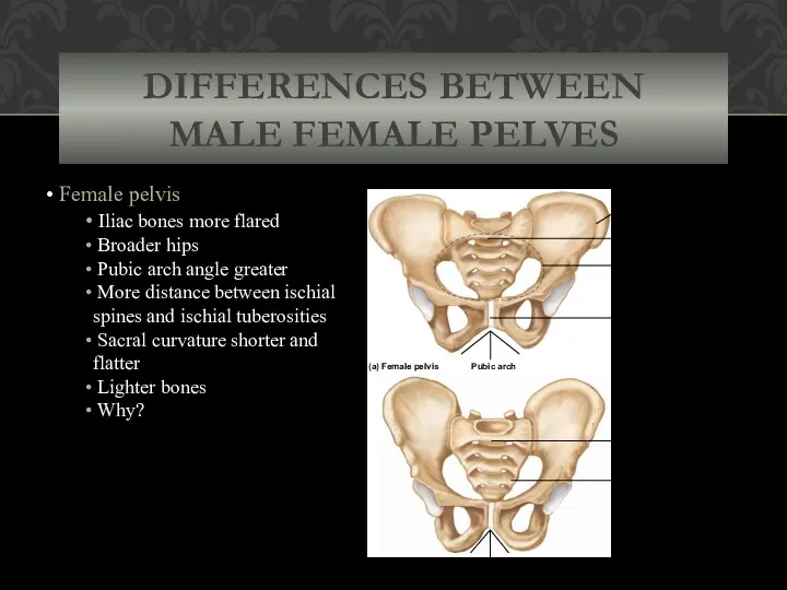
DIFFERENCES BETWEEN
MALE FEMALE PELVES
Female pelvis
Iliac bones more flared
DIFFERENCES BETWEEN
MALE FEMALE PELVES
Female pelvis
Iliac bones more flared
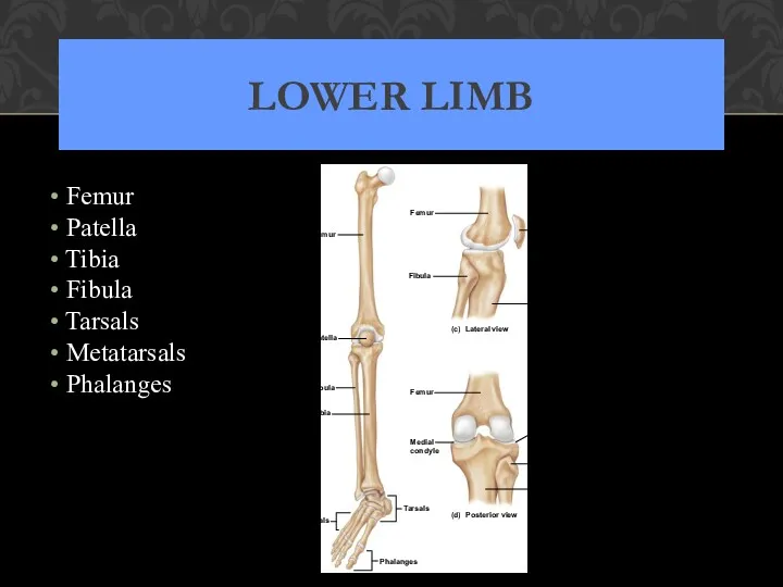
LOWER LIMB
Femur
Patella
Tibia
Fibula
Tarsals
Metatarsals
Phalanges
Metatarsals
Fibula
Tibia
T
ibia
Patella
Femur
Fibula
(c)
Lateral view
Fibula
T
ibia
Lateral
condyle
(d)
Posterior view
(b)
Medial
condyle
Femur
T
arsals
Phalanges
Femur
Patella
LOWER LIMB
Femur
Patella
Tibia
Fibula
Tarsals
Metatarsals
Phalanges
Metatarsals
Fibula
Tibia
T
ibia
Patella
Femur
Fibula
(c)
Lateral view
Fibula
T
ibia
Lateral
condyle
(d)
Posterior view
(b)
Medial
condyle
Femur
T
arsals
Phalanges
Femur
Patella
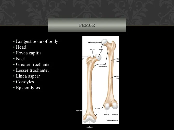
FEMUR
Longest bone of body
Head
Fovea capitis
Neck
Greater trochanter
FEMUR
Longest bone of body
Head
Fovea capitis
Neck
Greater trochanter
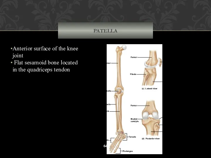
PATELLA
Anterior surface of the knee joint
Flat sesamoid bone located in
PATELLA
Anterior surface of the knee joint
Flat sesamoid bone located in
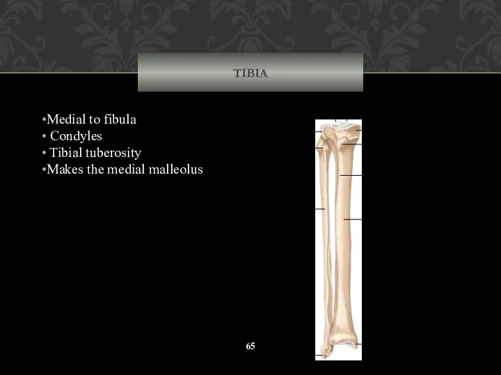
TIBIA
Medial to fibula
Condyles
Tibial tuberosity
Makes the medial malleolus
Tibia
Fibula
Medial
malleolus
Tibial
tuberosity
Anterior
crest
Medial
condyle
Intercondylar
eminence
Lateral
malleolus
Lateral
condyle
Head of
fibula
TIBIA
Medial to fibula
Condyles
Tibial tuberosity
Makes the medial malleolus
Tibia
Fibula
Medial
malleolus
Tibial
tuberosity
Anterior
crest
Medial
condyle
Intercondylar
eminence
Lateral
malleolus
Lateral
condyle
Head of
fibula
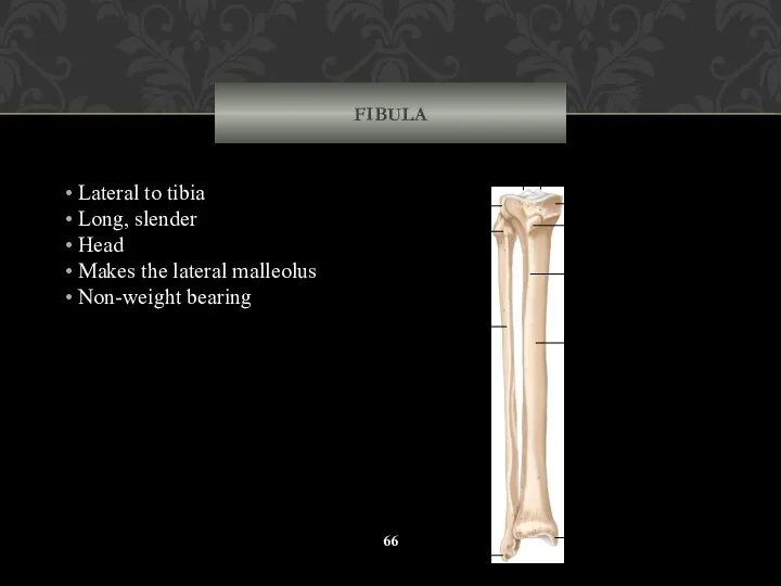
FIBULA
Lateral to tibia
Long, slender
Head
Makes the lateral malleolus
FIBULA
Lateral to tibia
Long, slender
Head
Makes the lateral malleolus
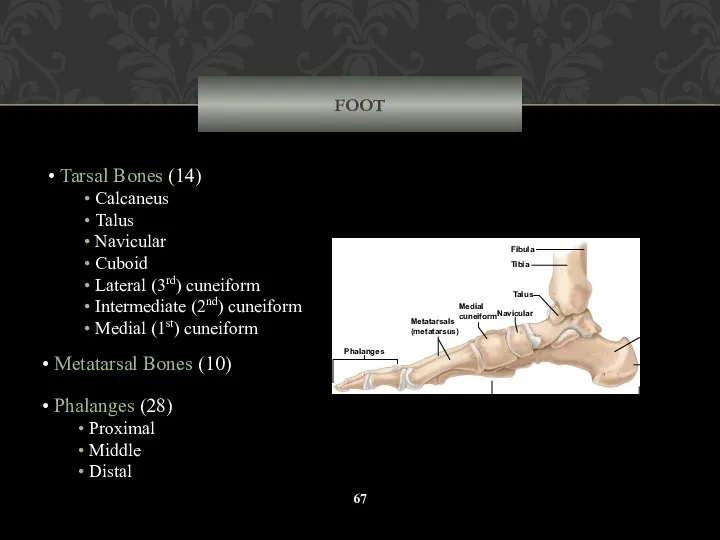
FOOT
Tarsal Bones (14)
Calcaneus
Talus
Navicular
Cuboid
Lateral (3rd) cuneiform
FOOT
Tarsal Bones (14)
Calcaneus
Talus
Navicular
Cuboid
Lateral (3rd) cuneiform
FOOT
Calcaneus
Talus
Navicular
Cuboid
Lateral cuneiform
Intermediate cuneiform
Medial cuneiform
Proximal phalanx
Middle phalanx
Distal phalanx
Phalanges
Metatarsals
(metatarsus)
Tarsals
(tarsus)
5
4
3
2
1
(a)
FOOT
Calcaneus
Talus
Navicular
Cuboid
Lateral cuneiform
Intermediate cuneiform
Medial cuneiform
Proximal phalanx
Middle phalanx
Distal phalanx
Phalanges
Metatarsals
(metatarsus)
Tarsals
(tarsus)
5
4
3
2
1
(a)
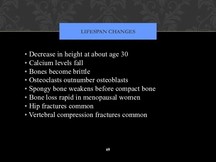
LIFESPAN CHANGES
Decrease in height at about age 30
Calcium levels
LIFESPAN CHANGES
Decrease in height at about age 30
Calcium levels
Joints
Slide 5.43
Copyright © 2003 Pearson Education, Inc. publishing as Benjamin Cummings
Articulations
Joints
Slide 5.43
Copyright © 2003 Pearson Education, Inc. publishing as Benjamin Cummings
Articulations
Functional Classification of Joints
Slide 5.44
Synarthroses – immovable joints
Amphiarthroses – slightly moveable
Functional Classification of Joints
Slide 5.44
Synarthroses – immovable joints
Amphiarthroses – slightly moveable
Structural Classification of Joints
Slide 5.45
Copyright © 2003 Pearson Education, Inc. publishing
Structural Classification of Joints
Slide 5.45
Copyright © 2003 Pearson Education, Inc. publishing
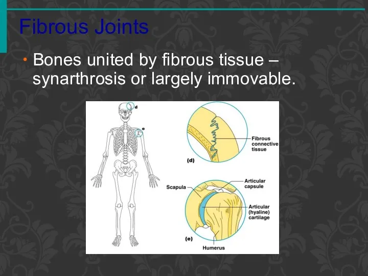
Fibrous Joints
Bones united by fibrous tissue – synarthrosis or largely immovable.
Fibrous Joints
Bones united by fibrous tissue – synarthrosis or largely immovable.
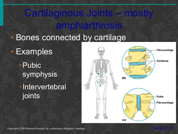
Cartilaginous Joints – mostly amphiarthrosis
Slide 5.47
Copyright © 2003 Pearson Education, Inc.
Cartilaginous Joints – mostly amphiarthrosis
Slide 5.47
Copyright © 2003 Pearson Education, Inc.
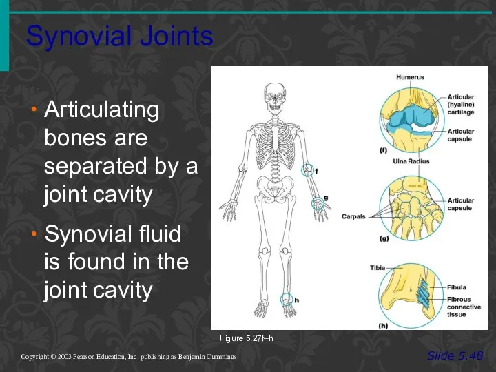
Synovial Joints
Slide 5.48
Copyright © 2003 Pearson Education, Inc. publishing as Benjamin
Synovial Joints
Slide 5.48
Copyright © 2003 Pearson Education, Inc. publishing as Benjamin
Features of Synovial Joints- Diarthroses
Articular cartilage (hyaline cartilage) covers the ends
Features of Synovial Joints- Diarthroses
Articular cartilage (hyaline cartilage) covers the ends
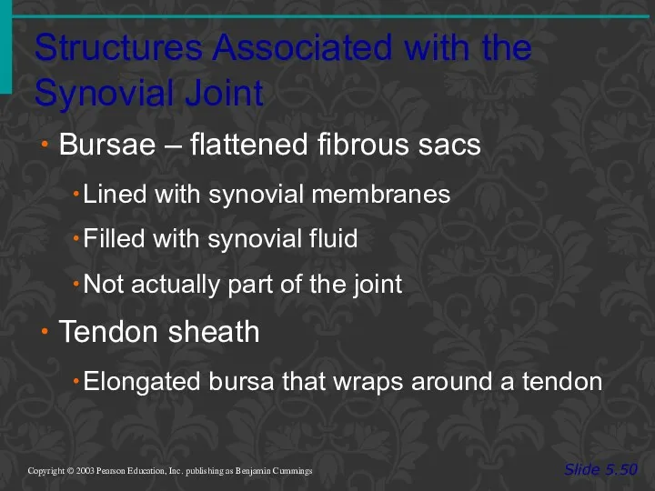
Structures Associated with the Synovial Joint
Slide 5.50
Copyright © 2003 Pearson Education,
Structures Associated with the Synovial Joint
Slide 5.50
Copyright © 2003 Pearson Education,
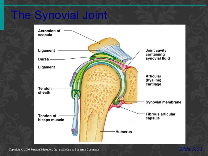
The Synovial Joint
Slide 5.51
Copyright © 2003 Pearson Education, Inc. publishing as
The Synovial Joint
Slide 5.51
Copyright © 2003 Pearson Education, Inc. publishing as
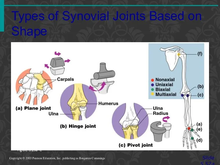
Types of Synovial Joints Based on Shape
Slide 5.52a
Copyright © 2003 Pearson
Types of Synovial Joints Based on Shape
Slide 5.52a
Copyright © 2003 Pearson
 Болезни и вредители цитрусовых
 Рыба речная: Окунь
 Витамины – понятие о гиповитаминозах, гипервитаминозах, авитаминозах
 Мышечные цепи. Взаимосвязи миофасциальной сети
 Вода. Роль воды в жизни человека
 Строение. Функции. Значение кожи
 Тип Хордовые класс Млекопитающие
 Профилактика употреблениия ПАВ через преподавание биологии
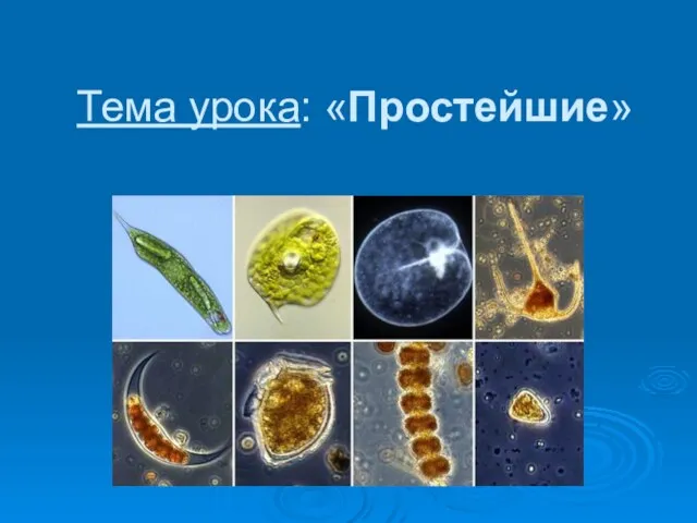 Простейшие
 Наружные и внутренние мужские и женские половые органы
 Процессы жизнеобеспечения в организме человека. Эндокринные железы (железы внутренней секреции)
 Применение проектной технологии на уроках биологии и во внеурочной деятельности
 Ядовитые змеи мира
 Пластичность клеток разных тканей
 الزواحف
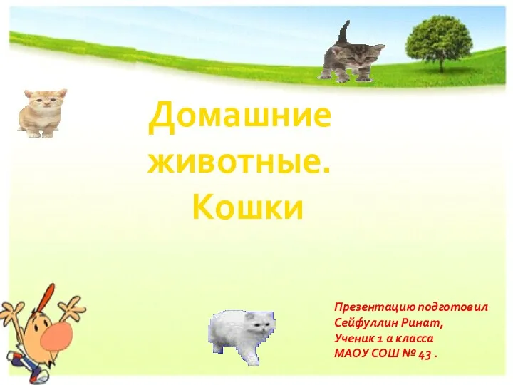 Домашние животные. Кошки
 Биоритмы. Характеристика хронологических типов человека
 Рост и развитие волос
 Биотехнология түсініктері, даму тарихы, негізгі әдістері
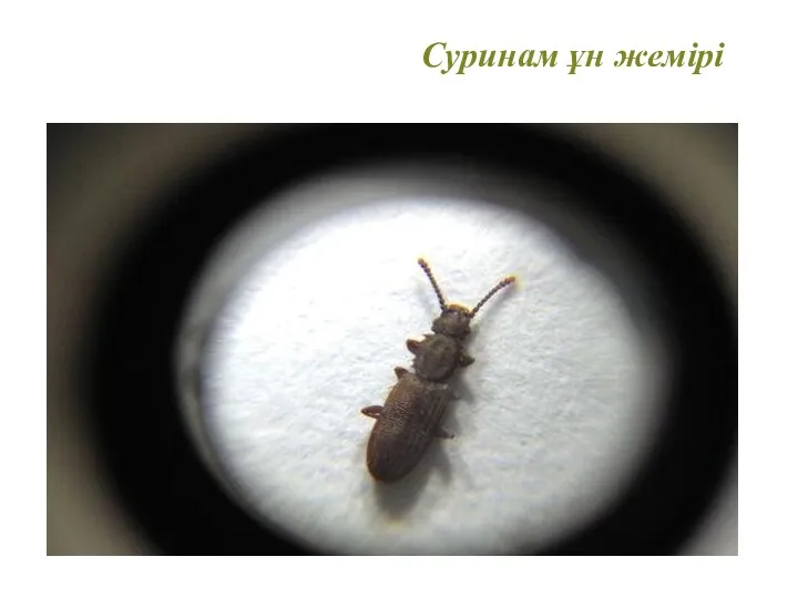 Суринам ұн жемірі
 Використання тваринами знарядь праці
 Гүл, оның құрылысы мен маңызы
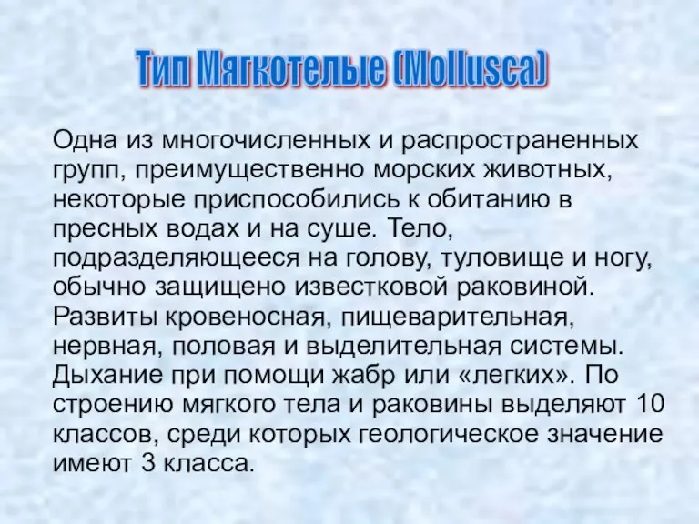 Морские животные. Тип мягкотелые
 Экология и природопользование. Экосистемы
 Передвижение веществ в организме растения
 Транспорт веществ в организме. 6 класс
 Класс Пресмыкающиеся, или Рептилии
 Методы иммуноанализа с применением различных меток