Carbohydrate Metabolism I: Aerobic oxidation of glucose. Anaerobic Glycolysis. Gluconeogenesis презентация
- Carbohydrate Metabolism I: Aerobic oxidation of glucose. Anaerobic Glycolysis. Gluconeogenesis

Содержание
- 2. OBJECTIVES in Carbohydrate Metabolism Consider the main metabolic pathways (the intermediates, enzymes, cofactors and regulation) for
- 3. Glucose Structure
- 5. The most significant fates for glucose Glucose 6-phosphate Ribose 5-phosphate Glycogen Pyruvate Pentose phosphate pathway Glucose
- 6. Carbohydrate Metabolism Processes that Yield Energy 1. Tissue respiration (with oxygen ): Break down 6C sugars
- 7. Tissue Respiration (Aerobic Oxidation) for Glucose Consists of 3 Main Phases:
- 8. Aerobic Glycolysis Definition: Aerobic Glycolysis is the metabolic pathway in which monosaccharides (mainly glucose) are split
- 9. Functions of aerobic Glycolysis : 1) to convert glucose to pyruvate which can be: - burned
- 10. Glycolysis reactions: overview Add phosphoryl groups to activate glucose Convert the phosphorylated intermediates into high energy
- 11. Preparatory Phase Step 1: Phosphorylation of Glucose Hexokinase (HK) ATP ADP Mg++ Glucose Glucose 6-phosphate Phosphorylation
- 12. Yeast hexokinase Binding of glucose (purple) causes a large conformational change
- 13. Hexokinase characteristics There are four important mammalian hexokinase isozymes. They are designated hexokinases I, II, III,
- 14. Step 2: Conversion of glucose 6-phosphate to fructose 6-phosphate Phosphohexose Isomerase G6-P F 6-P
- 15. Step 3: Phosphorylation of fructose 6-phosphate to fructose 1,6-bisphosphate Phosphofructokinase 1 Mg++ F 6-P F1,6-bisP ATP
- 16. Step 4: Cleavage of fructose 1,6-bisphosphate Aldolase A Dihydroxyacetone phosphate (DHAP) Glyceraldehyde-3-phosphate (GAP)
- 17. Step 5: Interconversion of the triose phosphates Triosephosphate isomerase A rapid equilibrium allows GAP to be
- 18. Step 6: Oxidation of glyceraldehyde 3-phosphate to 1, 3-bisphosphoglycerate Glyceraldehyde-3-phosphate dehydrogenase NAD+ +Pi NADH 1,3 bisPGl
- 19. Step 7: Phosphoryl transfer from 1,3-bisphosphoglycerate to ADP First ATP generation step Phosphoglycerate Kinase 1,3 bisP-Gl
- 20. 3 P-Gl 2 P-Gl Step 8: Conversion of 3-phosphoglycerate to 2-phosphoglycerate Phosphoglycerate mutase Mg++
- 21. Step 9: Dehydration of 2-phosphoglycerate to phosphoenolpyruvate Enolase 2 P-Gl Phosphoenol pyruvate H2O H2O
- 22. Step 10: Transfer of the phosphoryl group from phosphoenolpyruvate to ADP Second ATP generation step Pyruvate
- 23. Oxidizing power of NAD+ must be recycled 2 2 2 6 2 2 2 2 8
- 24. I. The metabolic fate of pyruvate in aerobic conditions Pyruvate dehydrogenase complex (PDC) transforms pyruvate into
- 25. Cut-away model of the fully assembled PDC It consists of a total of 96 subunits, organized
- 26. Mechanism of PDC action (see in a text-book)
- 27. The metabolic fate of NADH in aerobic conditions. Shuttle systems: Malate-aspartate shuttle (liver, heart) Malate Oxaloacetate
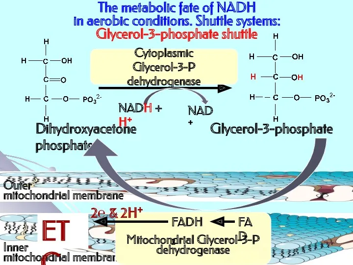
- 28. The metabolic fate of NADH in aerobic conditions. Shuttle systems: Glycerol-3-phosphate shuttle Dihydroxyacetone phosphate Glycerol-3-phosphate NADH
- 29. II. The metabolic fate of pyruvate in anaerobic conditions. Anaerobic glycolysis Definition: Anaerobic Glycolysis is the
- 30. Functions of anaerobic Glycolysis : - ATP production - 2,3 bisphosphoglycerate as powerful effector of O2
- 31. Anaerobic glycolysis last step Lactate dehydrogenase (LDH) Functional LDH are homo or hetero tetramers composed of
- 32. II. The metabolic fate of pyruvate in anaerobic conditions in yeast Alcoholic fermentation: Glucose → 2
- 33. Comparative characteristics of aerobic oxidation of glucose (to CO2&H2O) and anaerobic glycolysis energy balance Aerobic oxidation
- 34. III. Krebs cycle (2 acetyl CoA enter) stage: + 18 ATP (due to utilization of 6
- 35. Regulation
- 36. Glycolysis is regulated at 3 steps involving non equilibrium reactions Step 1: Hexokinase Step 3: Phosphofructokinase
- 37. Specific effectors of Glycolysis
- 38. Regulation of PDC PDC is inhibited when one or more of the three following ratios are
- 39. Gluconeogenesis Definition: Gluconeogenesis is an anabolic pathway whereby non-carbohydrate precursors are converted to glucose Functions: -
- 40. Gluconeogenesis Location in the body : Glucose is synthesized between almost nil and perhaps 200 g/day
- 41. Gluconeogenesis Substrates: Lactate ( produced in RBC, muscles) Glycerol (produced in adipocytes due to lipolysis) Glucogenic
- 42. The major metabolic product produced under normal circumstances by erythrocytes and by muscle cells during intense
- 43. Gluconeogenesis reactions Synthesis of glucose from pyruvate utilizes many of the same enzymes as Glycolysis. Gluconeogenesis
- 44. Bypass of Hexokinase reaction G 6-Pase enzyme is embedded in the endoplasmic reticulum (ER) membrane in
- 45. Bypass of PFK 1 reaction PFKase 1 (Glycolysis) F 1,6-bisPase (Gluconeogenesis) catalyzes:
- 46. Bypass of Pyruvate Kinase reaction Pyruvate Kinase (last step of Glycolysis) Pyruvate Carboxylase (PC) Phosphoenolpyruvate Carboxykinase
- 47. Energy balance for 1 mole of glucose synthesis from 2 moles of pyruvate PC reaction –
- 48. Gluconeogenesis regulation: mitochondrial step NADH, ATP + NADH, ATP Acetyl CoA is allosteric activator of Pyruvate
- 49. To prevent the waste of a futile cycle, Glycolysis (producing 2 ATP) & Gluconeogenesis (consuming 4
- 50. Global Control in liver cells It includes reciprocal effects of a cyclic AMP cascade, triggered by
- 51. Global Control in liver cells Enzymes relevant to these pathways that are phosphorylated by Protein Kinase
- 52. PFK2 domain FBP2 domain PFK2 domain FBP2 domain
- 53. Reciprocal hormonal regulation through F-2,6-bisP
- 54. Phosphofructokinase (PFK) characteristics Mammalian PFK1: - catalyzes the irreversible transformation of F6P to F1,6bisP; - is
- 55. Specific and common effectors for Glycolysis & Gluconeogenesis (in liver)
- 57. Скачать презентацию
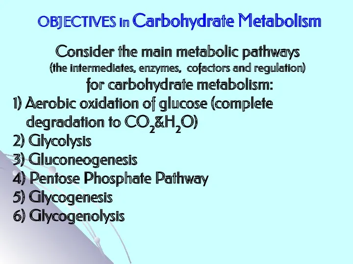
OBJECTIVES in Carbohydrate Metabolism
Consider the main metabolic pathways
(the intermediates,
OBJECTIVES in Carbohydrate Metabolism
Consider the main metabolic pathways
(the intermediates,
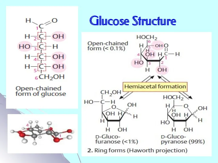
Glucose Structure
Glucose Structure
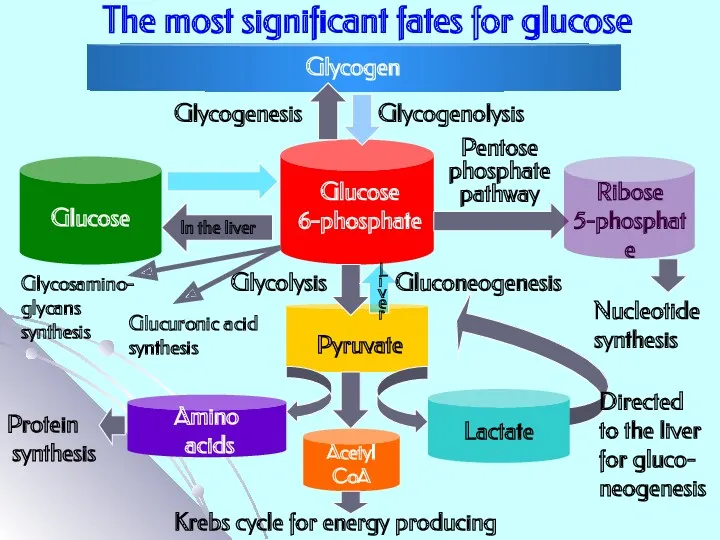
The most significant fates for glucose
Glucose 6-phosphate
Ribose 5-phosphate
Glycogen
Pyruvate
Pentose
phosphate
pathway
Glucose
The most significant fates for glucose
Glucose 6-phosphate
Ribose 5-phosphate
Glycogen
Pyruvate
Pentose
phosphate
pathway
Glucose
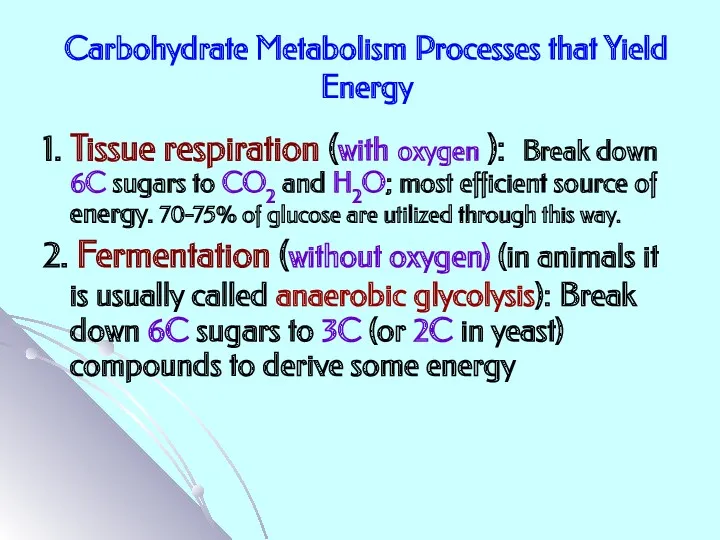
Carbohydrate Metabolism Processes that Yield Energy
1. Tissue respiration (with oxygen ):
Carbohydrate Metabolism Processes that Yield Energy
1. Tissue respiration (with oxygen ):
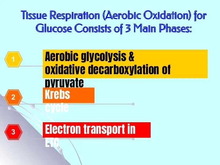
Tissue Respiration (Aerobic Oxidation) for Glucose Consists of 3 Main Phases:
Tissue Respiration (Aerobic Oxidation) for Glucose Consists of 3 Main Phases:
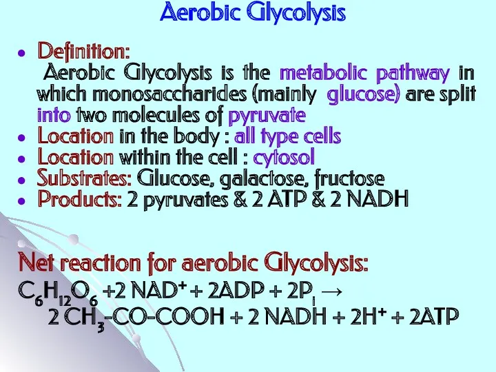
Aerobic Glycolysis
Definition:
Aerobic Glycolysis is the metabolic pathway in which
Aerobic Glycolysis
Definition:
Aerobic Glycolysis is the metabolic pathway in which
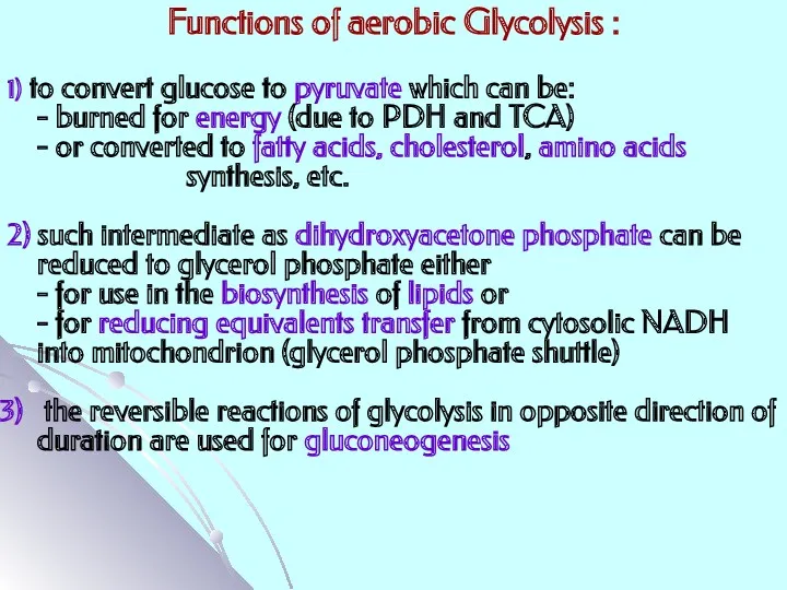
Functions of aerobic Glycolysis :
1) to convert glucose to pyruvate
Functions of aerobic Glycolysis :
1) to convert glucose to pyruvate
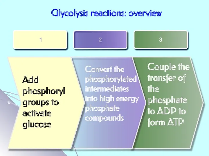
Glycolysis reactions: overview
Add phosphoryl groups to activate glucose
Convert the phosphorylated intermediates
Glycolysis reactions: overview
Add phosphoryl groups to activate glucose
Convert the phosphorylated intermediates
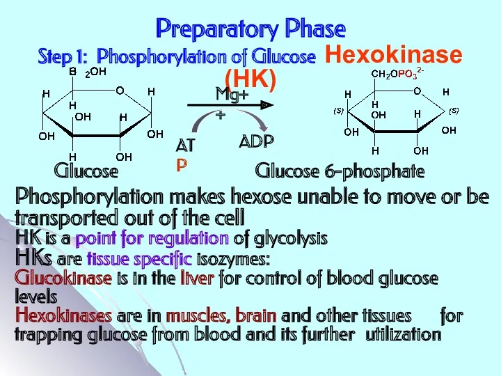
Preparatory Phase
Step 1: Phosphorylation of Glucose Hexokinase (HK)
ATP
ADP
Mg++
Glucose Glucose 6-phosphate
Phosphorylation
Preparatory Phase
Step 1: Phosphorylation of Glucose Hexokinase (HK)
ATP
ADP
Mg++
Glucose Glucose 6-phosphate
Phosphorylation
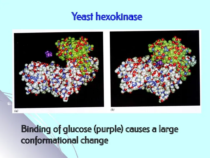
Yeast hexokinase
Binding of glucose (purple) causes a large conformational change
Yeast hexokinase
Binding of glucose (purple) causes a large conformational change
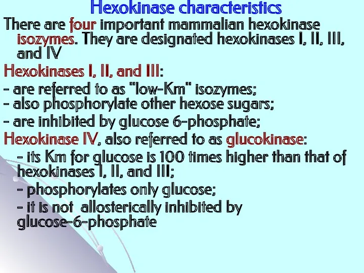
Hexokinase characteristics
There are four important mammalian hexokinase isozymes. They are designated
Hexokinase characteristics
There are four important mammalian hexokinase isozymes. They are designated
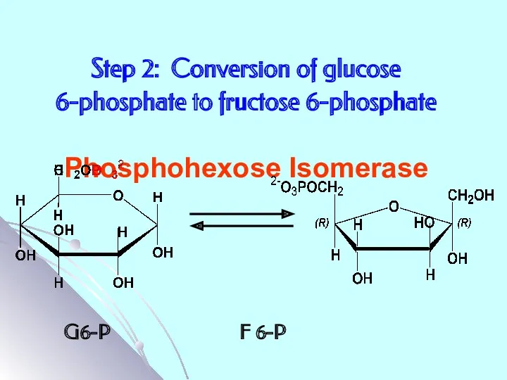
Step 2: Conversion of glucose 6-phosphate to fructose 6-phosphate
Phosphohexose Isomerase
G6-P
Step 2: Conversion of glucose 6-phosphate to fructose 6-phosphate
Phosphohexose Isomerase
G6-P
Step 3: Phosphorylation of fructose 6-phosphate to fructose 1,6-bisphosphate
Phosphofructokinase 1
Mg++
F
Step 3: Phosphorylation of fructose 6-phosphate to fructose 1,6-bisphosphate
Phosphofructokinase 1
Mg++
F
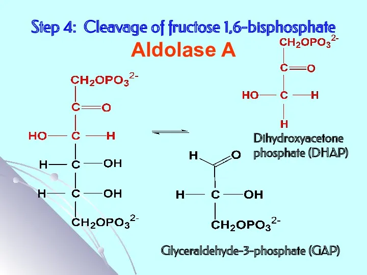
Step 4: Cleavage of fructose 1,6-bisphosphate
Aldolase A
Dihydroxyacetone
phosphate (DHAP)
Glyceraldehyde-3-phosphate (GAP)
Step 4: Cleavage of fructose 1,6-bisphosphate
Aldolase A
Dihydroxyacetone
phosphate (DHAP)
Glyceraldehyde-3-phosphate (GAP)
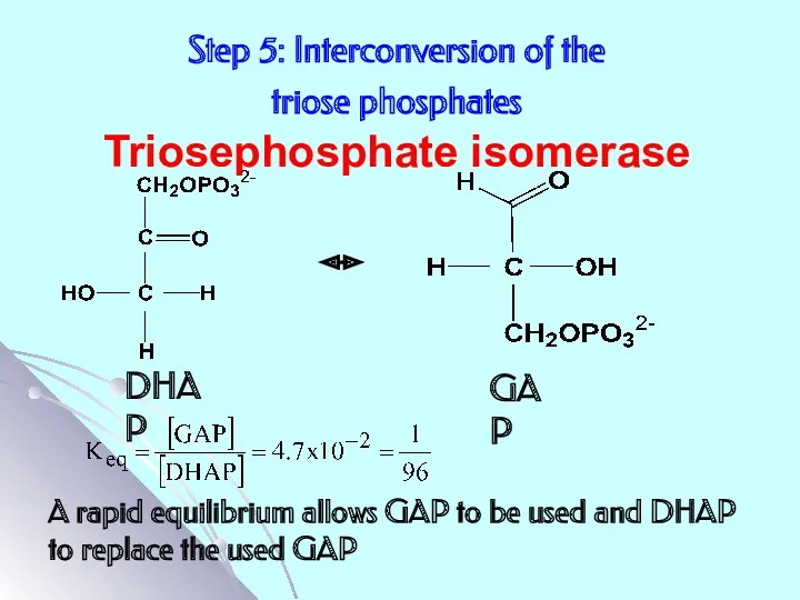
Step 5: Interconversion of the
triose phosphates
Triosephosphate isomerase
A rapid equilibrium
Step 5: Interconversion of the
triose phosphates
Triosephosphate isomerase
A rapid equilibrium
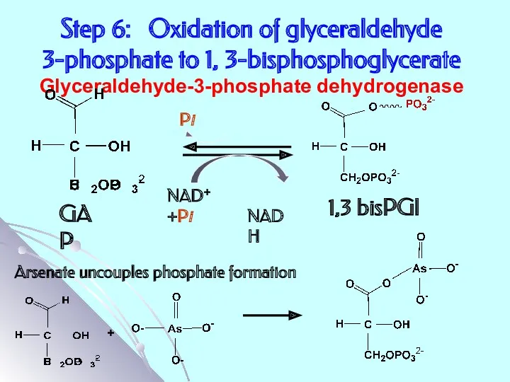
Step 6: Oxidation of glyceraldehyde 3-phosphate to 1, 3-bisphosphoglycerate
Glyceraldehyde-3-phosphate dehydrogenase
NAD+
Step 6: Oxidation of glyceraldehyde 3-phosphate to 1, 3-bisphosphoglycerate
Glyceraldehyde-3-phosphate dehydrogenase
NAD+
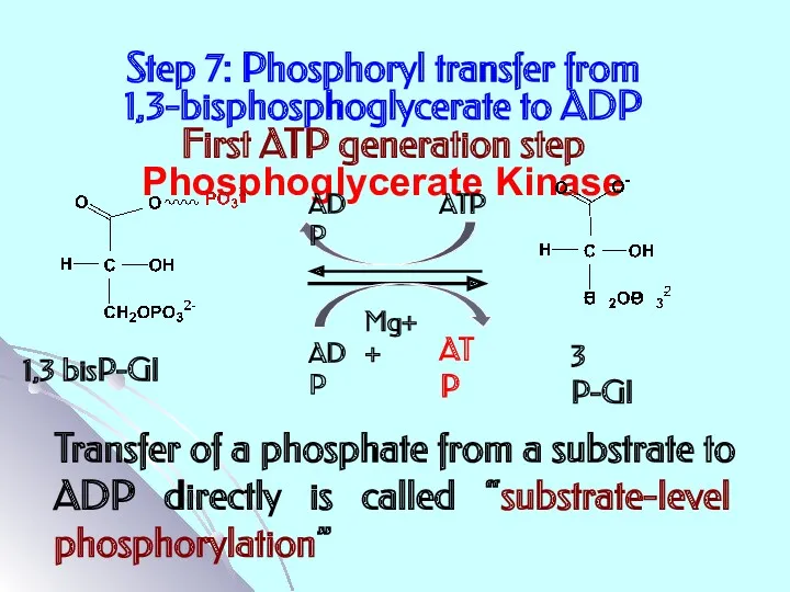
Step 7: Phosphoryl transfer from
1,3-bisphosphoglycerate to ADP
First ATP generation
Step 7: Phosphoryl transfer from 1,3-bisphosphoglycerate to ADP First ATP generation
3 P-Gl
2 P-Gl
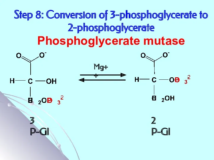
Step 8: Conversion of 3-phosphoglycerate to 2-phosphoglycerate
Phosphoglycerate mutase
Mg++
3 P-Gl
2 P-Gl
Step 8: Conversion of 3-phosphoglycerate to 2-phosphoglycerate
Phosphoglycerate mutase
Mg++
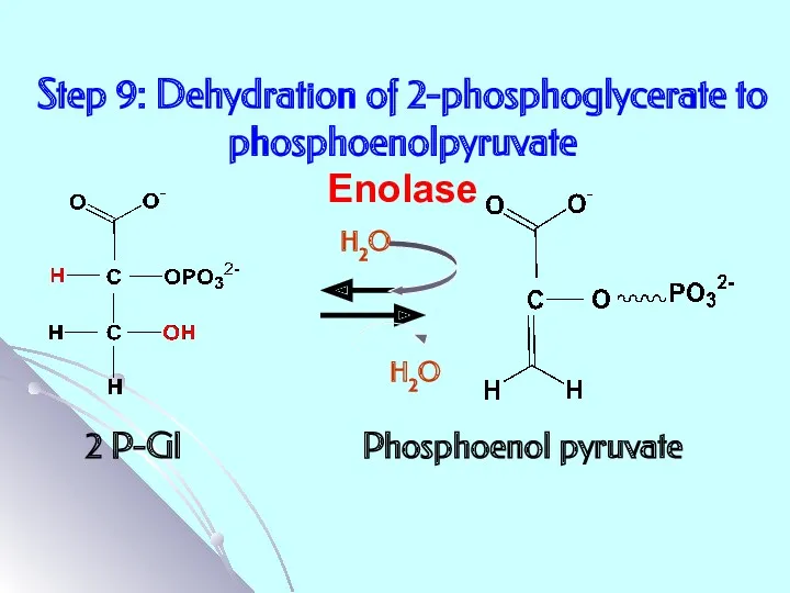
Step 9: Dehydration of 2-phosphoglycerate to phosphoenolpyruvate
Enolase
2 P-Gl Phosphoenol pyruvate
H2O
H2O
Step 9: Dehydration of 2-phosphoglycerate to phosphoenolpyruvate
Enolase
2 P-Gl Phosphoenol pyruvate
H2O
H2O
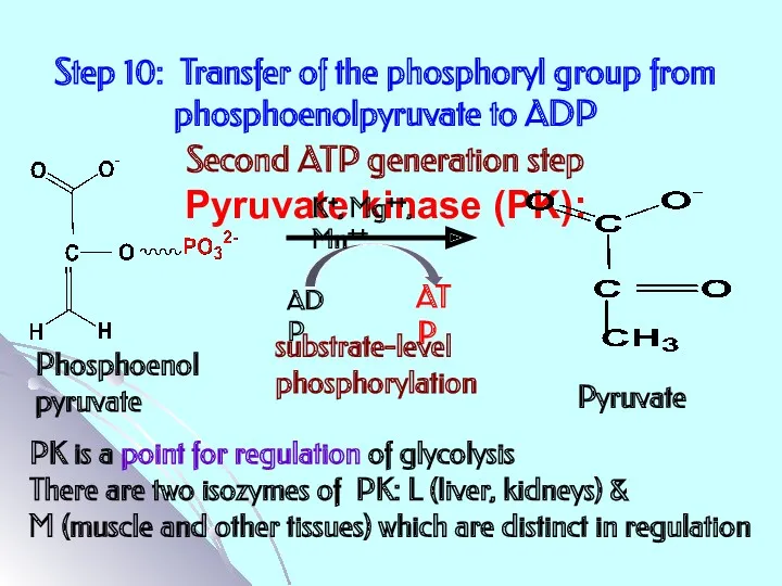
Step 10: Transfer of the phosphoryl group from phosphoenolpyruvate to ADP
Step 10: Transfer of the phosphoryl group from phosphoenolpyruvate to ADP
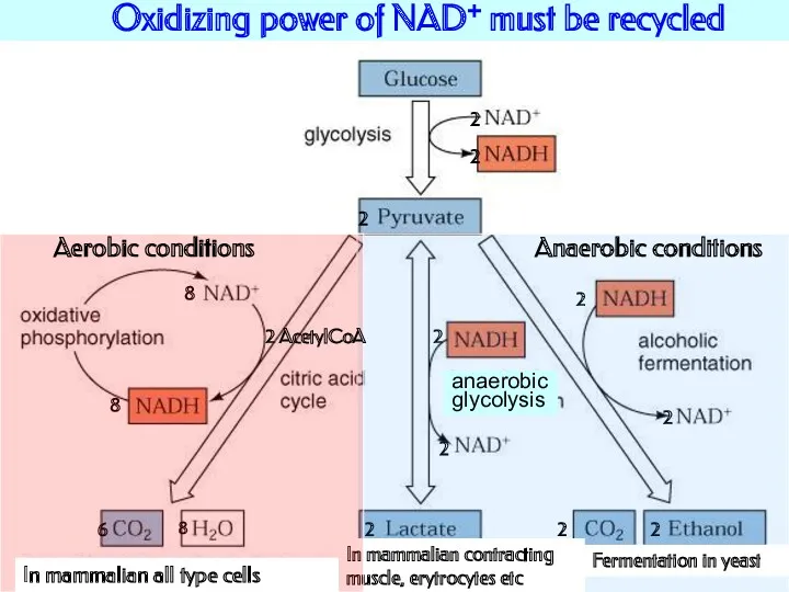
Oxidizing power of NAD+ must be recycled
2
2
2
6
2
2
2
2
8
8
2
2
2
Anaerobic conditions
Fermentation in yeast
In mammalian
Oxidizing power of NAD+ must be recycled
2
2
2
6
2
2
2
2
8
8
2
2
2
Anaerobic conditions
Fermentation in yeast
In mammalian
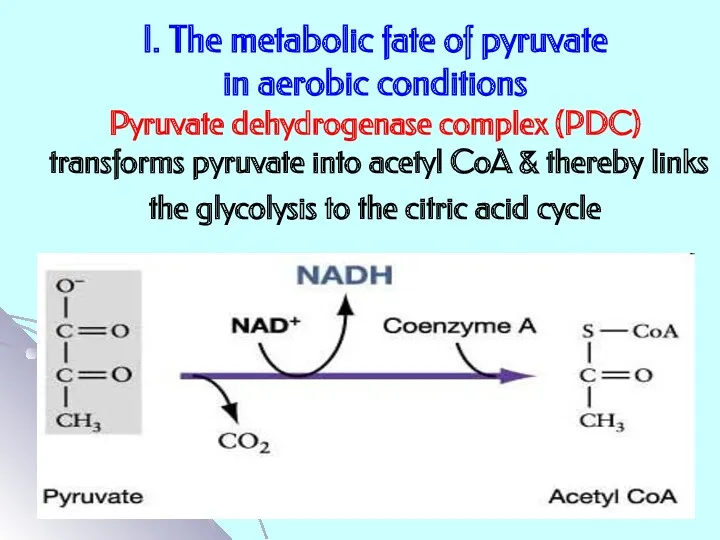
I. The metabolic fate of pyruvate
in aerobic conditions
Pyruvate dehydrogenase complex
I. The metabolic fate of pyruvate in aerobic conditions Pyruvate dehydrogenase complex
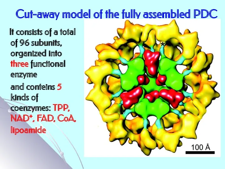
Cut-away model of the fully assembled PDC
It consists of a
Cut-away model of the fully assembled PDC
It consists of a
Mechanism of PDC action
(see in a text-book)
Mechanism of PDC action
(see in a text-book)
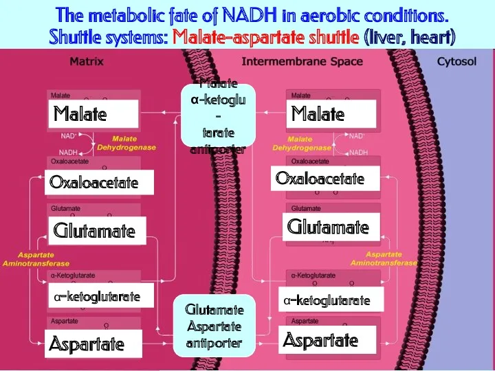
The metabolic fate of NADH in aerobic conditions.
Shuttle systems: Malate-aspartate
The metabolic fate of NADH in aerobic conditions. Shuttle systems: Malate-aspartate
The metabolic fate of NADH
in aerobic conditions. Shuttle systems:
Glycerol-3-phosphate shuttle
Dihydroxyacetone
phosphate
Glycerol-3-phosphate
NADH
The metabolic fate of NADH
in aerobic conditions. Shuttle systems:
Glycerol-3-phosphate shuttle
Dihydroxyacetone
phosphate
Glycerol-3-phosphate
NADH

II. The metabolic fate of pyruvate
in anaerobic conditions.
Anaerobic glycolysis
Definition:
II. The metabolic fate of pyruvate
in anaerobic conditions.
Anaerobic glycolysis
Definition:
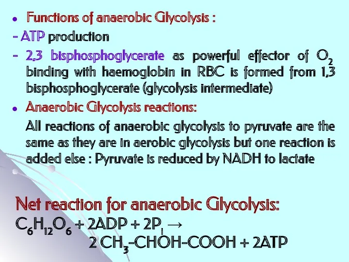
Functions of anaerobic Glycolysis :
- ATP production
- 2,3 bisphosphoglycerate as
Functions of anaerobic Glycolysis :
- ATP production
- 2,3 bisphosphoglycerate as
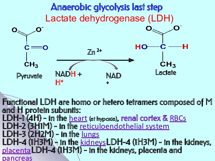
Anaerobic glycolysis last step
Lactate dehydrogenase (LDH)
Functional LDH are homo or
Anaerobic glycolysis last step
Lactate dehydrogenase (LDH)
Functional LDH are homo or
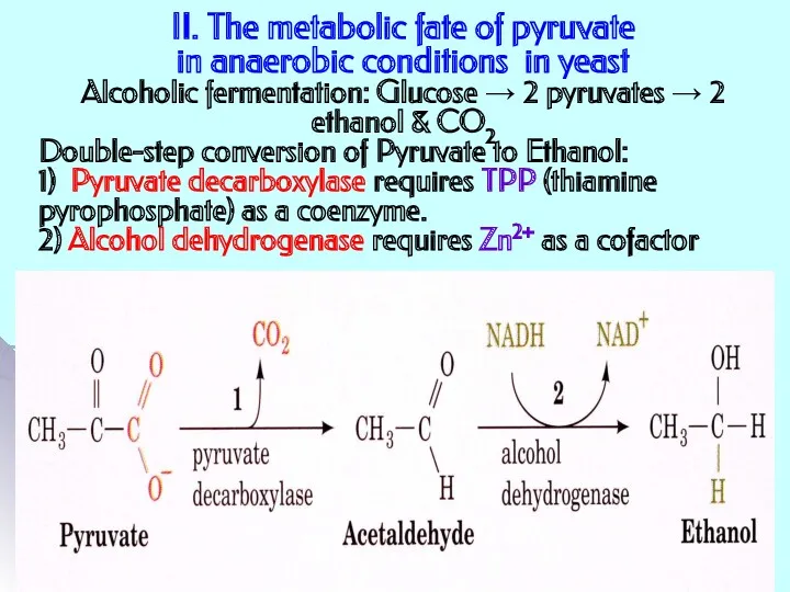
II. The metabolic fate of pyruvate
in anaerobic conditions in yeast
Alcoholic
II. The metabolic fate of pyruvate in anaerobic conditions in yeast Alcoholic
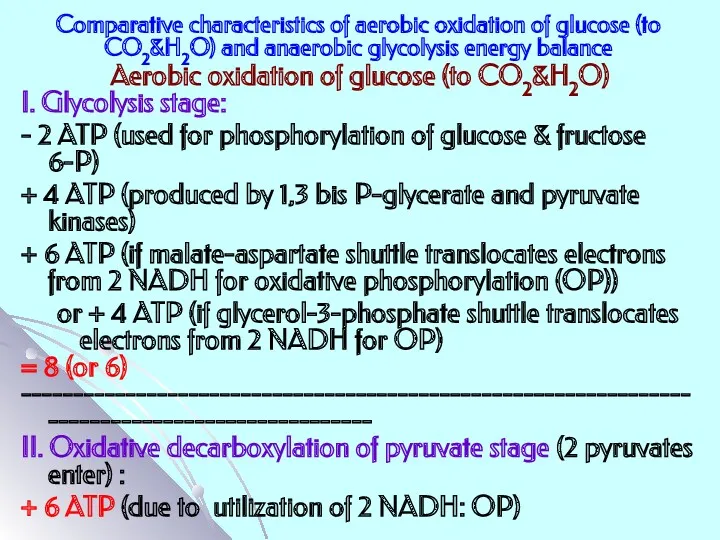
Comparative characteristics of aerobic oxidation of glucose (to CO2&H2O) and anaerobic
Comparative characteristics of aerobic oxidation of glucose (to CO2&H2O) and anaerobic
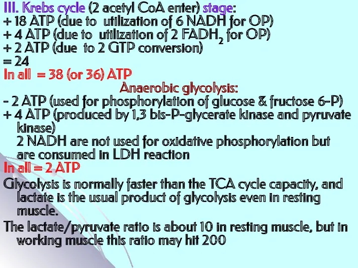
III. Krebs cycle (2 acetyl CoA enter) stage:
+ 18 ATP (due
III. Krebs cycle (2 acetyl CoA enter) stage:
+ 18 ATP (due
Regulation
Regulation
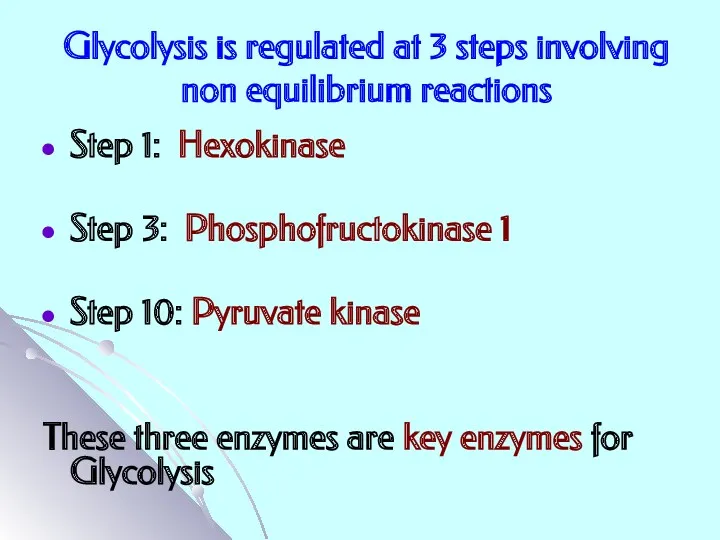
Glycolysis is regulated at 3 steps involving non equilibrium reactions
Step 1:
Glycolysis is regulated at 3 steps involving non equilibrium reactions
Step 1:
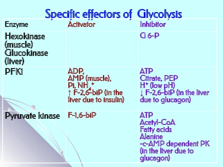
Specific effectors of Glycolysis
Specific effectors of Glycolysis
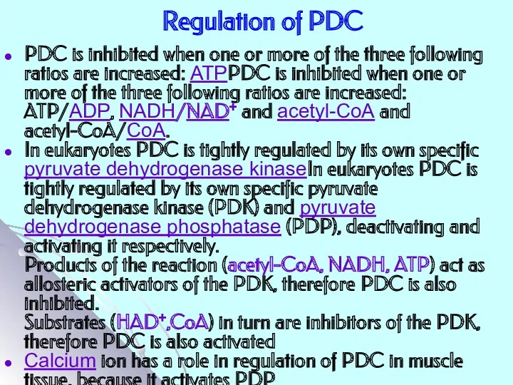
Regulation of PDC
PDC is inhibited when one or more of the
Regulation of PDC
PDC is inhibited when one or more of the
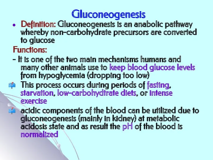
Gluconeogenesis
Definition: Gluconeogenesis is an anabolic pathway whereby non-carbohydrate precursors are converted
Gluconeogenesis
Definition: Gluconeogenesis is an anabolic pathway whereby non-carbohydrate precursors are converted
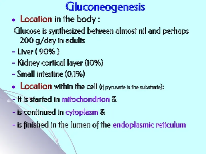
Gluconeogenesis
Location in the body :
Glucose is synthesized between almost
Gluconeogenesis
Location in the body :
Glucose is synthesized between almost
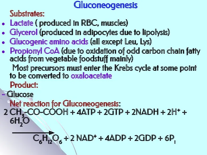
Gluconeogenesis
Substrates:
Lactate ( produced in RBC, muscles)
Glycerol (produced in adipocytes due
Gluconeogenesis
Substrates:
Lactate ( produced in RBC, muscles)
Glycerol (produced in adipocytes due
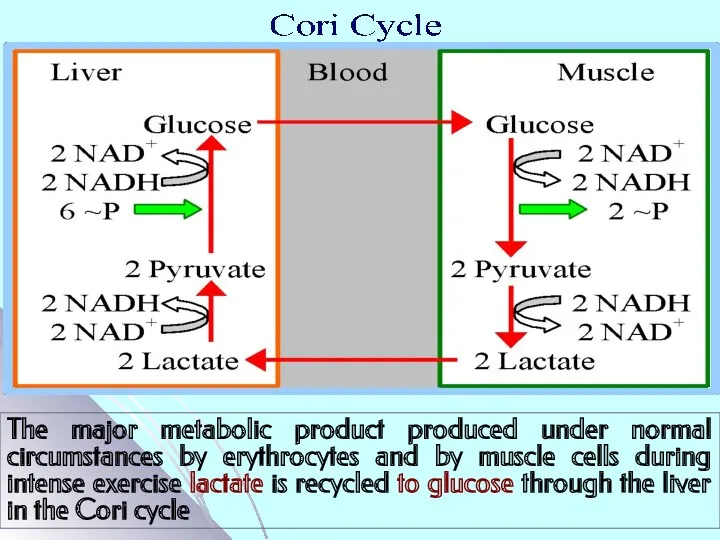
The major metabolic product produced under normal circumstances by erythrocytes and
The major metabolic product produced under normal circumstances by erythrocytes and
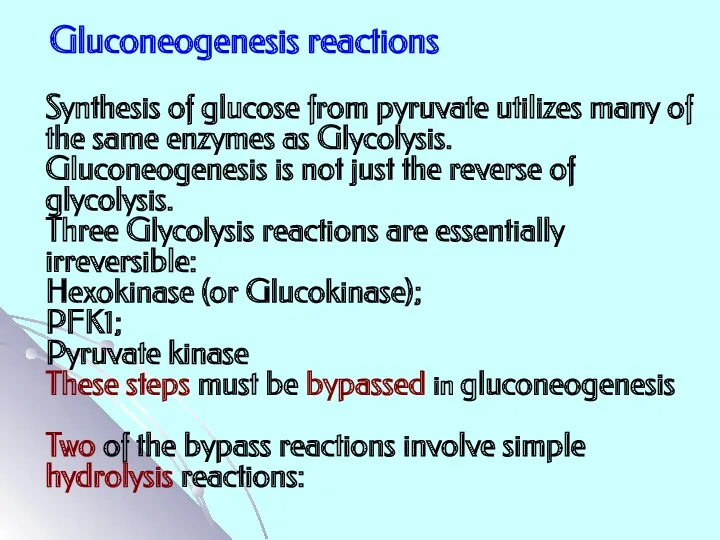
Gluconeogenesis reactions
Synthesis of glucose from pyruvate utilizes many of the same
Gluconeogenesis reactions
Synthesis of glucose from pyruvate utilizes many of the same
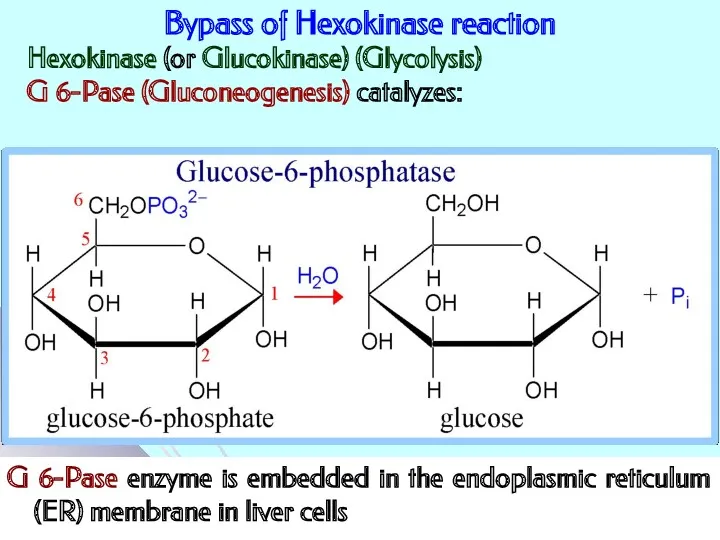
Bypass of Hexokinase reaction
G 6-Pase enzyme is embedded in the endoplasmic
Bypass of Hexokinase reaction
G 6-Pase enzyme is embedded in the endoplasmic
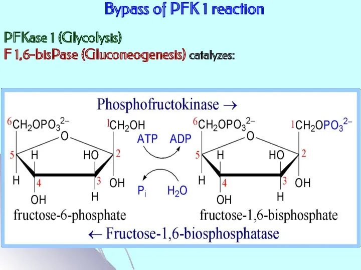
Bypass of PFK 1 reaction
PFKase 1 (Glycolysis)
F 1,6-bisPase (Gluconeogenesis) catalyzes:
Bypass of PFK 1 reaction
PFKase 1 (Glycolysis)
F 1,6-bisPase (Gluconeogenesis) catalyzes:
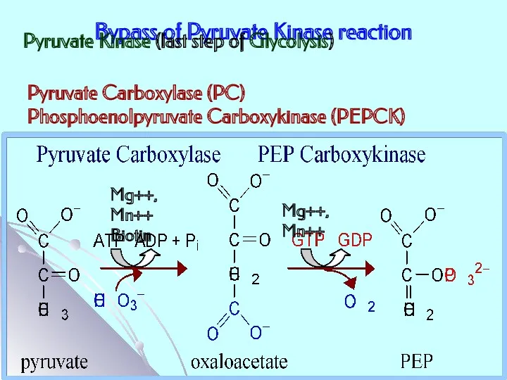
Bypass of Pyruvate Kinase reaction
Pyruvate Kinase (last step of Glycolysis)
Pyruvate Carboxylase
Bypass of Pyruvate Kinase reaction
Pyruvate Kinase (last step of Glycolysis)
Pyruvate Carboxylase
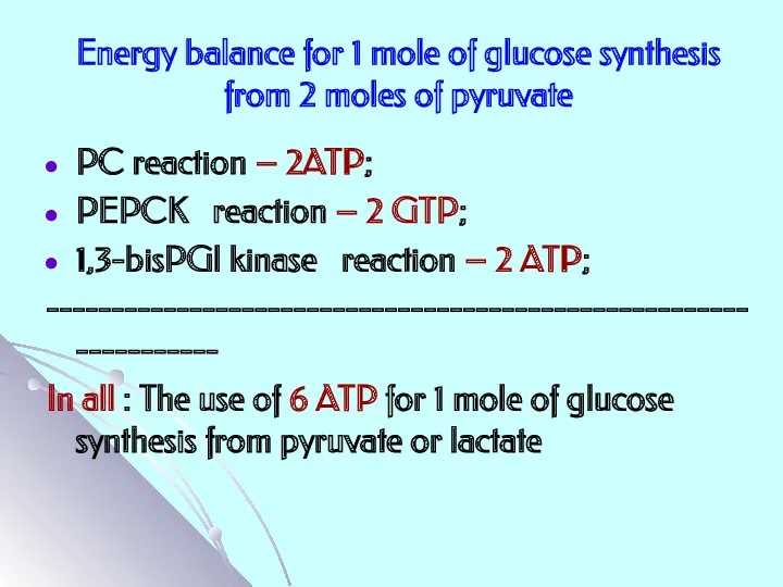
Energy balance for 1 mole of glucose synthesis from 2 moles
Energy balance for 1 mole of glucose synthesis from 2 moles
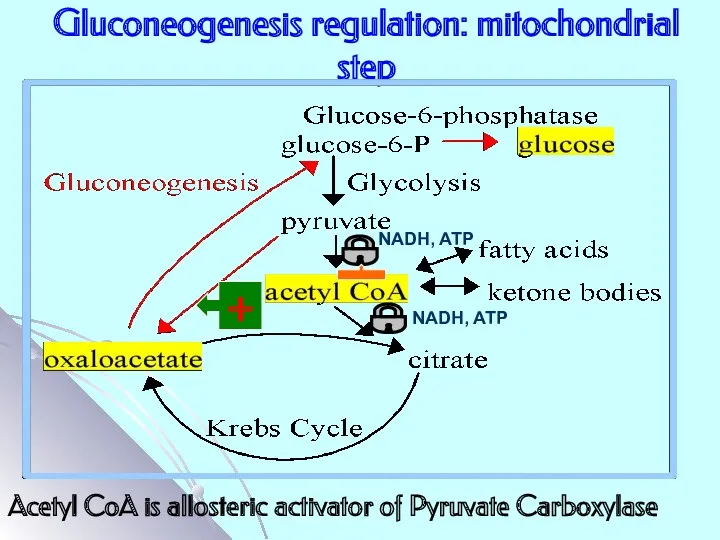
Gluconeogenesis regulation: mitochondrial step
NADH, ATP
+
NADH, ATP
Acetyl CoA is allosteric activator of
Gluconeogenesis regulation: mitochondrial step
NADH, ATP
+
NADH, ATP
Acetyl CoA is allosteric activator of
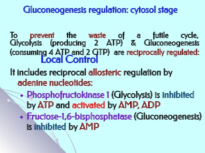
To prevent the waste of a futile cycle,
Glycolysis (producing 2
To prevent the waste of a futile cycle, Glycolysis (producing 2
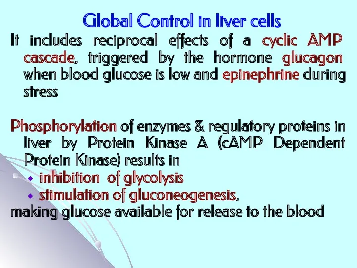
Global Control in liver cells
It includes reciprocal effects of a cyclic
Global Control in liver cells
It includes reciprocal effects of a cyclic
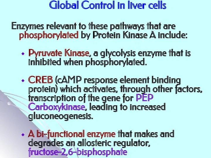
Global Control in liver cells
Enzymes relevant to these pathways that are
Global Control in liver cells
Enzymes relevant to these pathways that are
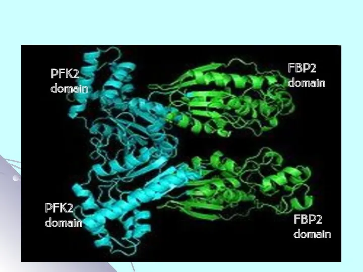
PFK2
domain
FBP2
domain
PFK2
domain
FBP2
domain
PFK2
domain
FBP2
domain
PFK2
domain
FBP2
domain
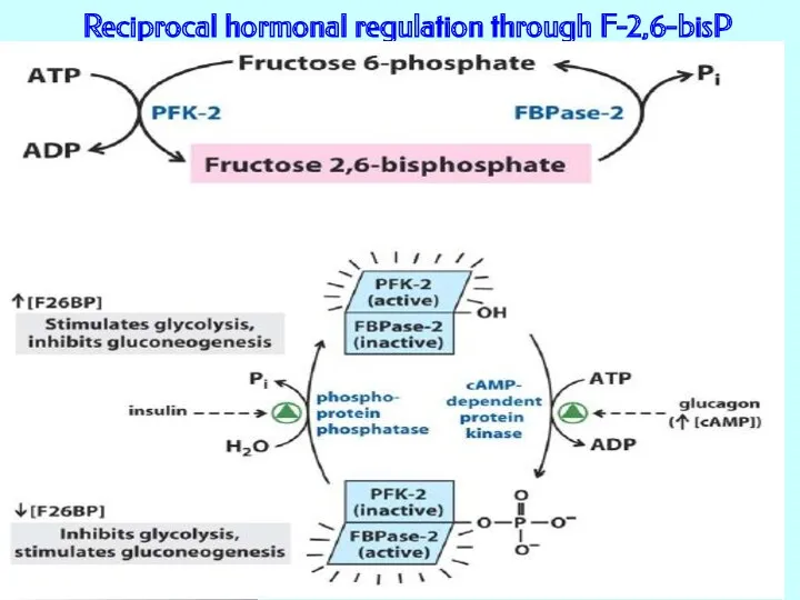
Reciprocal hormonal regulation through F-2,6-bisP
Reciprocal hormonal regulation through F-2,6-bisP
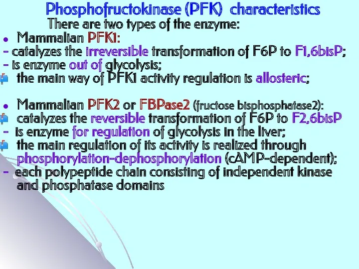
Phosphofructokinase (PFK) characteristics
Mammalian PFK1:
- catalyzes the irreversible transformation of F6P to
Phosphofructokinase (PFK) characteristics
Mammalian PFK1:
- catalyzes the irreversible transformation of F6P to
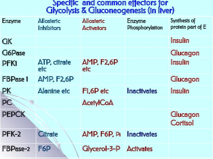
Specific and common effectors for
Glycolysis & Gluconeogenesis (in liver)
Specific and common effectors for
Glycolysis & Gluconeogenesis (in liver)
Похожие презентации
 Общая характеристика обмена веществ
 Класс млекопитающие
 Бесполое размножение
 Сообщества живых организмов
 Антропогенез. Гипотезы возникновения человека. Сходство и различия человека и животных. Часть 4
 Клеточный уровень
 Sound effects on plants
 Окружающий мир Мир растений. 3 класс
 Заповедник Нургуш - жемчужина Вятского края
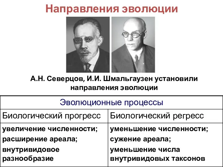 Пути и направления эволюции
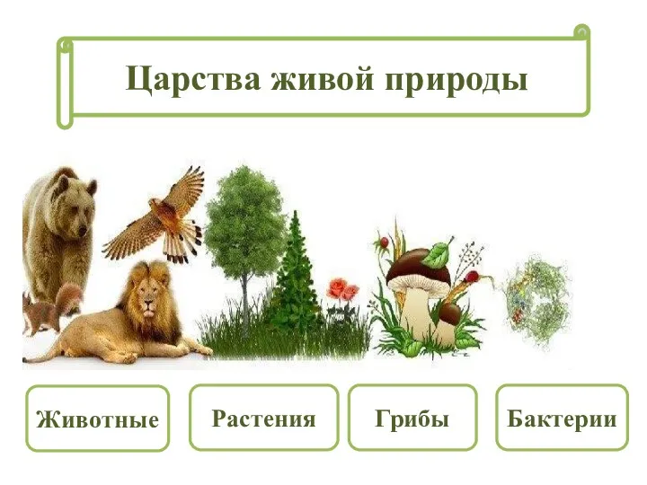 Царства живой природы
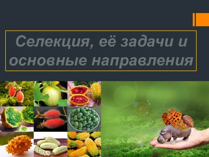 Селекция, ее задачи и основные направления
 Биологическая изменчивость людей в связи с биогеографическими особенностями среды. Формирование адаптивных экотипов людей
 Хромосоми. Каріотип
 бактерии Диск
 Всемирный день защиты животных
 Заболевания органов пищеварения и их профилактика.
 Қалқаншамаңы бездері,тимус(айырша без) және бүйрек үсті бездері
 Повреждение клетки. Патология клетки
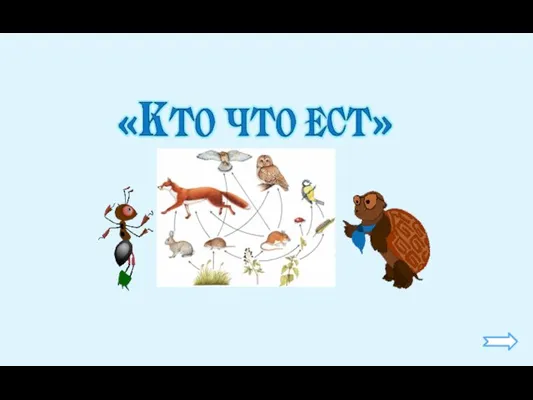 Классификация животных по способу питания (часть 1)
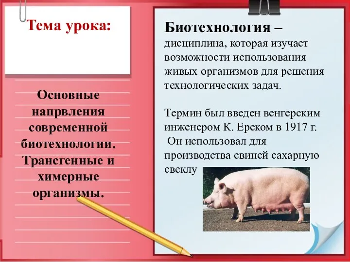 Основные напрвления современной биотехнологии
 Подготовка учащихся к ЕГЭ по биологии
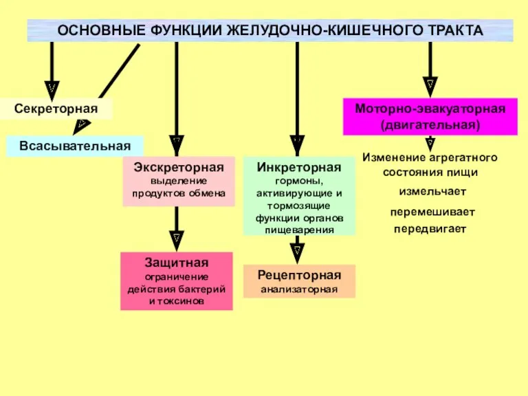 Основные функции желудочно-кишечного тракта
 Вид. Критерии вида. 9 класс
 Неживая и живая природа
 Игра по биологии Звездопад
 Наследование признаков, сцепленных с полом. (Задачи № 325, 336, 358)