- Cell Biology

Содержание
- 2. Introduction to Cell By Arnat Balabiyev PhD student Arizona State University
- 3. 1.0 Unity and diversity of cells
- 4. What defines “Life”? Are highly organized Homeostasis Reproduce themselves Grow and develop Use the energy from
- 5. Cells come in a variety of shapes and sizes Nerve cell Paramecium Plant tissue Bacterial cell
- 6. Living cells all have a similar basic chemistry Same biological molecules Evolved from common ancestor Homolog
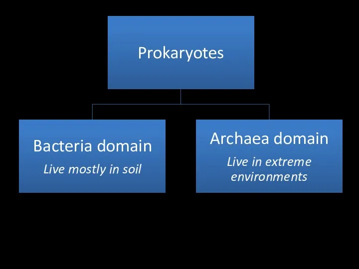
- 7. Prokaryotic cell Have simplest structure No organelles No nucleus, just naked DNA “Pro”- before, “karyo”-nucleus Different
- 8. Different size and shapes of bacteria
- 9. Some other features of bacteria Have cell wall- may differ upon peptidoglycan content: gram positive and
- 10. Prokaryotes are the most diverse and numerous cells on Earth Can be single celled and form
- 11. E.coli as a model organism
- 12. Some bacteria are photosynthetic Anabaena cylindrica H: structure that fix N2 S: structure that become spores
- 14. The eukaryotic cells Bigger in size Elaborate lots of forms: unicellular and multicellular Have nucleus and
- 15. The nucleus is the information store of the cell
- 16. Chromosomes become visible when a cell is about to divide
- 17. Mitochondria generate usable energy from food to power the cell
- 18. Mitochondria probably evolved from bacteria
- 19. Chloroplasts capture energy from sunlight
- 20. The same story with chloroplasts
- 21. ER-the factory of many structures phospholipid Membrane bound proteins Post translational modification Place of lipid synthesis
- 22. Golgi Apparatus Proteins are further mod Ified in GA Stack of membrane Vesicles Cis: ER facing
- 23. Membrane enclosed organelles
- 24. Enter and exit the cell
- 25. Cytoskeleton Actin filaments Cell crawling Muscle contraction Cell shape Microtubules Cell division Cell movement Intercellular transport
- 26. Eukaryotic Cells may have originated as predators
- 27. Model organisms E.coli Simple structure (small genome size) Easy to grow (37C) in agar media 20
- 28. Yeast cells Short doubling time Unicellular Eukaryotic cell Many conserved genes Easy to grow Easy to
- 29. C. elegans: nematode First animal genome sequenced Fixed number of cells Developmental stage is clear Easy
- 30. Arabidopsis Fast growing plant Easy to grow and maintain Good model organism to study plants
- 31. Drosophila melanogaster Great model to study animals Insects are the most numerous Conserved genes Easy to
- 32. Zebra fish First developmental stages are transparent Good model to study vertebrate development Easy to grow
- 33. Mouse model Easy to breed. Many conserved genes with human genome. Easy to manipulate
- 34. Cell lines Fibroblasts Nerve cells Epithelial cells
- 35. Genome information
- 37. Скачать презентацию
Introduction to Cell
By Arnat Balabiyev
PhD student
Arizona State University
Introduction to Cell
By Arnat Balabiyev
PhD student
Arizona State University
1.0 Unity and diversity of cells
1.0 Unity and diversity of cells
What defines “Life”?
Are highly organized
Homeostasis
Reproduce themselves
Grow and develop
Use the energy from
What defines “Life”?
Are highly organized
Homeostasis
Reproduce themselves
Grow and develop
Use the energy from
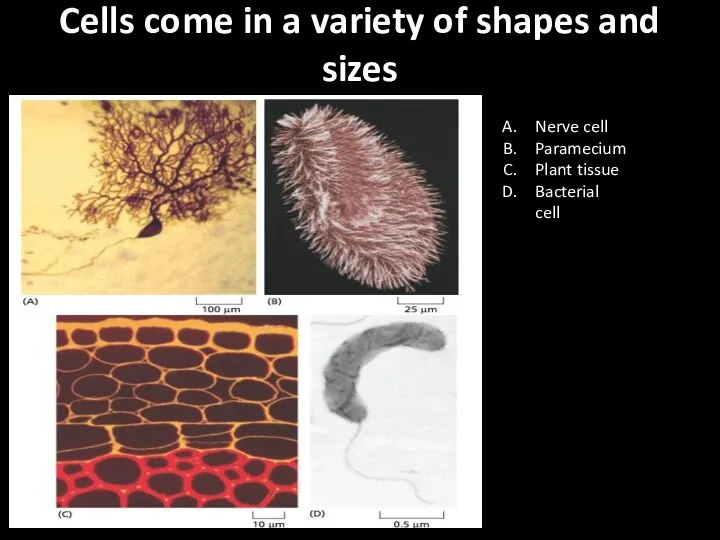
Cells come in a variety of shapes and sizes
Nerve cell
Paramecium
Plant
Cells come in a variety of shapes and sizes
Nerve cell
Paramecium
Plant
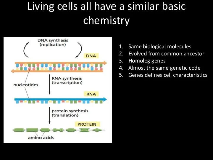
Living cells all have a similar basic chemistry
Same biological molecules
Evolved
Living cells all have a similar basic chemistry
Same biological molecules
Evolved
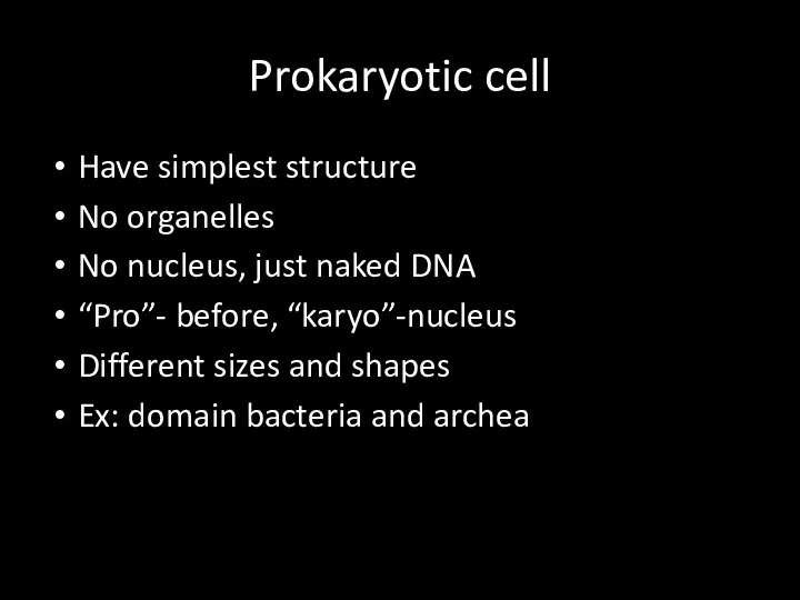
Prokaryotic cell
Have simplest structure
No organelles
No nucleus, just naked DNA
“Pro”- before, “karyo”-nucleus
Different
Prokaryotic cell
Have simplest structure
No organelles
No nucleus, just naked DNA
“Pro”- before, “karyo”-nucleus
Different
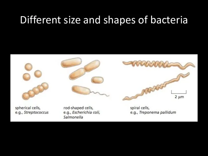
Different size and shapes of bacteria
Different size and shapes of bacteria
Some other features of bacteria
Have cell wall- may differ upon peptidoglycan
Some other features of bacteria
Have cell wall- may differ upon peptidoglycan
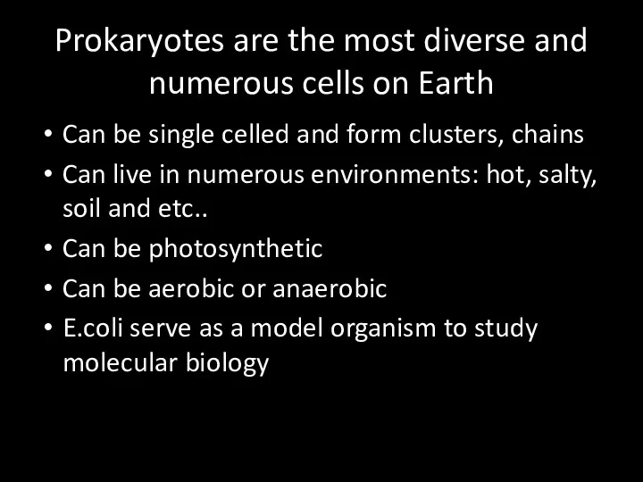
Prokaryotes are the most diverse and numerous cells on Earth
Can be
Prokaryotes are the most diverse and numerous cells on Earth
Can be
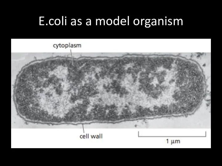
E.coli as a model organism
E.coli as a model organism
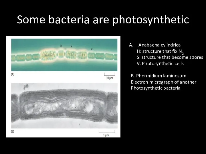
Some bacteria are photosynthetic
Anabaena cylindrica
H: structure that fix N2
S:
Some bacteria are photosynthetic
Anabaena cylindrica
H: structure that fix N2
S:
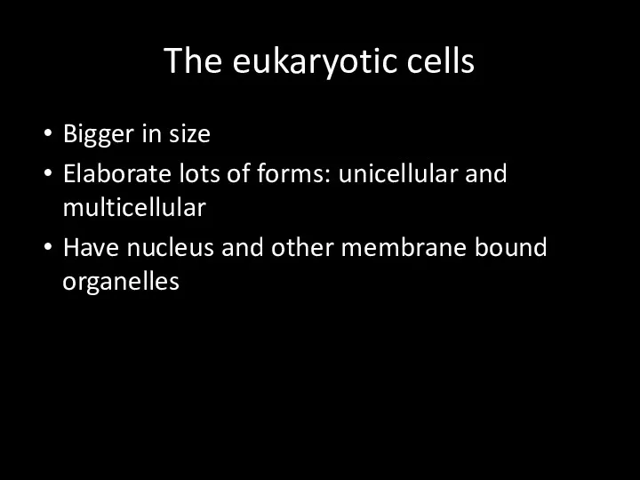
The eukaryotic cells
Bigger in size
Elaborate lots of forms: unicellular and multicellular
The eukaryotic cells
Bigger in size
Elaborate lots of forms: unicellular and multicellular
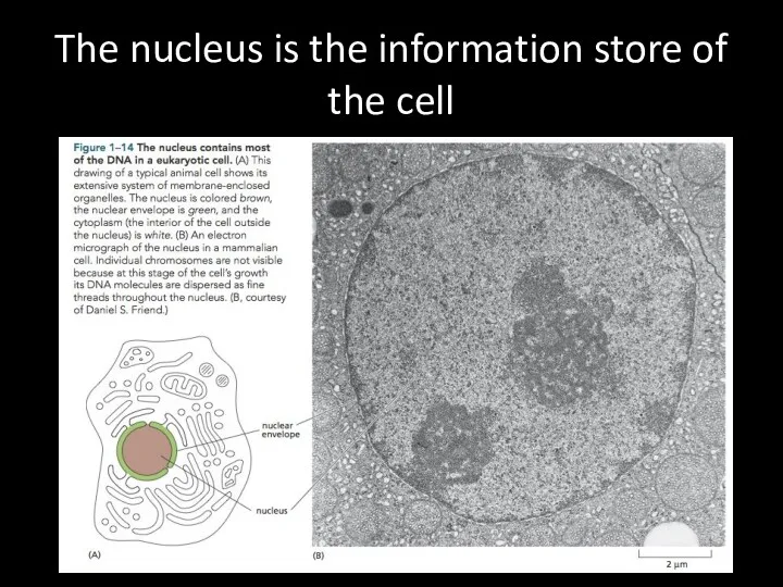
The nucleus is the information store of the cell
The nucleus is the information store of the cell
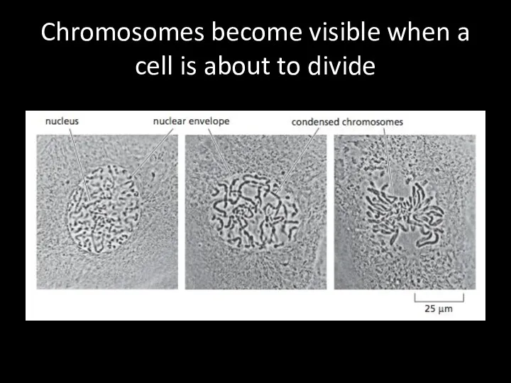
Chromosomes become visible when a cell is about to divide
Chromosomes become visible when a cell is about to divide
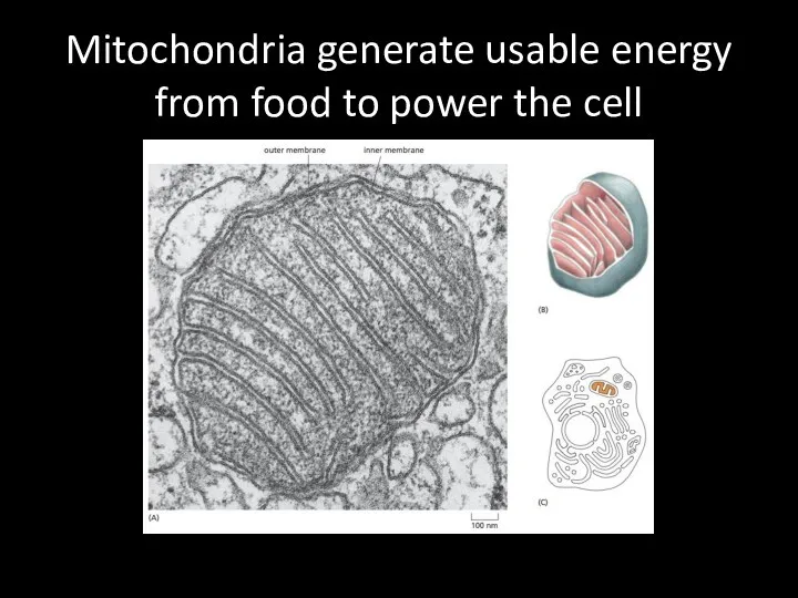
Mitochondria generate usable energy from food to power the cell
Mitochondria generate usable energy from food to power the cell
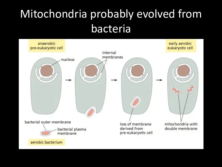
Mitochondria probably evolved from bacteria
Mitochondria probably evolved from bacteria
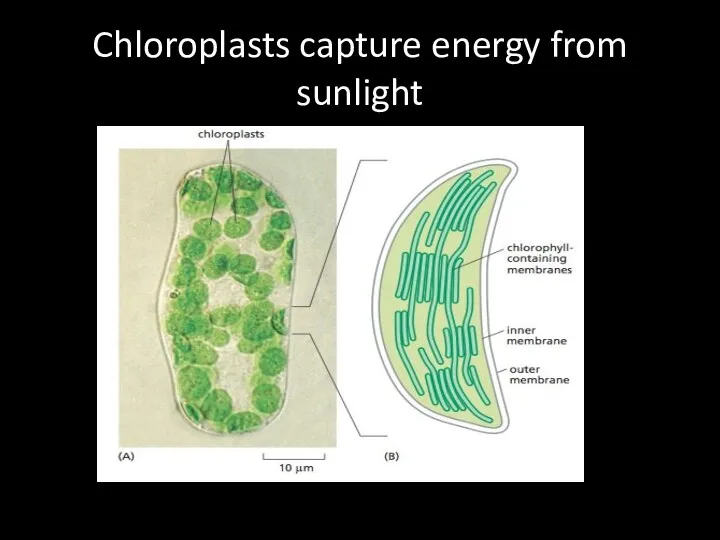
Chloroplasts capture energy from sunlight
Chloroplasts capture energy from sunlight
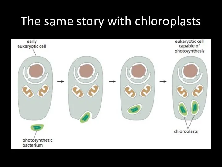
The same story with chloroplasts
The same story with chloroplasts
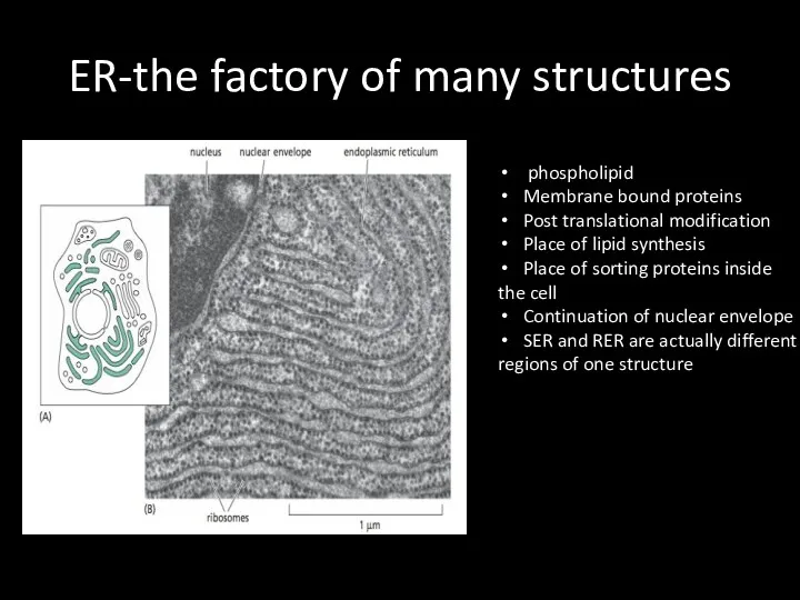
ER-the factory of many structures
phospholipid
Membrane bound proteins
Post translational modification
Place
ER-the factory of many structures
phospholipid
Membrane bound proteins
Post translational modification
Place
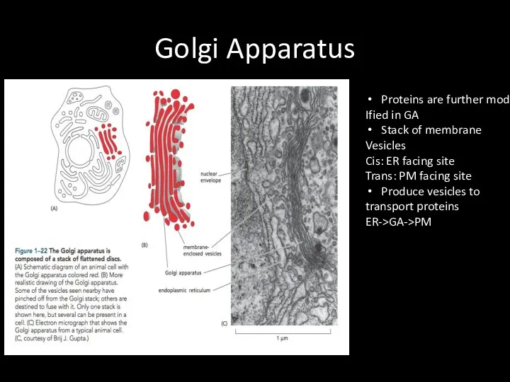
Golgi Apparatus
Proteins are further mod
Ified in GA
Stack of membrane
Vesicles
Cis: ER
Golgi Apparatus
Proteins are further mod
Ified in GA
Stack of membrane
Vesicles
Cis: ER
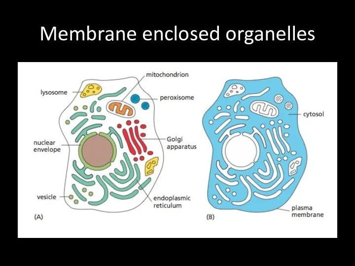
Membrane enclosed organelles
Membrane enclosed organelles
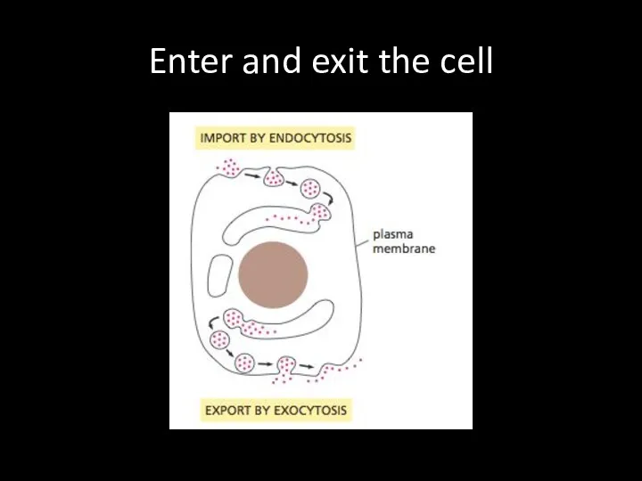
Enter and exit the cell
Enter and exit the cell
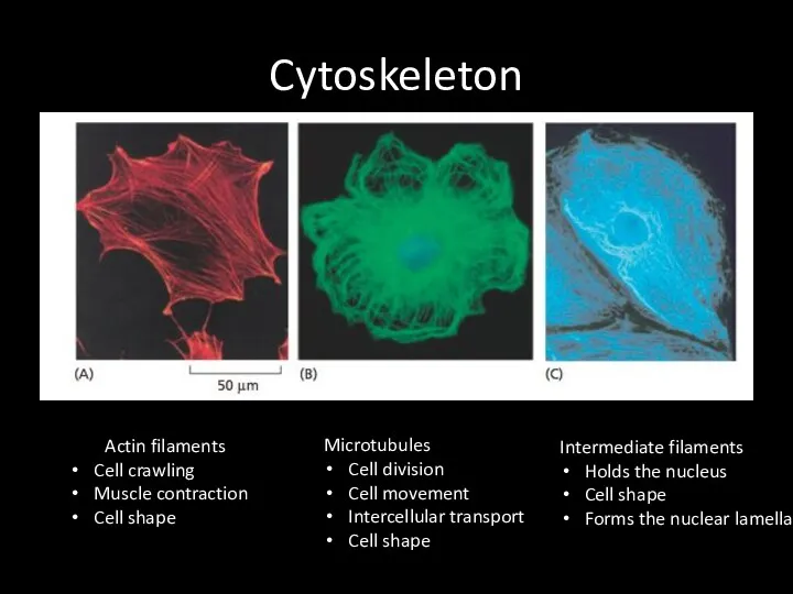
Cytoskeleton
Actin filaments
Cell crawling
Muscle contraction
Cell shape
Microtubules
Cell division
Cell movement
Intercellular transport
Cell shape
Intermediate filaments
Holds
Cytoskeleton
Actin filaments
Cell crawling
Muscle contraction
Cell shape
Microtubules
Cell division
Cell movement
Intercellular transport
Cell shape
Intermediate filaments
Holds
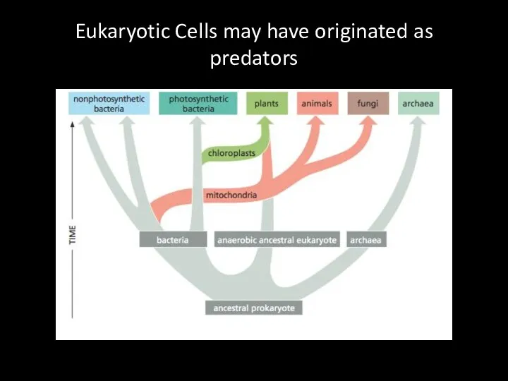
Eukaryotic Cells may have originated as predators
Eukaryotic Cells may have originated as predators
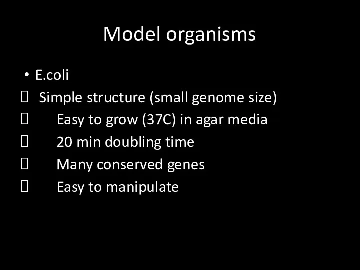
Model organisms
E.coli
Simple structure (small genome size)
Easy to grow (37C)
Model organisms
E.coli
Simple structure (small genome size)
Easy to grow (37C)
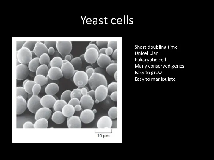
Yeast cells
Short doubling time
Unicellular
Eukaryotic cell
Many conserved genes
Easy to grow
Easy to manipulate
Yeast cells
Short doubling time
Unicellular
Eukaryotic cell
Many conserved genes
Easy to grow
Easy to manipulate
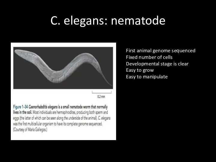
C. elegans: nematode
First animal genome sequenced
Fixed number of cells
Developmental stage is
C. elegans: nematode
First animal genome sequenced
Fixed number of cells
Developmental stage is
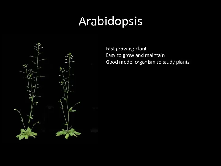
Arabidopsis
Fast growing plant
Easy to grow and maintain
Good model organism to study
Arabidopsis
Fast growing plant
Easy to grow and maintain
Good model organism to study
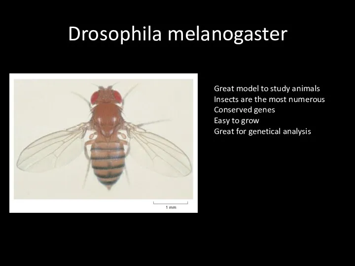
Drosophila melanogaster
Great model to study animals
Insects are the most numerous
Conserved genes
Easy
Drosophila melanogaster
Great model to study animals
Insects are the most numerous
Conserved genes
Easy
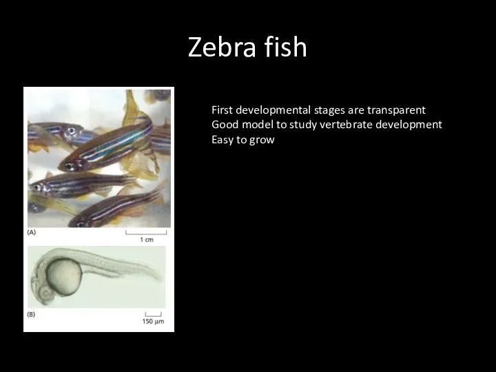
Zebra fish
First developmental stages are transparent
Good model to study vertebrate development
Easy
Zebra fish
First developmental stages are transparent
Good model to study vertebrate development
Easy
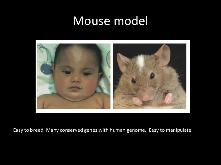
Mouse model
Easy to breed. Many conserved genes with human genome. Easy
Mouse model
Easy to breed. Many conserved genes with human genome. Easy
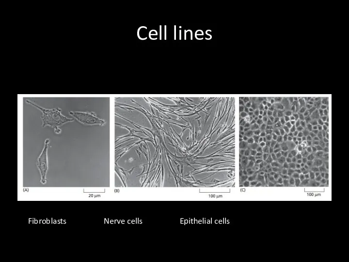
Cell lines
Fibroblasts Nerve cells Epithelial cells
Cell lines
Fibroblasts Nerve cells Epithelial cells
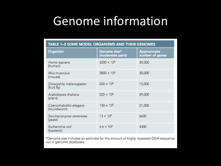
Genome information
Genome information
 Загадочный мир океана
 Что такое биоинформатика? Банк SwissProt
 Продление рода. Органы размножения
 Популяционные волны
 Органы чувств. Анализаторы
 5 Natural Ways To Get Rid of House Crickets
 Самые редкие и необычные породы кошек
 Интересные факты о животном мире
 Всероссийская проверочная работа по окружающему миру. Опыты (4 класс)
 Санитарлық-көрсеткіш микроағзалар. Стерилдеу мен залалсыздандыру
 Отряд Непарнокопытные
 4 стихии зарождения жизни
 Методическая разработка урока-игры по биологии в 10 классе по теме: Путешествие по эукариотической клетке
 Растения и животные леса
 Микробиота тела человека. Роль микроорганизмов в возникновении инфекций. Способы передачи инфекций
 α-Aminoacids, peptides, proteins
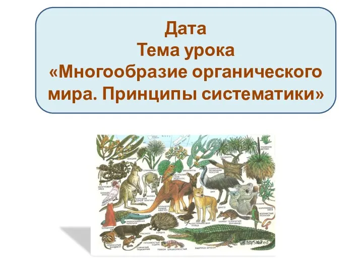 Многообразие органического мира. Принципы систематики
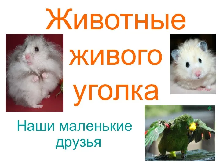 Животные живого уголка
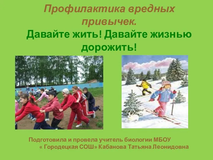 Профилактика вредных привычек.Давайте жить! Давайте жизнью дорожить!
 Что такое метаболизм и как его измерить
 оплодотворение
 Pielea, organ tactil, termic, dureros și de presiune. (Lectie 11)
 Древнейшие люди
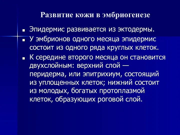 Развитие кожи в эмбриогенезе
 Обмен веществ и энергии. Фотосинтез
 Тип Губки
 Птичий переполох (игра)
 Самые опасные насекомые в мире