- Cellular neurophysiology

Содержание
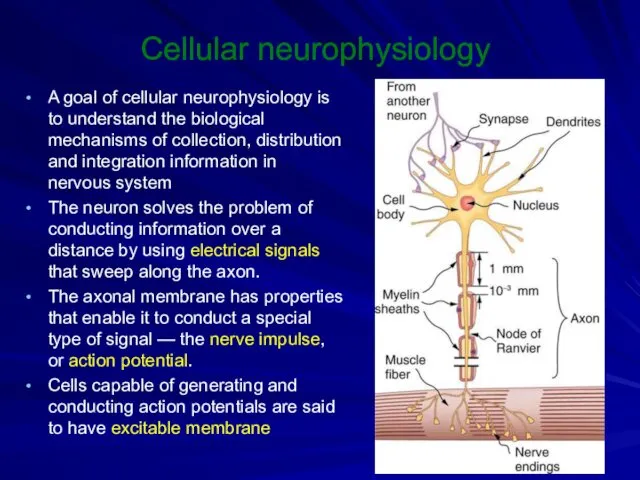
- 2. Cellular neurophysiology A goal of cellular neurophysiology is to understand the biological mechanisms of collection, distribution
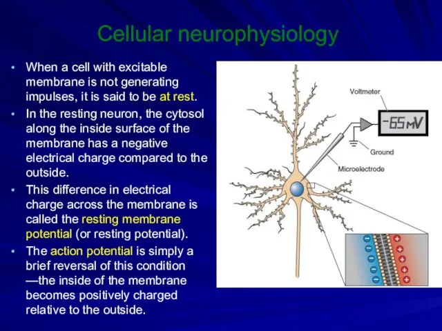
- 3. Cellular neurophysiology When a cell with excitable membrane is not generating impulses, it is said to
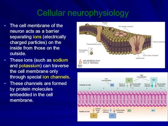
- 4. Cellular neurophysiology The cell membrane of the neuron acts as a barrier separating ions (electrically charged
- 5. Cellular neurophysiology In specific configurations, ion channels create a passageway that allows ions to flow in
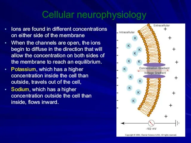
- 6. Cellular neurophysiology Ions are found in different concentrations on either side of the membrane When the
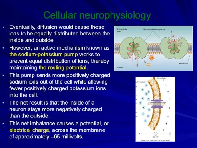
- 7. Cellular neurophysiology Eventually, diffusion would cause these ions to be equally distributed between the inside and
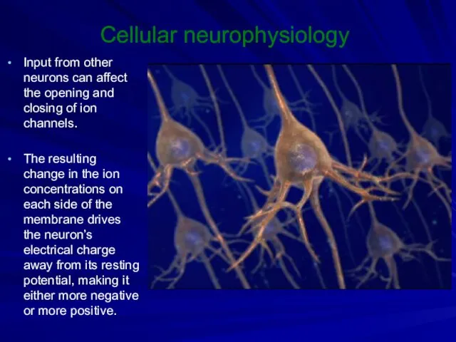
- 8. Cellular neurophysiology Input from other neurons can affect the opening and closing of ion channels. The
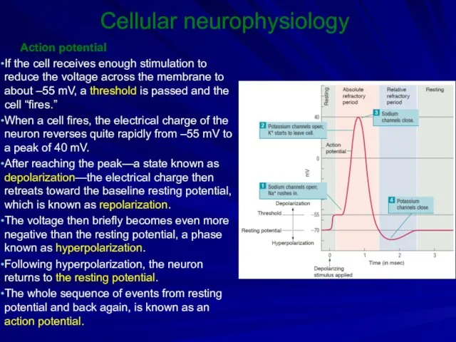
- 9. Cellular neurophysiology Action potential If the cell receives enough stimulation to reduce the voltage across the
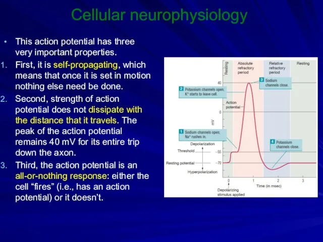
- 10. Cellular neurophysiology This action potential has three very important properties. First, it is self-propagating, which means
- 11. Cellular neurophysiology The action potential is first produced at a specific part of the neuron near
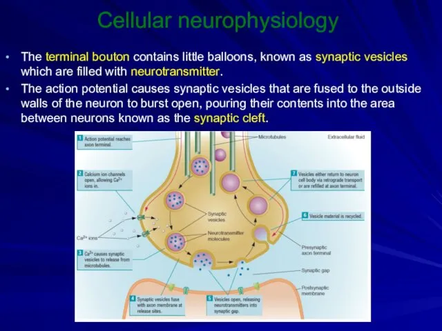
- 12. Cellular neurophysiology The terminal bouton contains little balloons, known as synaptic vesicles which are filled with
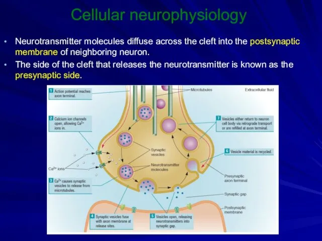
- 13. Cellular neurophysiology Neurotransmitter molecules diffuse across the cleft into the postsynaptic membrane of neighboring neuron. The
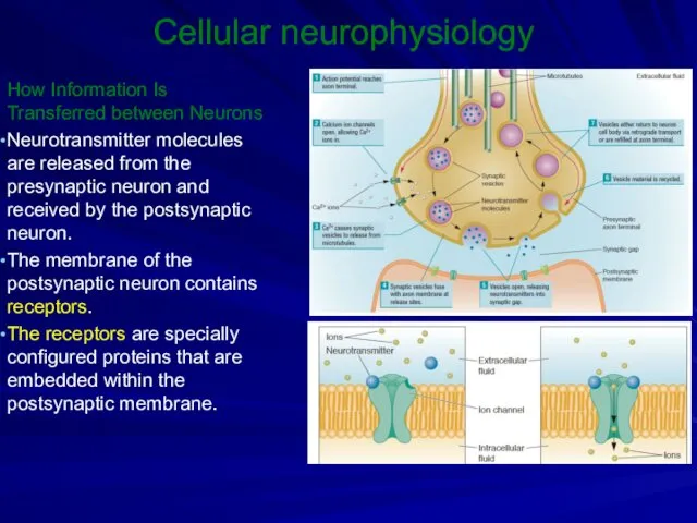
- 14. Cellular neurophysiology How Information Is Transferred between Neurons Neurotransmitter molecules are released from the presynaptic neuron
- 15. Cellular neurophysiology When neurotransmitter reaches the postsynaptic membrane, it fits into a specific region of the
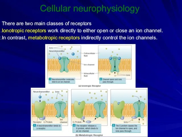
- 16. Cellular neurophysiology There are two main classes of receptors Ionotropic receptors work directly to either open
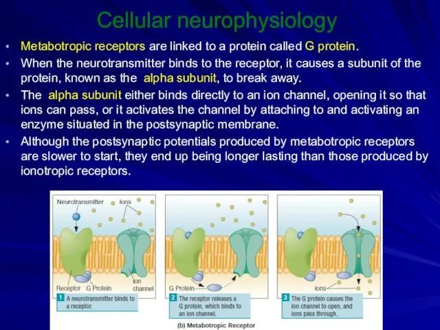
- 17. Cellular neurophysiology Metabotropic receptors are linked to a protein called G protein. When the neurotransmitter binds
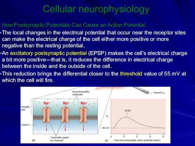
- 18. Cellular neurophysiology How Postsynaptic Potentials Can Cause an Action Potential The local changes in the electrical
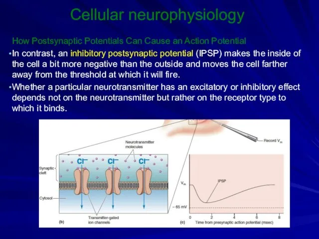
- 19. Cellular neurophysiology How Postsynaptic Potentials Can Cause an Action Potential In contrast, an inhibitory postsynaptic potential
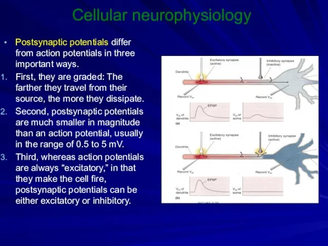
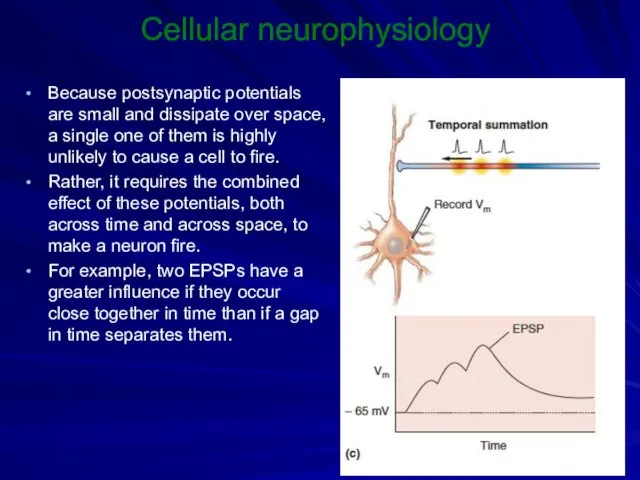
- 20. Cellular neurophysiology Postsynaptic potentials differ from action potentials in three important ways. First, they are graded:
- 21. Cellular neurophysiology Because postsynaptic potentials are small and dissipate over space, a single one of them
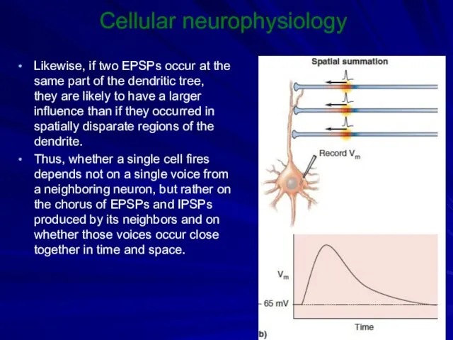
- 22. Cellular neurophysiology Likewise, if two EPSPs occur at the same part of the dendritic tree, they
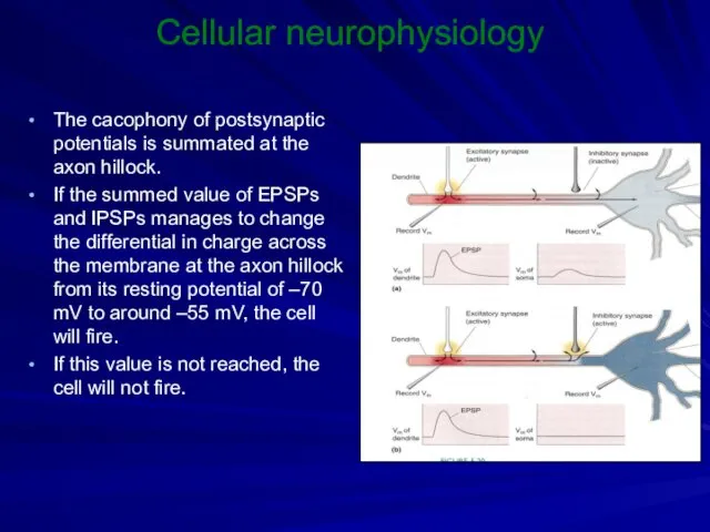
- 23. Cellular neurophysiology The cacophony of postsynaptic potentials is summated at the axon hillock. If the summed
- 24. Cellular neurophysiology The cacophony of postsynaptic potentials is summated at the axon hillock. If the summed
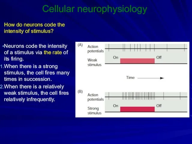
- 25. Cellular neurophysiology How do neurons code the intensity of stimulus? Neurons code the intensity of a
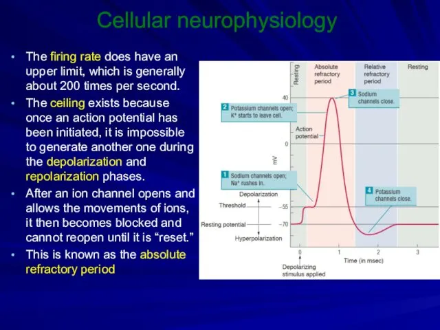
- 26. Cellular neurophysiology The firing rate does have an upper limit, which is generally about 200 times
- 28. Скачать презентацию
Cellular neurophysiology
A goal of cellular neurophysiology is to understand the biological
Cellular neurophysiology
A goal of cellular neurophysiology is to understand the biological
Cellular neurophysiology
When a cell with excitable membrane is not generating impulses,
Cellular neurophysiology
When a cell with excitable membrane is not generating impulses,
Cellular neurophysiology
The cell membrane of the neuron acts as a barrier
Cellular neurophysiology
The cell membrane of the neuron acts as a barrier

Cellular neurophysiology
In specific configurations, ion channels create a passageway that allows
Cellular neurophysiology
In specific configurations, ion channels create a passageway that allows
Cellular neurophysiology
Ions are found in different concentrations on either side of
Cellular neurophysiology
Ions are found in different concentrations on either side of
Cellular neurophysiology
Eventually, diffusion would cause these ions to be equally distributed
Cellular neurophysiology
Eventually, diffusion would cause these ions to be equally distributed
Cellular neurophysiology
Input from other neurons can affect the opening and closing
Cellular neurophysiology
Input from other neurons can affect the opening and closing
Cellular neurophysiology
Action potential
If the cell receives enough stimulation to reduce
Cellular neurophysiology
Action potential
If the cell receives enough stimulation to reduce
Cellular neurophysiology
This action potential has three very important properties.
First, it is
Cellular neurophysiology
This action potential has three very important properties.
First, it is

Cellular neurophysiology
The action potential is first produced at a specific part
Cellular neurophysiology
The action potential is first produced at a specific part
Cellular neurophysiology
The terminal bouton contains little balloons, known as synaptic vesicles
Cellular neurophysiology
The terminal bouton contains little balloons, known as synaptic vesicles
Cellular neurophysiology
Neurotransmitter molecules diffuse across the cleft into the postsynaptic membrane
Cellular neurophysiology
Neurotransmitter molecules diffuse across the cleft into the postsynaptic membrane
Cellular neurophysiology
How Information Is Transferred between Neurons
Neurotransmitter molecules are released from
Cellular neurophysiology
How Information Is Transferred between Neurons
Neurotransmitter molecules are released from

Cellular neurophysiology
When neurotransmitter reaches the postsynaptic membrane, it fits into a
Cellular neurophysiology
When neurotransmitter reaches the postsynaptic membrane, it fits into a
Cellular neurophysiology
There are two main classes of receptors
Ionotropic receptors work directly
Cellular neurophysiology
There are two main classes of receptors
Ionotropic receptors work directly
Cellular neurophysiology
Metabotropic receptors are linked to a protein called G protein.
Cellular neurophysiology
Metabotropic receptors are linked to a protein called G protein.
Cellular neurophysiology
How Postsynaptic Potentials Can Cause an Action Potential
The local changes
Cellular neurophysiology
How Postsynaptic Potentials Can Cause an Action Potential
The local changes
Cellular neurophysiology
How Postsynaptic Potentials Can Cause an Action Potential
In contrast, an
Cellular neurophysiology
How Postsynaptic Potentials Can Cause an Action Potential
In contrast, an
Cellular neurophysiology
Postsynaptic potentials differ from action potentials in three important ways.
Cellular neurophysiology
Postsynaptic potentials differ from action potentials in three important ways.
Cellular neurophysiology
Because postsynaptic potentials are small and dissipate over space, a
Cellular neurophysiology
Because postsynaptic potentials are small and dissipate over space, a
Cellular neurophysiology
Likewise, if two EPSPs occur at the same part of
Cellular neurophysiology
Likewise, if two EPSPs occur at the same part of
Cellular neurophysiology
The cacophony of postsynaptic potentials is summated at the axon
Cellular neurophysiology
The cacophony of postsynaptic potentials is summated at the axon

Cellular neurophysiology
The cacophony of postsynaptic potentials is summated at the axon
Cellular neurophysiology
The cacophony of postsynaptic potentials is summated at the axon
Cellular neurophysiology
How do neurons code the intensity of stimulus?
Neurons code the
Cellular neurophysiology
How do neurons code the intensity of stimulus?
Neurons code the
Cellular neurophysiology
The firing rate does have an upper limit, which is
Cellular neurophysiology
The firing rate does have an upper limit, which is
 Покровы тела животных
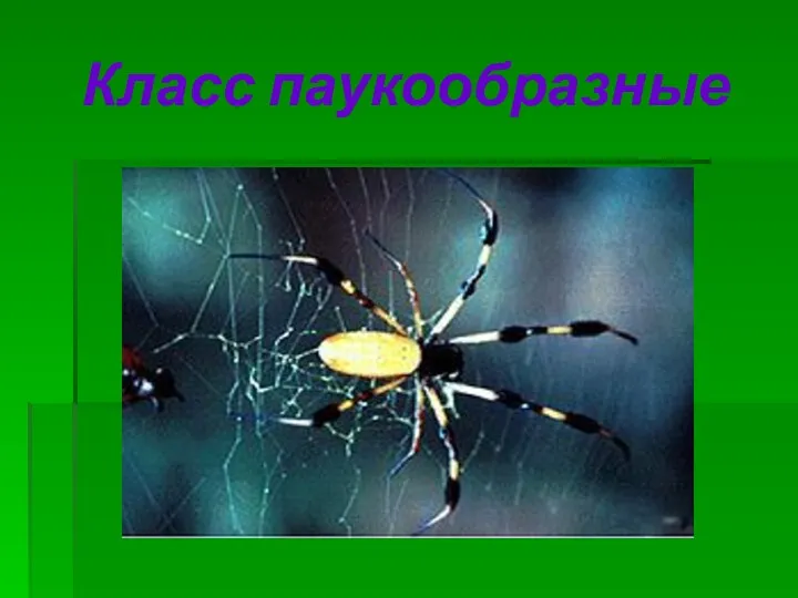 Класс Паукообразные
 Анатомия человека. Средний мозг. Промежуточный мозг
 Презентация Бактерии в нашей жизни
 Конспект урока для 6 класса Общая характеристика грибов
 Медицинская гельминтология. Трематодозы
 Дәрумендер. Дәрумендердің классификациясы
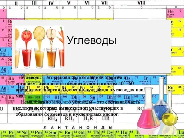 Углеводы. Классификация углеводов
 Презентация по биологии на тему Членистоногие 7 класс
 Класс Пресмыкающиеся. Класс Птицы. Класс Млекопитающие
 Эпителиальные ткани: морфофункциональная классификация и общая характеристика
 Ствол мозга
 Пищеварительная система
 Углеводы. Классификация углеводов. Глюкоза
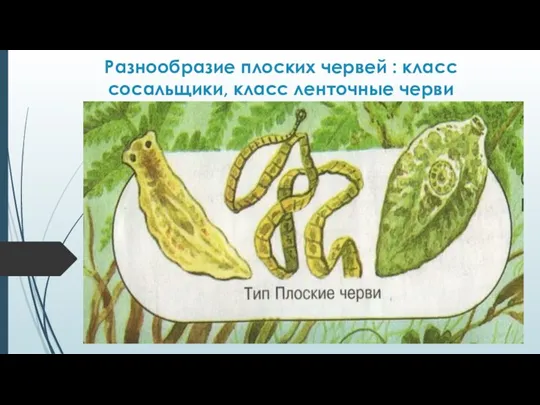 Презентация к уроку биологии в 6 классе
 Бактериофаги
 Ақуыздар
 Отряд Зайцеобразные
 Властивості та характеристики екосистем. Типи зв'язків між популяціями в екосистемі
 Многообразие и значение земноводных
 Стебель. Побег
 Экстерьер корпуса
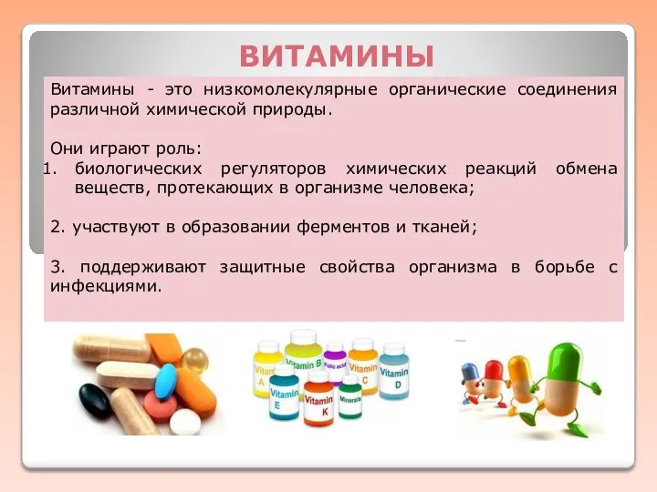 Витамины. Авитаминоз. Гиповитаминоз. Гипервитаминоз
 Дыхательная система в организме человека
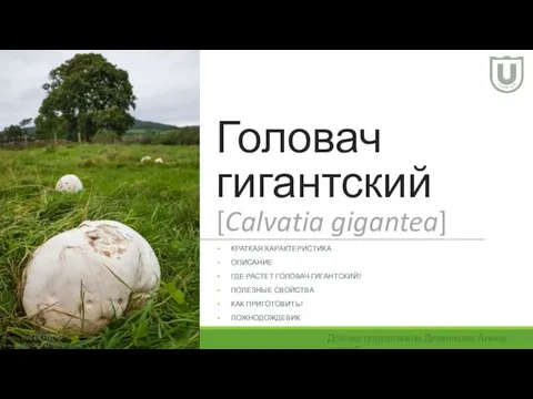 Головач гигантский
 Корень. Строение и функции
 Болотные птицы
 Отряды млекопитающих