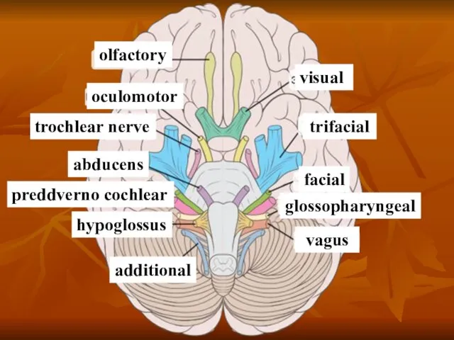
- Cranio-cerebral nerves

Содержание
- 2. Cranial - cerebral nerves are nerves walking away from a cerebrum or included in him. There
- 3. Distinguish: motoriuss (III, IV, VI, XI and XII of pair); mixed nerves (V, VII, IX and
- 4. trochlear nerve
- 5. Motor cranial nerves
- 6. Classification of motoriuss Motoriuss begin in the motive kernels of barrel. To mainly motive take the
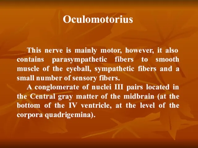
- 7. Oculomotorius This nerve is mainly motor, however, it also contains parasympathetic fibers to smooth muscle of
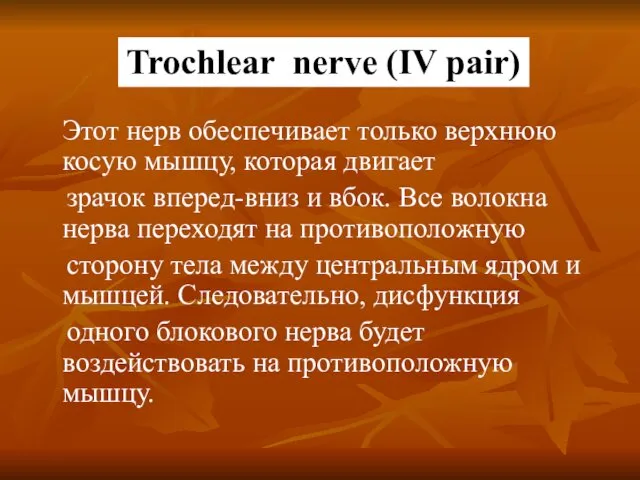
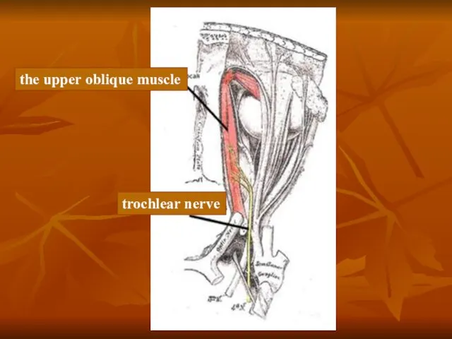
- 9. Этот нерв обеспечивает только верхнюю косую мышцу, которая двигает зрачок вперед-вниз и вбок. Все волокна нерва
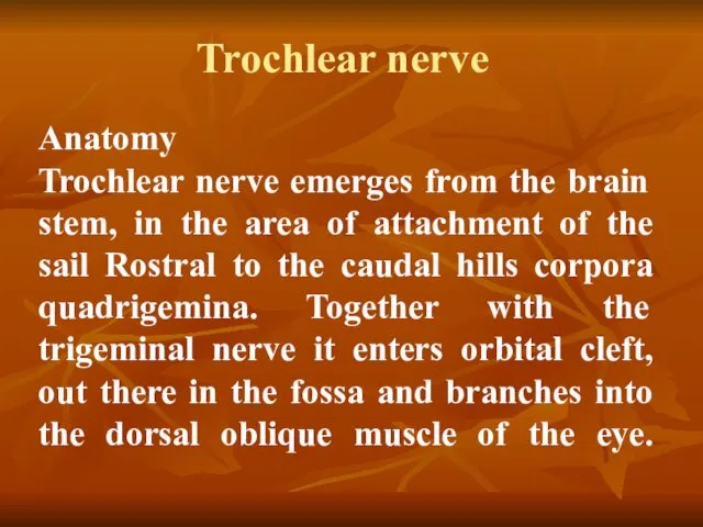
- 11. Trochlear nerve Anatomy Trochlear nerve emerges from the brain stem, in the area of attachment of
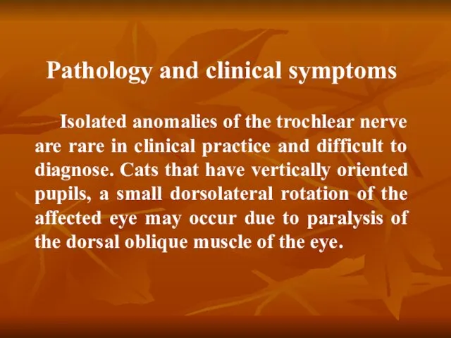
- 12. Pathology and clinical symptoms Isolated anomalies of the trochlear nerve are rare in clinical practice and
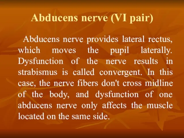
- 13. Abducens nerve (VI pair) Abducens nerve provides lateral rectus, which moves the pupil laterally. Dysfunction of
- 14. The trunk of the nerve exits the brain at the back edge of the bridge, between
- 15. Anatomy Nucleus abducens nerve are located on both sides of the median sulcus in the caudal
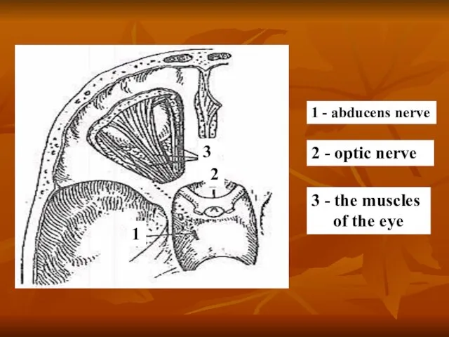
- 16. 1 - abducens nerve 2 - optic nerve 3 - the muscles of the eye 1
- 17. Hypoglossal nerve (XII pair) Formed by processes of nerve cells of the same nucleus, which is
- 18. Anatomy The neurons forming the hypoglossal nerve originate from the hypoglossal nerve centre in the medulla
- 19. Hypoglossal nerve and cervical (hyoid) loop: 1 - hypoglossal nerve; 2 - thyrohyoid branch; 3 -
- 20. Hypoglossal nerve Pathology and clinical symptoms Damage to hypoglossal nerve leads to the weakening of the
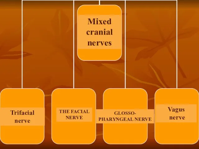
- 21. Mixed cranial nerves
- 22. Mixed cranial nerves THE FACIAL NERVE Trifacial nerve Vagus nerve GLOSSO- PHARYNGEAL NERVE
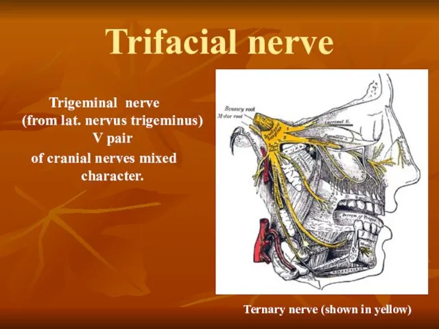
- 23. Trifacial nerve Trigeminal nerve (from lat. nervus trigeminus) V pair of cranial nerves mixed character. Ternary
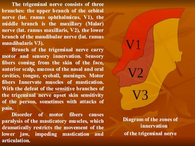
- 24. The trigeminal nerve consists of three branches: the upper branch of the orbital nerve (lat. ramus
- 25. Trigeminal nerve Anatomy The nerve center of the trigeminal nerve is weakly expressed anatomically, it is
- 26. Pathology Disease affecting the sensory and motor functions of the trigeminal nerve: infectious diseases; injuries; tumors;
- 27. Regeneration of the trigeminal nerve Neurology of the trigeminal nerve
- 28. The facial nerve The facial nerve enters the temporal bone through the internal auditory hole. Deep
- 30. The facial nerve (VII nerve) Anatomy The facial nerve is a mixed nerve, which unites the
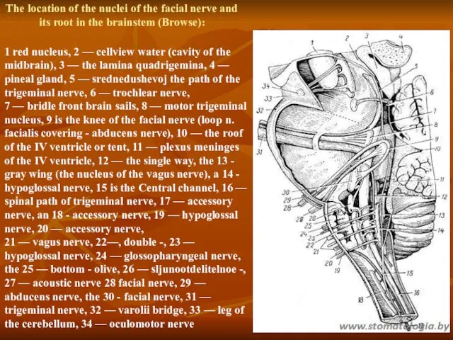
- 31. The location of the nuclei of the facial nerve and its root in the brainstem (Browse):
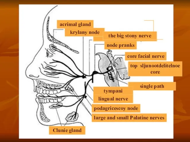
- 32. The divisions of the facial nerve In the facial canal the nerve divides into several branches:
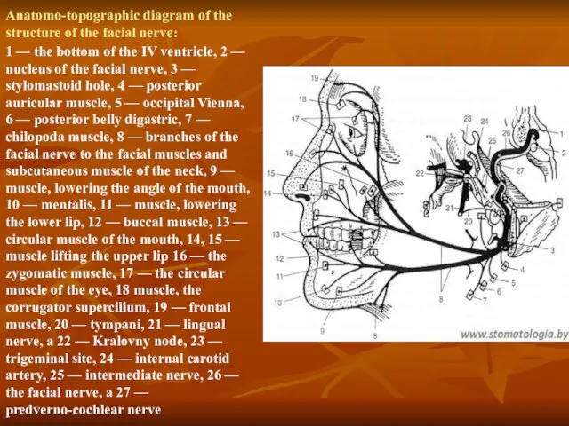
- 33. Anatomo-topographic diagram of the structure of the facial nerve: 1 — the bottom of the IV
- 34. Pathology and clinical symptoms Clinical symptoms depend on the level of the lesion. For example, if
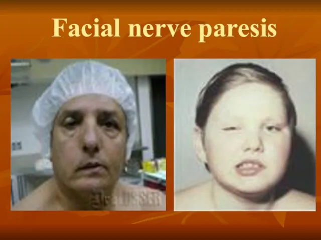
- 35. Facial nerve paresis
- 36. Diagnostic methods of neurology facial nerve Clinical neurological examination Instrumental methods Electromyography Doppler ultrasound with assessment
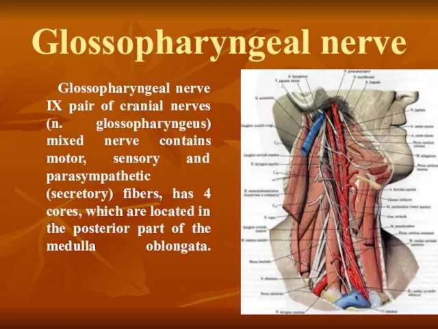
- 37. Glossopharyngeal nerve Glossopharyngeal nerve IX pair of cranial nerves (n. glossophaгyngeus) mixed nerve contains motor, sensory
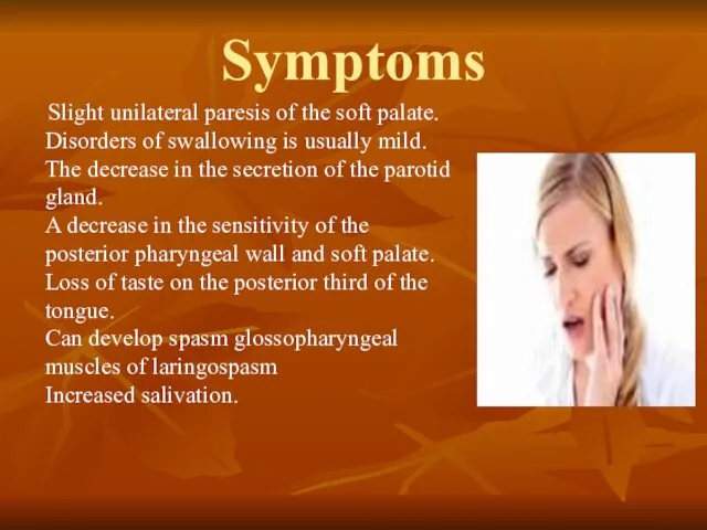
- 38. Symptoms Slight unilateral paresis of the soft palate. Disorders of swallowing is usually mild. The decrease
- 39. With the defeat of motor nuclei of the vagus nerve disturbances of swallowing, phonation, articulation, breathing,
- 40. Sensitive cranial nerves Anatomy of the Chemoreceptors of the nasal mucosa recognize various odors and transmit
- 42. Скачать презентацию
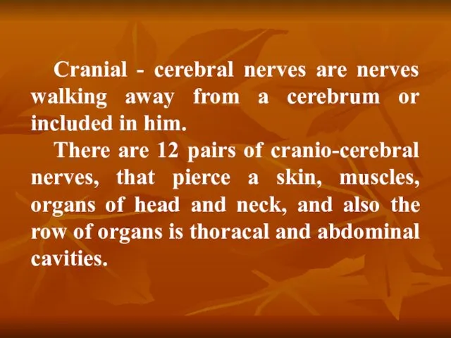
Cranial - cerebral nerves are nerves walking away from a cerebrum
Cranial - cerebral nerves are nerves walking away from a cerebrum
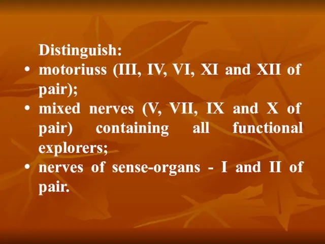
Distinguish:
motoriuss (III, IV, VI, XI and XII of pair);
mixed nerves
Distinguish:
motoriuss (III, IV, VI, XI and XII of pair);
mixed nerves
trochlear nerve
trochlear nerve
Motor cranial nerves
Motor cranial nerves
Classification of motoriuss
Motoriuss begin in the motive kernels of barrel.
To
Classification of motoriuss
Motoriuss begin in the motive kernels of barrel.
To
Oculomotorius
This nerve is mainly motor, however, it also contains parasympathetic fibers
Oculomotorius
This nerve is mainly motor, however, it also contains parasympathetic fibers
Этот нерв обеспечивает только верхнюю косую мышцу, которая двигает
зрачок вперед-вниз
Этот нерв обеспечивает только верхнюю косую мышцу, которая двигает
зрачок вперед-вниз
Trochlear nerve
Anatomy
Trochlear nerve emerges from the brain stem, in the area
Trochlear nerve
Anatomy Trochlear nerve emerges from the brain stem, in the area
Pathology and clinical symptoms
Isolated anomalies of the trochlear nerve are
Pathology and clinical symptoms
Isolated anomalies of the trochlear nerve are
Abducens nerve (VI pair)
Abducens nerve provides lateral rectus, which moves
Abducens nerve (VI pair)
Abducens nerve provides lateral rectus, which moves
The trunk of the nerve exits the brain at the
The trunk of the nerve exits the brain at the
Anatomy
Nucleus abducens nerve are located on both sides of the
Anatomy Nucleus abducens nerve are located on both sides of the
1 - abducens nerve
2 - optic nerve
3 - the muscles
1 - abducens nerve
2 - optic nerve
3 - the muscles
Hypoglossal nerve (XII pair)
Formed by processes of nerve cells of the
Hypoglossal nerve (XII pair)
Formed by processes of nerve cells of the
Anatomy
The neurons forming the hypoglossal nerve originate from the hypoglossal nerve
Anatomy The neurons forming the hypoglossal nerve originate from the hypoglossal nerve
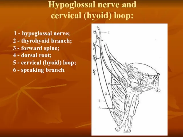
Hypoglossal nerve and
cervical (hyoid) loop:
1 - hypoglossal nerve;
2
Hypoglossal nerve and
cervical (hyoid) loop:
1 - hypoglossal nerve; 2
Hypoglossal nerve
Pathology and clinical symptoms
Damage to hypoglossal nerve leads to
Hypoglossal nerve
Pathology and clinical symptoms
Damage to hypoglossal nerve leads to
Mixed cranial nerves
Mixed cranial nerves
Mixed cranial
nerves
THE FACIAL
NERVE
Trifacial nerve
Vagus
nerve
GLOSSO-
PHARYNGEAL NERVE
Mixed cranial
nerves
THE FACIAL
NERVE
Trifacial nerve
Vagus
nerve
GLOSSO-
PHARYNGEAL NERVE
Trifacial nerve
Trigeminal nerve
(from lat. nervus trigeminus)
V pair
of cranial nerves
Trifacial nerve
Trigeminal nerve
(from lat. nervus trigeminus)
V pair
of cranial nerves
The trigeminal nerve consists of three branches: the upper branch of
The trigeminal nerve consists of three branches: the upper branch of
Trigeminal nerve
Anatomy
The nerve center of the trigeminal nerve is weakly expressed
Trigeminal nerve
Anatomy
The nerve center of the trigeminal nerve is weakly expressed
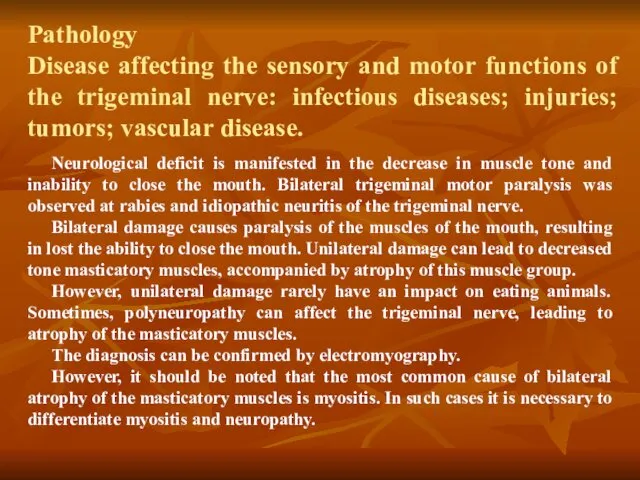
Pathology
Disease affecting the sensory and motor functions of the trigeminal
Pathology Disease affecting the sensory and motor functions of the trigeminal
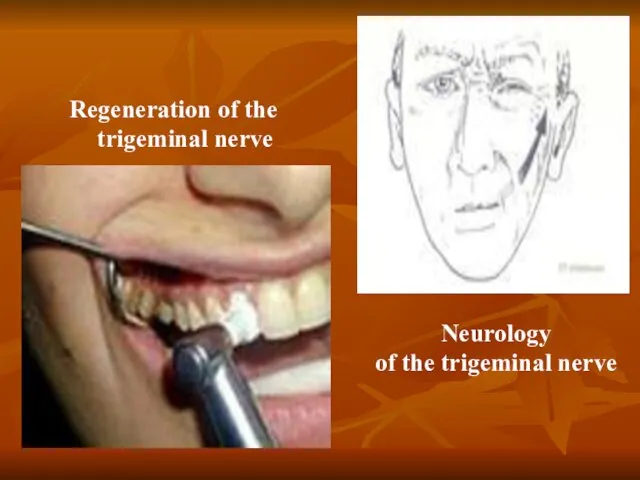
Regeneration of the trigeminal nerve
Neurology
of the trigeminal nerve
Regeneration of the trigeminal nerve
Neurology
of the trigeminal nerve
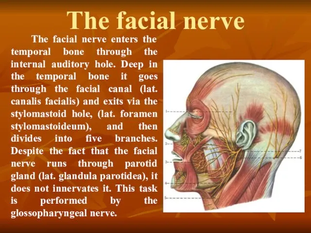
The facial nerve
The facial nerve enters the temporal bone through
The facial nerve
The facial nerve enters the temporal bone through
The facial nerve (VII nerve)
Anatomy
The facial nerve is a mixed
The facial nerve (VII nerve)
Anatomy
The facial nerve is a mixed
The location of the nuclei of the facial nerve and its
The location of the nuclei of the facial nerve and its
The divisions of the facial nerve
In the facial canal the nerve
The divisions of the facial nerve
In the facial canal the nerve
Anatomo-topographic diagram of the structure of the facial nerve:
1 — the
Anatomo-topographic diagram of the structure of the facial nerve:
1 — the
Pathology and clinical symptoms
Clinical symptoms depend on the level of
Pathology and clinical symptoms
Clinical symptoms depend on the level of
Facial nerve paresis
Facial nerve paresis
Diagnostic methods of neurology facial nerve
Clinical neurological examination
Instrumental methods
Electromyography
Doppler
Diagnostic methods of neurology facial nerve
Clinical neurological examination
Instrumental methods
Electromyography
Doppler
Glossopharyngeal nerve
Glossopharyngeal nerve IX pair of cranial nerves (n. glossophaгyngeus)
Glossopharyngeal nerve
Glossopharyngeal nerve IX pair of cranial nerves (n. glossophaгyngeus)
Symptoms
Slight unilateral paresis of the soft palate.
Disorders of swallowing
Symptoms
Slight unilateral paresis of the soft palate. Disorders of swallowing
With the defeat of motor nuclei of the vagus nerve
With the defeat of motor nuclei of the vagus nerve
Sensitive cranial nerves
Anatomy of the Chemoreceptors of the nasal
Sensitive cranial nerves
Anatomy of the Chemoreceptors of the nasal
 WWF = World Wide Fund for Nature
 Подготовка к ОГЭ по биологии
 Покормите птиц зимой
 Многообразие организмов. Значение работ К. Линнея и Ж.Б. Ламарка. Основные систематические категории
 Некоторые растения и животные, включённые в Красную книгу Мурманской области
 Немного о собаках
 Генетическая инженерия. Тема №1
 Доклад на тему Круговорот азота
 Бионика. Биология – техника
 Ткани. 4 типа тканей
 Первично чувствующие органы чувств: орган зрения и орган обоняния
 Физиология нервов и нервно-мышечного синапса
 ГОСТы на посадочный материал
 Катаболизм. Энергетический обмен и всё, что с ним связано
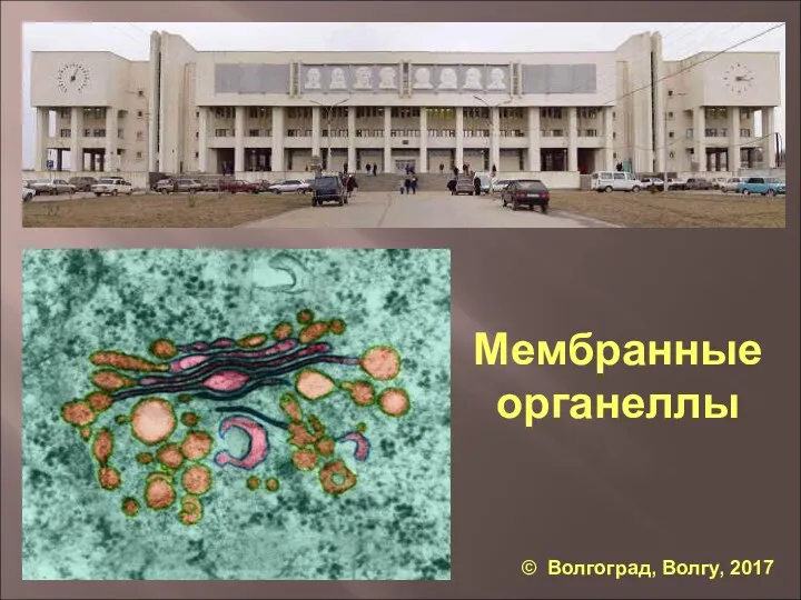 Мембранные органеллы
 Строение клетки
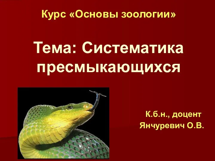 Систематика пресмыкающихся
 Определение пола в животном мире. 9 класс
 Глюкоза. Молекулярна формула. Фізичні та хімічні властивості глюкози. Поширення в природі
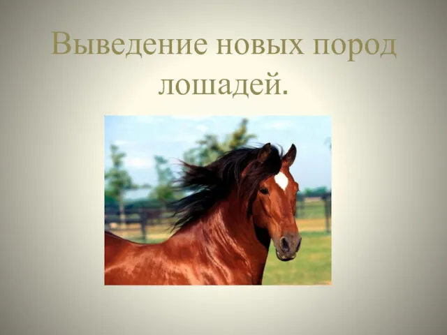 Выведение новых пород лошадей
 Методи очистки води в домашніх умовах
 Высшая нервная деятельность человека
 Визначення особливостей зовнішньої будови хребетних тварин у зв’язку з пристосуванням до різних умов існування. ЛР №2
 Царство Бактерии
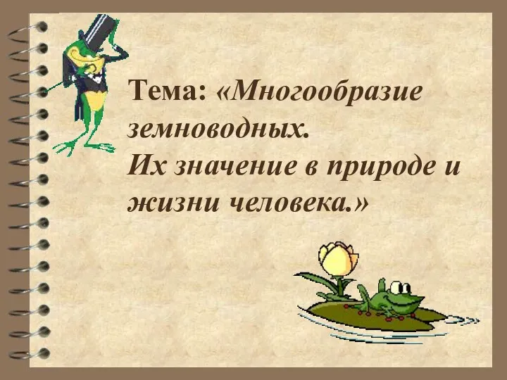 Многообразие земноводных
 Дыхание растений и животных
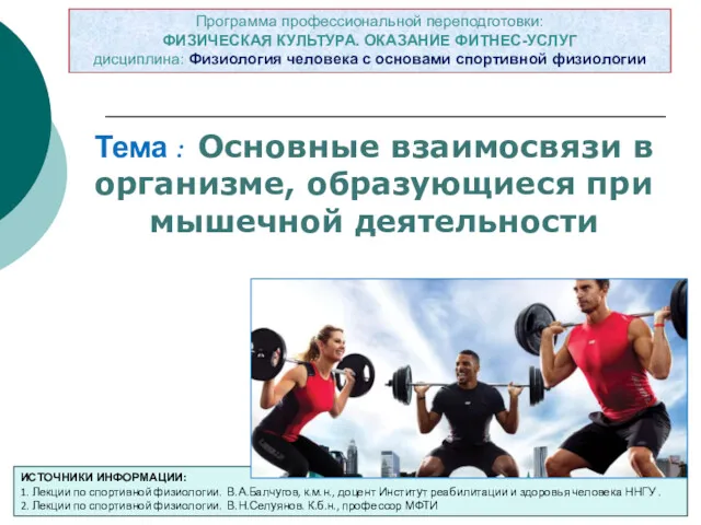 Основные взаимосвязи в организме, образующиеся при мышечной деятельности. Тема 1
 Презентация к уроку Факторы эволюции