- Central nervous system

Содержание
- 2. THE HUMAN BRAIN Complex 1.4 kg in weight Pre frontal cortex 2% of body weight 20%
- 3. THE BLOOD BRAIN BARRIER Brain protection system The BBB is both; A physical barrier that restricts
- 4. DIVISIONS OF CNS CNS - central nervous system: consists of brain and spinal cord • Nerves
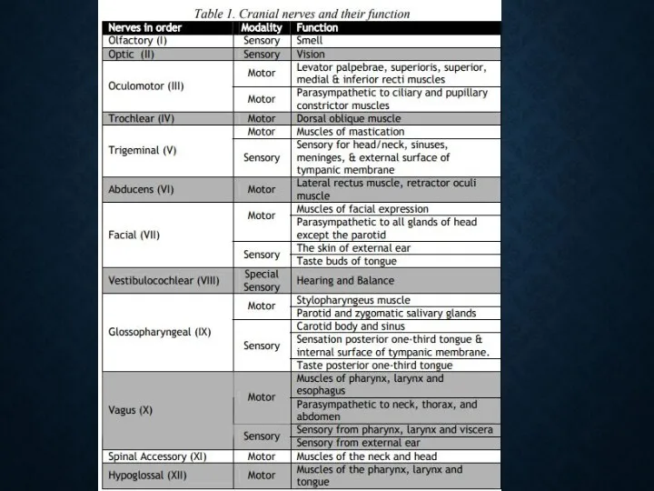
- 5. A. CRANIAL NERVES 12 pairs & their branches Some responsible for special senses: sight, hearing, taste,
- 7. C. CEREBRUM Largest section of the brain Responsible for: reasoning, thought, memory, speaking, sensstion, sight, hearing,
- 8. D. CEREBELLUM Responsible for: coordination of muscles, balance, posture, & muscle tone
- 9. E. MIDBRAIN Responsible for: conducting impulses between brain parts certain eye reflexes
- 10. PONS Responsible for: conducting messages to other parts of the brain Reflex actions such as chewing,
- 11. G. MEDULLA OBLONGATA Lowest part of brain stem Connects to the spinal cord Responsible for: regulating
- 12. 2. SPINAL CORD Goes down back of body from Medulla Oblongata Surrounded and protected by vertebrae
- 13. 3. MENINGES Consists of 3 membranes Covers and protects the brain and spinal cord
- 14. THREE MEMBRANES C. Dura mater thick, tough outer layer D. Arachnoid membrane middle delicate weblike layer
- 15. 4. VENTRICLES Four hallow spaces located in the middle of the brain. Connected to each other
- 16. CEREBROSPINAL FLUID Circulates continuously Serves as shock absorber to protect brain and spinal cord Carries nurients
- 17. B. SPINAL NERVES 31 pairs & their branches carries messages to & from the spinal cord
- 18. 3. AUTONOMIC NERVOUS SYSTEM Autonomic nervous system • It is further subdivided into sympathetic and parasympathetic
- 19. NEUROTRANSMISSION Neurotransmission • A nerve impulse is an electric current that passes along an axon to
- 20. NEUROTRANSMISSION and choline by acetylcholine esterase (AchE), found principally in neurons and neuromuscular junctions . •
- 21. NEUROTRANSMISSION Adrenergic receptors : Alpha receptors are mainly subdivided into alpha-1 and alpha 2 receptors •
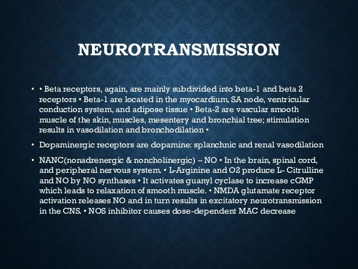
- 22. NEUROTRANSMISSION • Beta receptors, again, are mainly subdivided into beta-1 and beta 2 receptors • Beta-1
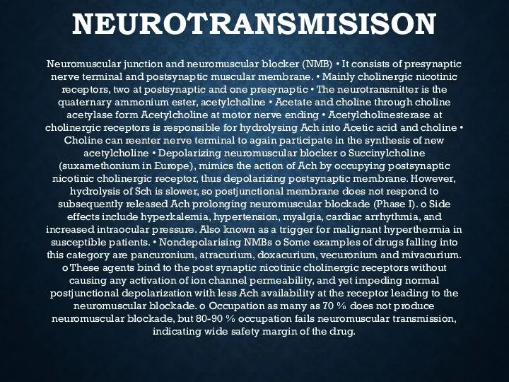
- 23. NEUROTRANSMISISON Neuromuscular junction and neuromuscular blocker (NMB) • It consists of presynaptic nerve terminal and postsynaptic
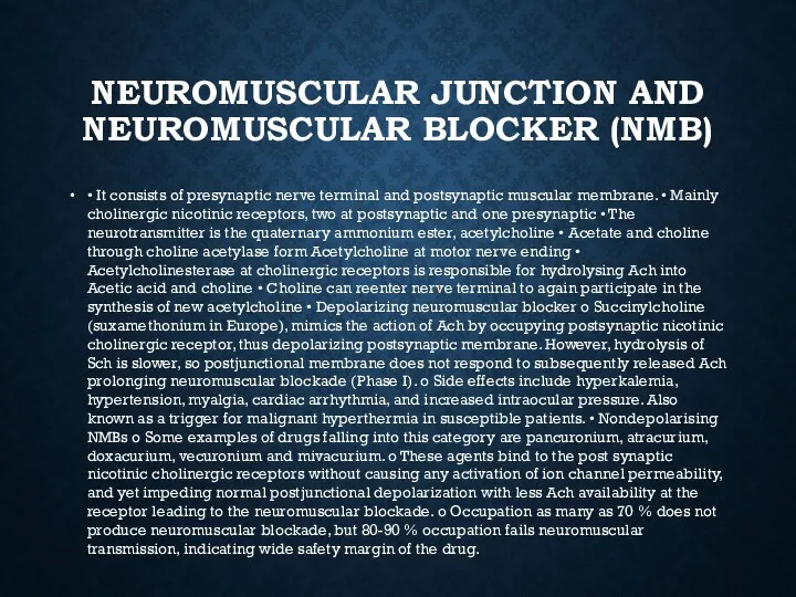
- 24. NEUROMUSCULAR JUNCTION AND NEUROMUSCULAR BLOCKER (NMB) • It consists of presynaptic nerve terminal and postsynaptic muscular
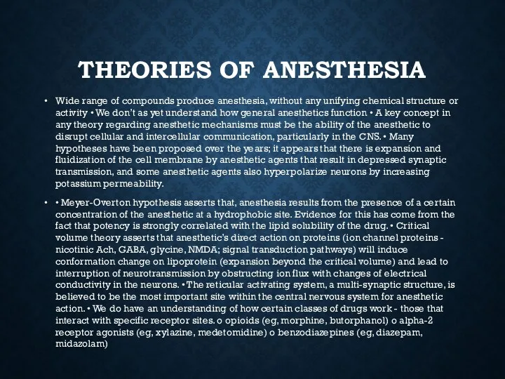
- 25. THEORIES OF ANESTHESIA Wide range of compounds produce anesthesia, without any unifying chemical structure or activity
- 27. Скачать презентацию
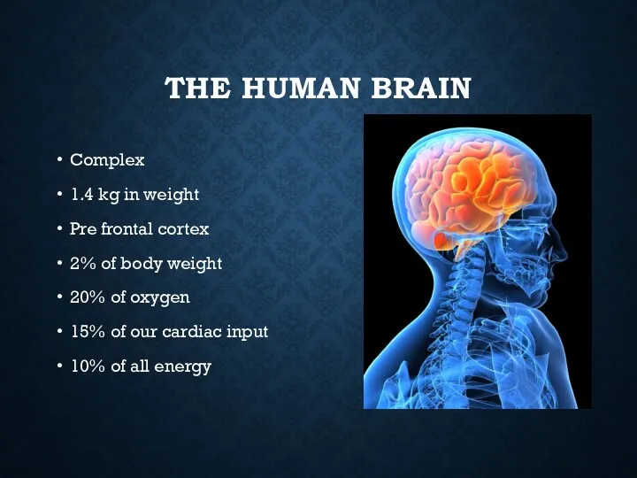
THE HUMAN BRAIN
Complex
1.4 kg in weight
Pre frontal cortex
2% of body weight
20%
THE HUMAN BRAIN
Complex
1.4 kg in weight
Pre frontal cortex
2% of body weight
20%
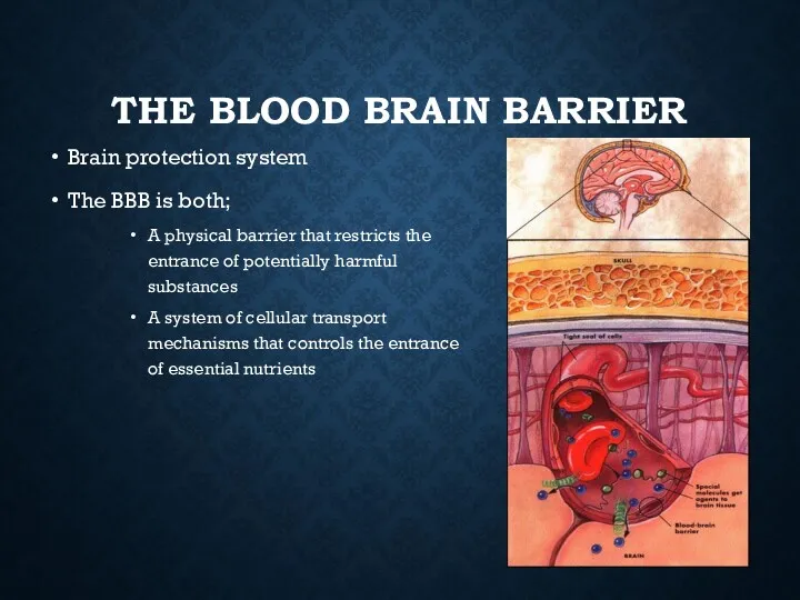
THE BLOOD BRAIN BARRIER
Brain protection system
The BBB is both;
A physical barrier
THE BLOOD BRAIN BARRIER
Brain protection system
The BBB is both;
A physical barrier
DIVISIONS OF CNS
CNS - central nervous system:
consists of brain and spinal
DIVISIONS OF CNS
CNS - central nervous system:
consists of brain and spinal
A. CRANIAL NERVES
12 pairs & their branches
Some responsible for special senses:
A. CRANIAL NERVES
12 pairs & their branches
Some responsible for special senses:
C. CEREBRUM
Largest section of the brain
Responsible for:
reasoning, thought, memory, speaking, sensstion,
C. CEREBRUM
Largest section of the brain
Responsible for:
reasoning, thought, memory, speaking, sensstion,
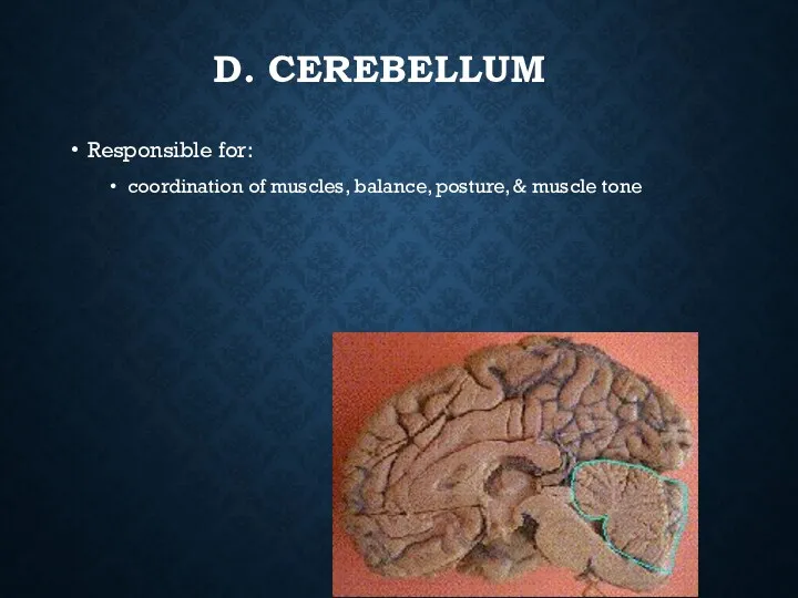
D. CEREBELLUM
Responsible for:
coordination of muscles, balance, posture, & muscle tone
D. CEREBELLUM
Responsible for:
coordination of muscles, balance, posture, & muscle tone
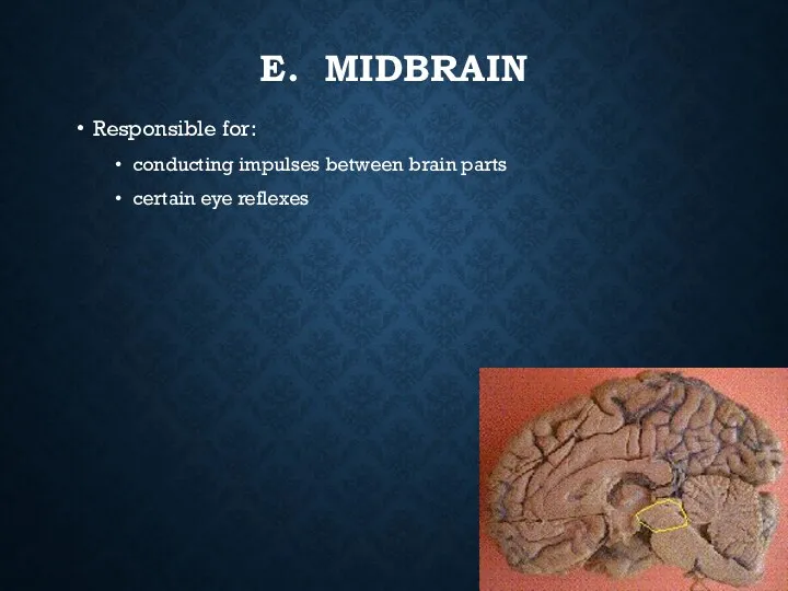
E. MIDBRAIN
Responsible for:
conducting impulses between brain parts
certain eye reflexes
E. MIDBRAIN
Responsible for:
conducting impulses between brain parts
certain eye reflexes
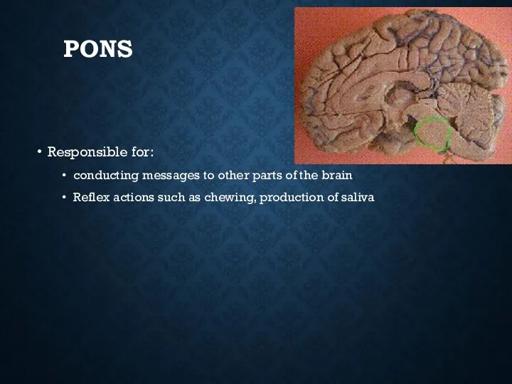
PONS
Responsible for:
conducting messages to other parts of the brain
Reflex actions such
PONS
Responsible for:
conducting messages to other parts of the brain
Reflex actions such
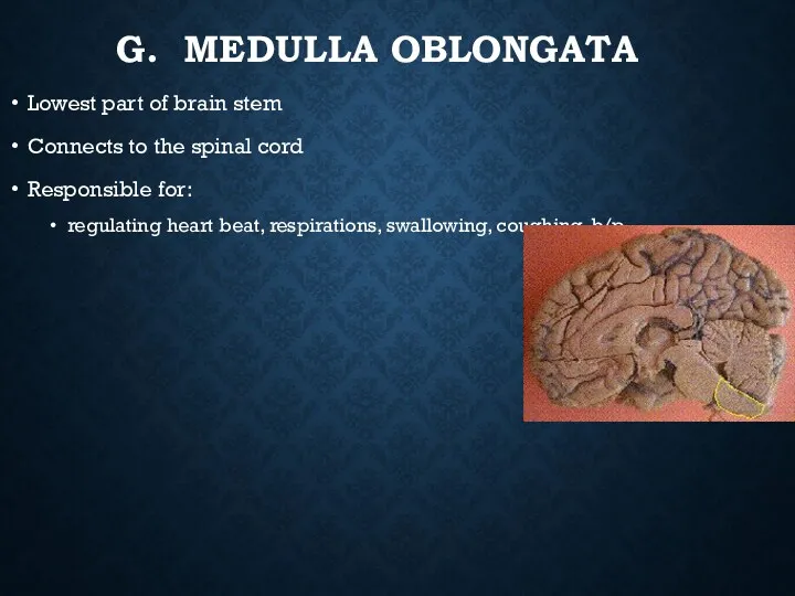
G. MEDULLA OBLONGATA
Lowest part of brain stem
Connects to the spinal cord
Responsible
G. MEDULLA OBLONGATA
Lowest part of brain stem
Connects to the spinal cord
Responsible
2. SPINAL CORD
Goes down back of body from Medulla Oblongata
Surrounded and
2. SPINAL CORD
Goes down back of body from Medulla Oblongata
Surrounded and
3. MENINGES
Consists of 3 membranes
Covers and protects the brain and spinal
3. MENINGES
Consists of 3 membranes
Covers and protects the brain and spinal
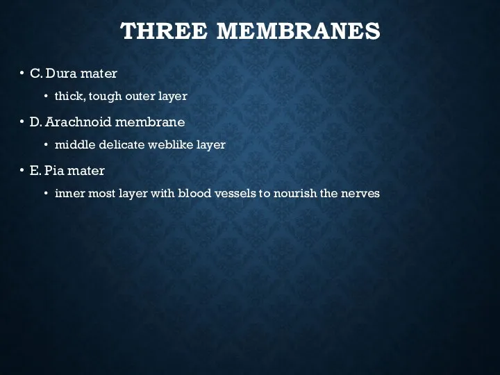
THREE MEMBRANES
C. Dura mater
thick, tough outer layer
D. Arachnoid membrane
middle delicate weblike
THREE MEMBRANES
C. Dura mater
thick, tough outer layer
D. Arachnoid membrane
middle delicate weblike
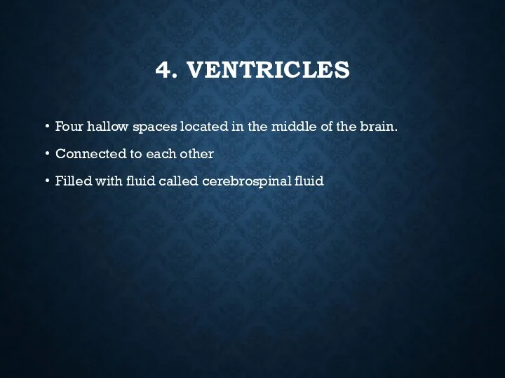
4. VENTRICLES
Four hallow spaces located in the middle of the brain.
Connected
4. VENTRICLES
Four hallow spaces located in the middle of the brain.
Connected
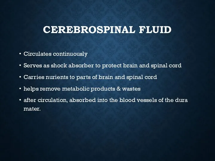
CEREBROSPINAL FLUID
Circulates continuously
Serves as shock absorber to protect brain and spinal
CEREBROSPINAL FLUID
Circulates continuously
Serves as shock absorber to protect brain and spinal
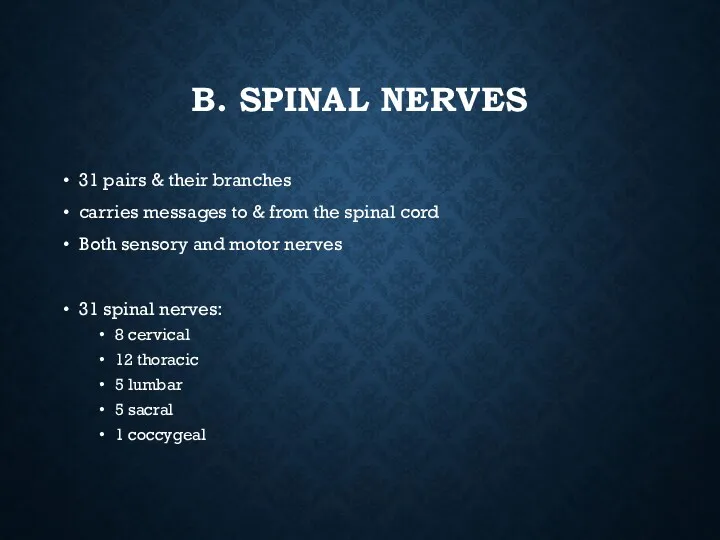
B. SPINAL NERVES
31 pairs & their branches
carries messages to & from
B. SPINAL NERVES
31 pairs & their branches
carries messages to & from
3. AUTONOMIC NERVOUS SYSTEM
Autonomic nervous system • It is further subdivided
3. AUTONOMIC NERVOUS SYSTEM
Autonomic nervous system • It is further subdivided
NEUROTRANSMISSION
Neurotransmission • A nerve impulse is an electric current that passes
NEUROTRANSMISSION
Neurotransmission • A nerve impulse is an electric current that passes
NEUROTRANSMISSION
and choline by acetylcholine esterase (AchE), found principally in neurons
NEUROTRANSMISSION
and choline by acetylcholine esterase (AchE), found principally in neurons
NEUROTRANSMISSION
Adrenergic receptors : Alpha receptors are mainly subdivided into alpha-1 and
NEUROTRANSMISSION
Adrenergic receptors : Alpha receptors are mainly subdivided into alpha-1 and
NEUROTRANSMISSION
• Beta receptors, again, are mainly subdivided into beta-1 and beta
NEUROTRANSMISSION
• Beta receptors, again, are mainly subdivided into beta-1 and beta
NEUROTRANSMISISON
Neuromuscular junction and neuromuscular blocker (NMB) • It consists of presynaptic
NEUROTRANSMISISON
Neuromuscular junction and neuromuscular blocker (NMB) • It consists of presynaptic
NEUROMUSCULAR JUNCTION AND NEUROMUSCULAR BLOCKER (NMB)
• It consists of presynaptic nerve
NEUROMUSCULAR JUNCTION AND NEUROMUSCULAR BLOCKER (NMB)
• It consists of presynaptic nerve
THEORIES OF ANESTHESIA
Wide range of compounds produce anesthesia, without any unifying
THEORIES OF ANESTHESIA
Wide range of compounds produce anesthesia, without any unifying
 Филогенез нервной системы. Онтогенез ЦНС у человека
 Терминологический контроль по биохимии. Базовые понятия
 Біогенні s-, р-, d-елементи. Біологічна роль та значення їх у медицині
 Основы морфологии микроорганизмов
 Нейропрорывы-2017/2018
 Мой первый гербарий. 4 класс
 Отдел формирования (школа саженцев)
 Цитологическое исследование выпотных жидкостей
 Способы размножения животных. Оплодотворение
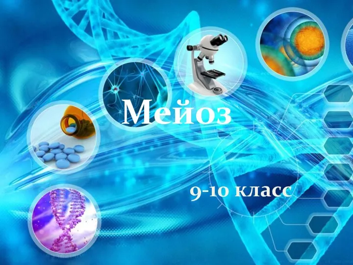 Мейоз. Биологическое значение мейоза. Мейоз в жизненном цикле организмов
 Цианобактерии. Лекция 2
 Внутрішнє середовище організму. Кров, її склад та функції
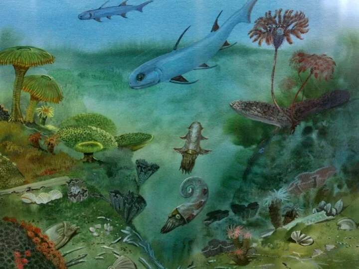 Простейшие. Кишечнополостные
 Биологическое и социальное в человеке
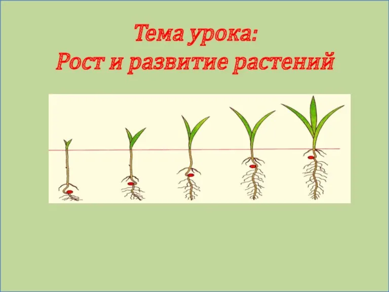 Рост и развитие растений
 Что такое жизнь с точки зрения биолога
 Тұқымқуалаушылық және өзгергіштік заңдылықтары
 Құстардың тыныс алуы және газ алмасуы
 Тип Хордовые. Ланцетник 7 класс
 Викторина Звери, птицы, лес и Я – вместе дружная семья
 Эволюция нервной, кровеносной и выделительной систем органов
 Рысь обыкновенная
 Что значит вода для человека
 Что нам делать с мусором?.
 Классификация животных
 Такие разные деревья
 Основы фитоценологии
 Nobel prize in physiology or medicine