- Enteric bacterial pathogens

Содержание
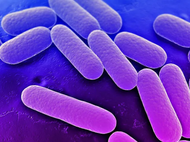
- 3. FAMILY ENTEROBACTERIACEAE are a large heterogeneous group of Gram«-» rods whose natural habitat is the intestinal
- 4. FAMILY ENTEROBACTERIACEAE Most of the members of Enterobacteriaceae are facultative anaerobes, ferment a wide range of
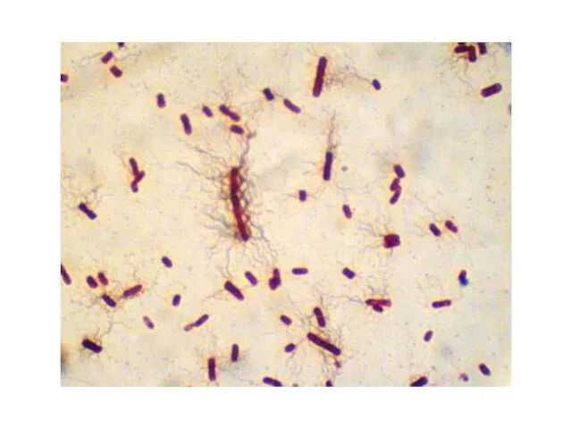
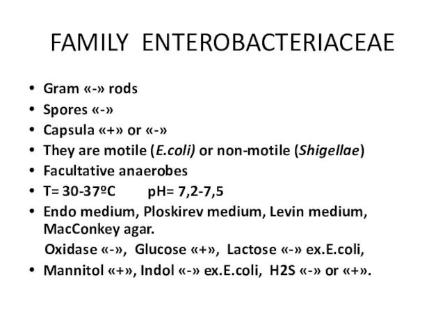
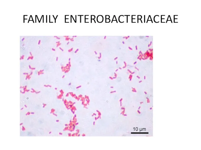
- 6. FAMILY ENTEROBACTERIACEAE Gram «-» rods Spores «-» Capsula «+» or «-» They are motile (E.coli) or
- 7. FAMILY ENTEROBACTERIACEAE
- 8. FAMILY ENTEROBACTERIACEAE The differential culture media that contain carbohydrates and special dyes (indicators) are used to
- 9. Endo Medium
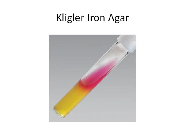
- 10. Kligler Iron Agar
- 11. Kligler Iron Agar Kligler iron agar is used for the differentiation of the Enterobacteriaceae members on
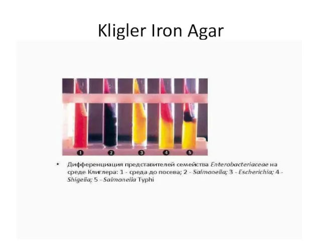
- 12. Kligler Iron Agar
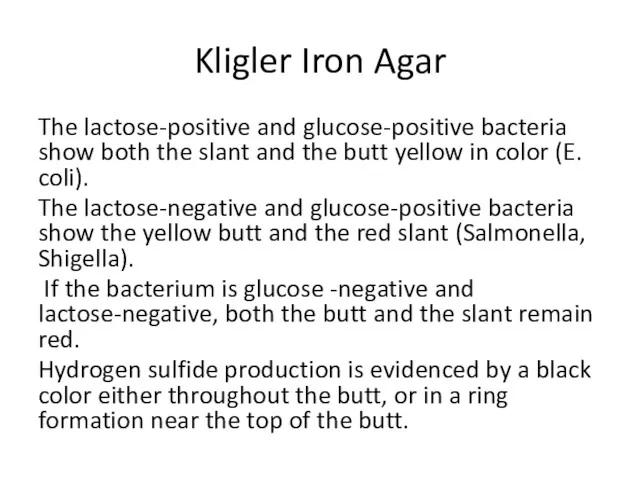
- 13. Kligler Iron Agar The lactose-positive and glucose-positive bacteria show both the slant and the butt yellow
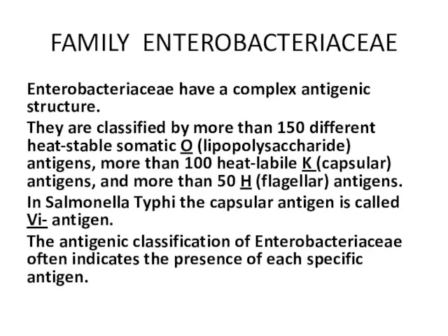
- 14. FAMILY ENTEROBACTERIACEAE Enterobacteriaceae have a complex antigenic structure. They are classified by more than 150 different
- 15. FAMILY ENTEROBACTERIACEAE Virulence factors: Fimbriae Enterotoxins Hemolysins Endotoxins
- 16. FAMILY ENTEROBACTERIACEAE Epidemiology: They are pathogenic for human and animals. They are transmitted by the fecal-oral
- 17. FAMILY ENTEROBACTERIACEAE Microbiological diagnosis. Specimens : feces, vomit, food,urine, blood. Methods: bacteriological, serological, biological.
- 18. Salmonellae Salmonella Family – Enterobacteriaceae Genus – Salmonellae Species - S. enterica Subspecies – S. typhi,
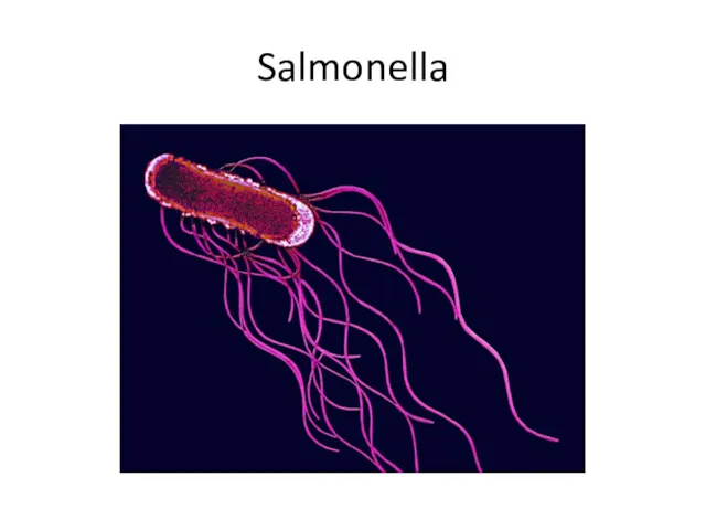
- 19. Salmonella
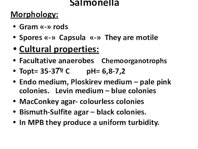
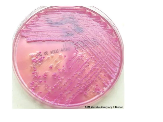
- 20. Salmonella Morphology: Gram «-» rods Spores «-» Capsula «-» They are motile Cultural properties: Facultative anaerobes
- 23. Bismuth Sulfite agar
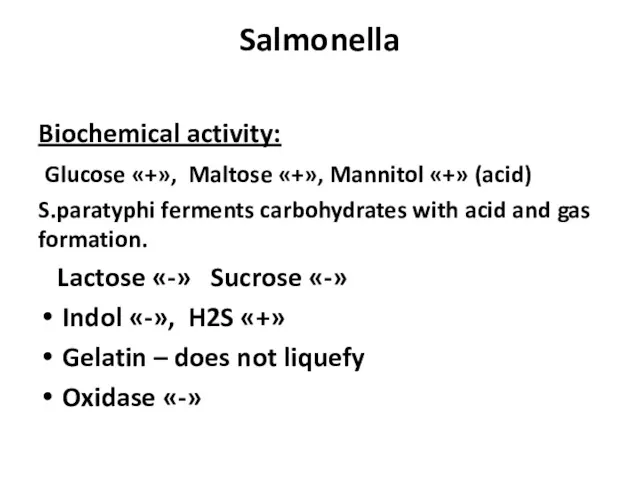
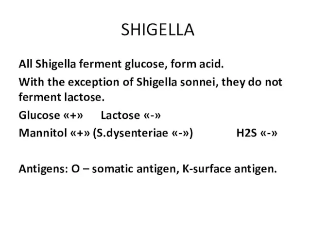
- 24. Salmonella Biochemical activity: Glucose «+», Maltose «+», Mannitol «+» (acid) S.paratyphi ferments carbohydrates with acid and
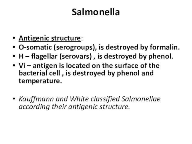
- 25. Salmonella Antigenic structure: O-somatic (serogroups), is destroyed by formalin. H – flagellar (serovars) , is destroyed
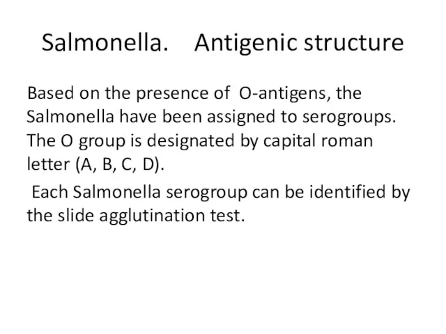
- 26. Salmonella. Antigenic structure Based on the presence of O-antigens, the Salmonella have been assigned to serogroups.
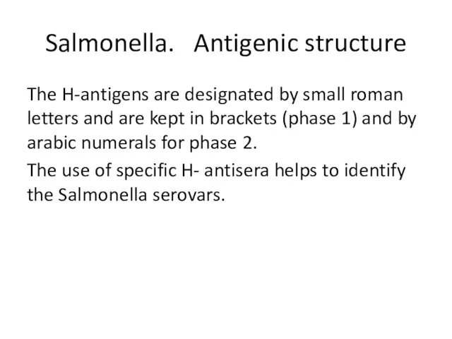
- 27. Salmonella. Antigenic structure The H-antigens are designated by small roman letters and are kept in brackets
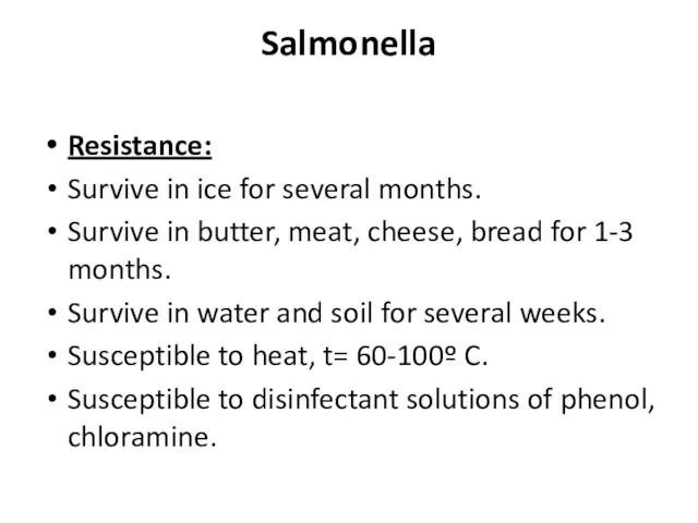
- 28. Salmonella Resistance: Survive in ice for several months. Survive in butter, meat, cheese, bread for 1-3
- 29. Salmonella Virulence Factors
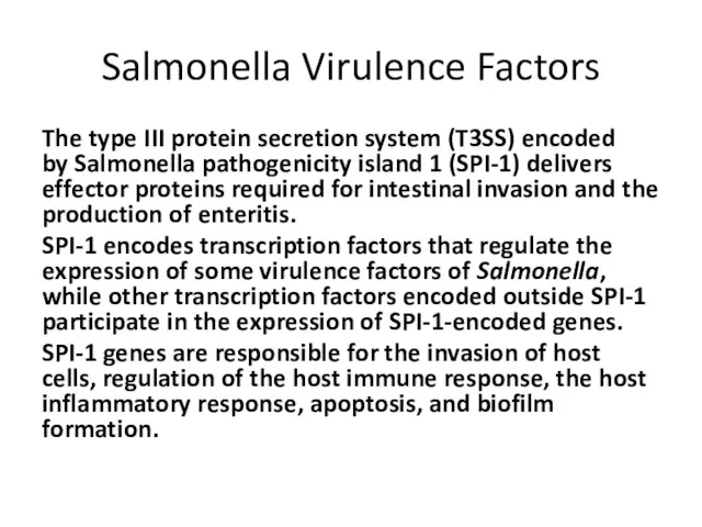
- 30. Salmonella Virulence Factors The type III protein secretion system (T3SS) encoded by Salmonella pathogenicity island 1
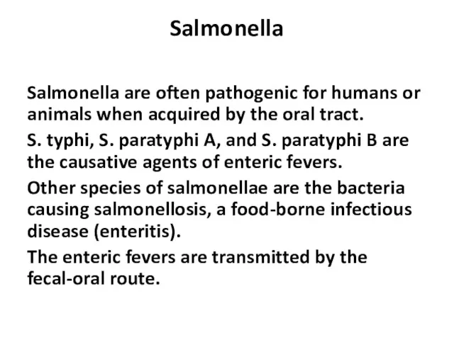
- 31. Salmonella Salmonella are often pathogenic for humans or animals when acquired by the oral tract. S.
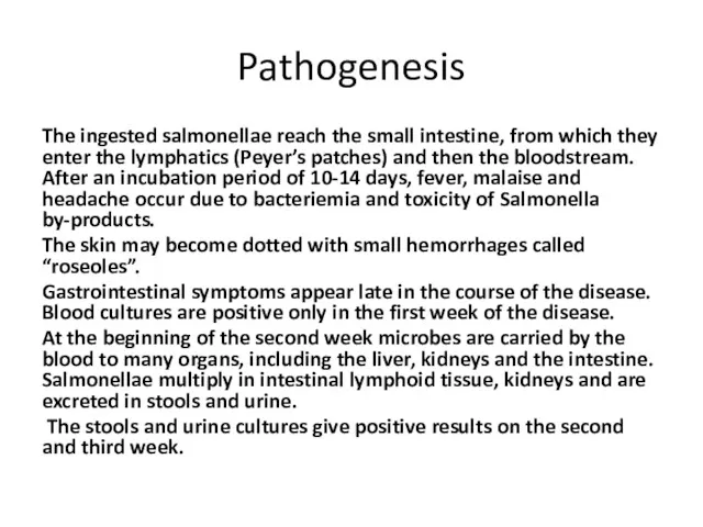
- 32. Pathogenesis The ingested salmonellae reach the small intestine, from which they enter the lymphatics (Peyer’s patches)
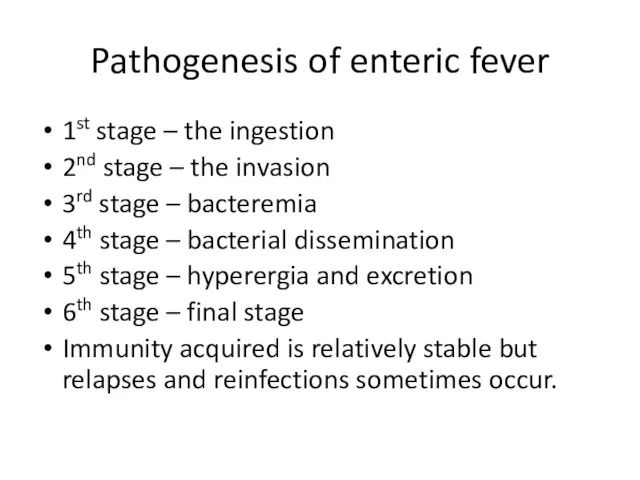
- 33. Pathogenesis of enteric fever 1st stage – the ingestion 2nd stage – the invasion 3rd stage
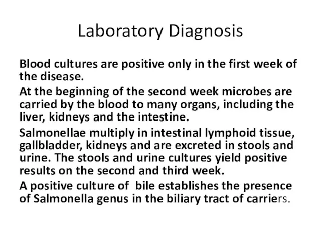
- 34. Laboratory Diagnosis Blood cultures are positive only in the first week of the disease. At the
- 35. Salmonella Infections. Treatment . Antimicrobial treatment. Replacement of fluids and electrolytes are essential. Susceptibility testing is
- 37. SHIGELLA The causative agent of dysentery was described in 1888 by A. Chantemesse and in 1891
- 38. SHIGELLA Shigellosis (or bacillary dysentery) is a clinical condition characterized by fever, bloody diarrhea, and fecal
- 39. Taxonomy Family – Enterobacteriaceae Genus – Shigella Species - S. dysenteriae, S. flexneri, S. boydii, S.
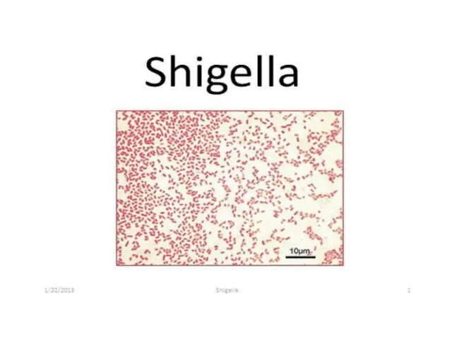
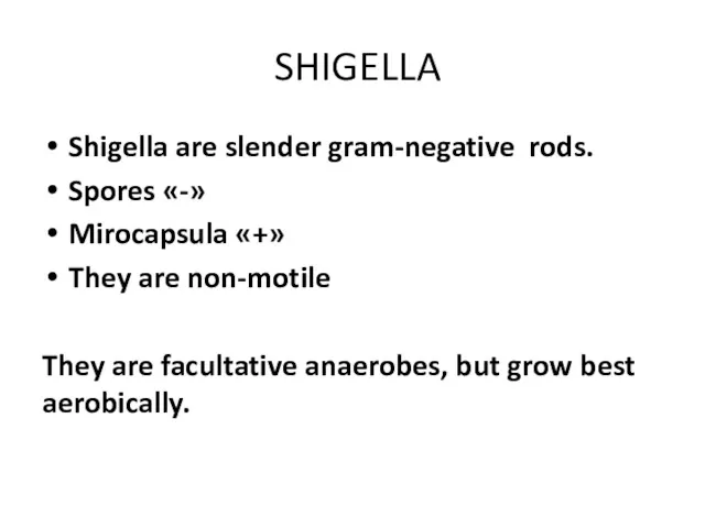
- 41. SHIGELLA Shigella are slender gram-negative rods. Spores «-» Mirocapsula «+» They are non-motile They are facultative
- 42. Ploskirev agar
- 43. Endo agar
- 44. Levine Eosin Methylene Blue agar
- 45. SHIGELLA All Shigella ferment glucose, form acid. With the exception of Shigella sonnei, they do not
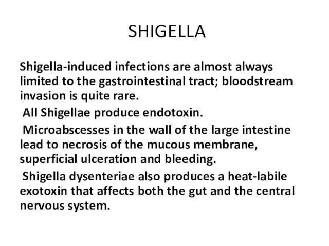
- 46. SHIGELLA Shigella-induced infections are almost always limited to the gastrointestinal tract; bloodstream invasion is quite rare.
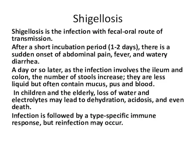
- 47. Shigellosis Shigellosis is the infection with fecal-oral route of transmission. After a short incubation period (1-2
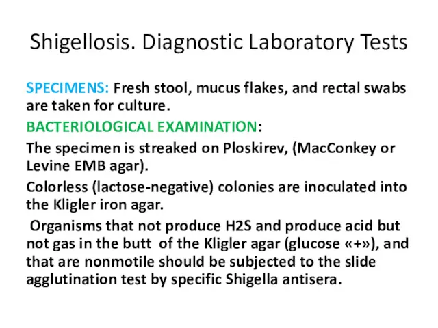
- 48. Shigellosis. Diagnostic Laboratory Tests SPECIMENS: Fresh stool, mucus flakes, and rectal swabs are taken for culture.
- 50. Скачать презентацию
FAMILY ENTEROBACTERIACEAE
are a large heterogeneous group of
Gram«-» rods whose natural
FAMILY ENTEROBACTERIACEAE
are a large heterogeneous group of
Gram«-» rods whose natural
FAMILY ENTEROBACTERIACEAE
Most of the members of Enterobacteriaceae are facultative anaerobes, ferment
FAMILY ENTEROBACTERIACEAE
Most of the members of Enterobacteriaceae are facultative anaerobes, ferment
FAMILY ENTEROBACTERIACEAE
Gram «-» rods
Spores «-»
Capsula «+» or «-»
They are motile (E.coli)
FAMILY ENTEROBACTERIACEAE
Gram «-» rods
Spores «-»
Capsula «+» or «-»
They are motile (E.coli)
FAMILY ENTEROBACTERIACEAE
FAMILY ENTEROBACTERIACEAE
FAMILY ENTEROBACTERIACEAE
The differential culture media that contain carbohydrates and special dyes
FAMILY ENTEROBACTERIACEAE
The differential culture media that contain carbohydrates and special dyes
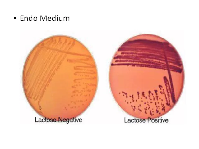
Endo Medium
Endo Medium
Kligler Iron Agar
Kligler Iron Agar
Kligler Iron Agar
Kligler iron agar is used for the differentiation of
Kligler Iron Agar
Kligler iron agar is used for the differentiation of
Kligler Iron Agar
Kligler Iron Agar
Kligler Iron Agar
The lactose-positive and glucose-positive bacteria show both the slant
Kligler Iron Agar
The lactose-positive and glucose-positive bacteria show both the slant
FAMILY ENTEROBACTERIACEAE
Enterobacteriaceae have a complex antigenic structure.
They are classified by
FAMILY ENTEROBACTERIACEAE
Enterobacteriaceae have a complex antigenic structure.
They are classified by
FAMILY ENTEROBACTERIACEAE
Virulence factors:
Fimbriae
Enterotoxins
Hemolysins
Endotoxins
FAMILY ENTEROBACTERIACEAE
Virulence factors:
Fimbriae
Enterotoxins
Hemolysins
Endotoxins
FAMILY ENTEROBACTERIACEAE
Epidemiology:
They are pathogenic for human and animals.
They are transmitted by
FAMILY ENTEROBACTERIACEAE
Epidemiology:
They are pathogenic for human and animals.
They are transmitted by
FAMILY ENTEROBACTERIACEAE
Microbiological diagnosis.
Specimens : feces, vomit, food,urine, blood.
Methods: bacteriological,
FAMILY ENTEROBACTERIACEAE
Microbiological diagnosis.
Specimens : feces, vomit, food,urine, blood.
Methods: bacteriological,
Salmonellae
Salmonella
Family – Enterobacteriaceae
Genus – Salmonellae
Species - S. enterica
Subspecies – S.
Salmonellae
Salmonella
Family – Enterobacteriaceae
Genus – Salmonellae
Species - S. enterica
Subspecies – S.
Salmonella
Salmonella
Salmonella
Morphology:
Gram «-» rods
Spores «-» Capsula «-» They are motile
Cultural
Salmonella
Morphology:
Gram «-» rods
Spores «-» Capsula «-» They are motile
Cultural

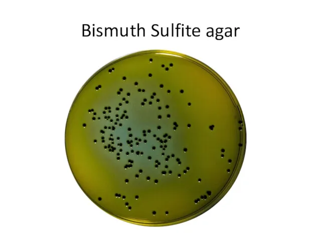
Bismuth Sulfite agar
Bismuth Sulfite agar
Salmonella
Biochemical activity:
Glucose «+», Maltose «+», Mannitol «+» (acid)
S.paratyphi ferments
Salmonella
Biochemical activity:
Glucose «+», Maltose «+», Mannitol «+» (acid)
S.paratyphi ferments
Salmonella
Antigenic structure:
O-somatic (serogroups), is destroyed by formalin.
H – flagellar (serovars) ,
Salmonella
Antigenic structure:
O-somatic (serogroups), is destroyed by formalin.
H – flagellar (serovars) ,
Salmonella. Antigenic structure
Based on the presence of O-antigens, the Salmonella have
Salmonella. Antigenic structure
Based on the presence of O-antigens, the Salmonella have
Salmonella. Antigenic structure
The H-antigens are designated by small roman letters and
Salmonella. Antigenic structure
The H-antigens are designated by small roman letters and
Salmonella
Resistance:
Survive in ice for several months.
Survive in butter, meat, cheese, bread
Salmonella
Resistance:
Survive in ice for several months.
Survive in butter, meat, cheese, bread
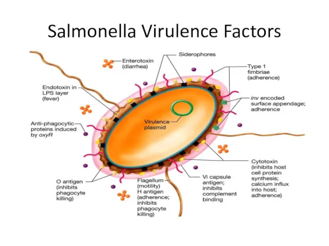
Salmonella Virulence Factors
Salmonella Virulence Factors
Salmonella Virulence Factors
The type III protein secretion system (T3SS) encoded by Salmonella
Salmonella Virulence Factors
The type III protein secretion system (T3SS) encoded by Salmonella
Salmonella
Salmonella are often pathogenic for humans or animals when acquired by
Salmonella
Salmonella are often pathogenic for humans or animals when acquired by
Pathogenesis
The ingested salmonellae reach the small intestine, from which they enter
Pathogenesis
The ingested salmonellae reach the small intestine, from which they enter
Pathogenesis of enteric fever
1st stage – the ingestion
2nd stage – the
Pathogenesis of enteric fever
1st stage – the ingestion
2nd stage – the
Laboratory Diagnosis
Blood cultures are positive only in the first week of
Laboratory Diagnosis
Blood cultures are positive only in the first week of
Salmonella Infections. Treatment .
Antimicrobial treatment.
Replacement of fluids and electrolytes are essential.
Salmonella Infections. Treatment .
Antimicrobial treatment.
Replacement of fluids and electrolytes are essential.
SHIGELLA
The causative agent of dysentery was described in 1888
SHIGELLA
The causative agent of dysentery was described in 1888
SHIGELLA
Shigellosis (or bacillary dysentery) is a clinical condition characterized
SHIGELLA
Shigellosis (or bacillary dysentery) is a clinical condition characterized
Taxonomy
Family – Enterobacteriaceae
Genus – Shigella
Species - S. dysenteriae, S. flexneri, S.
Taxonomy
Family – Enterobacteriaceae
Genus – Shigella
Species - S. dysenteriae, S. flexneri, S.
SHIGELLA
Shigella are slender gram-negative rods.
Spores «-»
Mirocapsula «+»
They are
SHIGELLA
Shigella are slender gram-negative rods.
Spores «-»
Mirocapsula «+»
They are
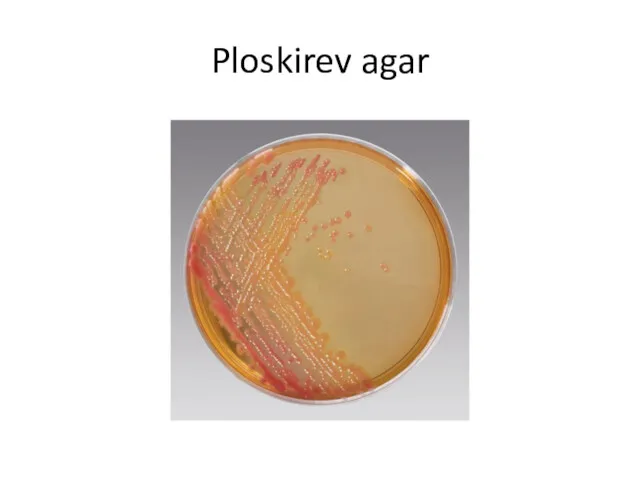
Ploskirev agar
Ploskirev agar
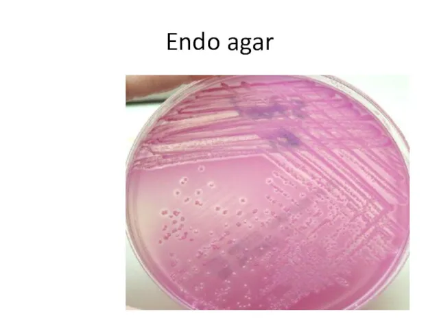
Endo agar
Endo agar
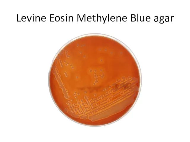
Levine Eosin Methylene Blue agar
Levine Eosin Methylene Blue agar
SHIGELLA
All Shigella ferment glucose, form acid.
With the exception of
SHIGELLA
All Shigella ferment glucose, form acid.
With the exception of
SHIGELLA
Shigella-induced infections are almost always limited to the gastrointestinal tract;
SHIGELLA
Shigella-induced infections are almost always limited to the gastrointestinal tract;
Shigellosis
Shigellosis is the infection with fecal-oral route of transmission.
After a short
Shigellosis
Shigellosis is the infection with fecal-oral route of transmission.
After a short
Shigellosis. Diagnostic Laboratory Tests
SPECIMENS: Fresh stool, mucus flakes, and rectal swabs
Shigellosis. Diagnostic Laboratory Tests
SPECIMENS: Fresh stool, mucus flakes, and rectal swabs
 Загадочный мир океана
 Что такое биоинформатика? Банк SwissProt
 Продление рода. Органы размножения
 Популяционные волны
 Органы чувств. Анализаторы
 5 Natural Ways To Get Rid of House Crickets
 Самые редкие и необычные породы кошек
 Интересные факты о животном мире
 Всероссийская проверочная работа по окружающему миру. Опыты (4 класс)
 Санитарлық-көрсеткіш микроағзалар. Стерилдеу мен залалсыздандыру
 Отряд Непарнокопытные
 4 стихии зарождения жизни
 Методическая разработка урока-игры по биологии в 10 классе по теме: Путешествие по эукариотической клетке
 Растения и животные леса
 Микробиота тела человека. Роль микроорганизмов в возникновении инфекций. Способы передачи инфекций
 α-Aminoacids, peptides, proteins
 Многообразие органического мира. Принципы систематики
 Животные живого уголка
 Профилактика вредных привычек.Давайте жить! Давайте жизнью дорожить!
 Что такое метаболизм и как его измерить
 оплодотворение
 Pielea, organ tactil, termic, dureros și de presiune. (Lectie 11)
 Древнейшие люди
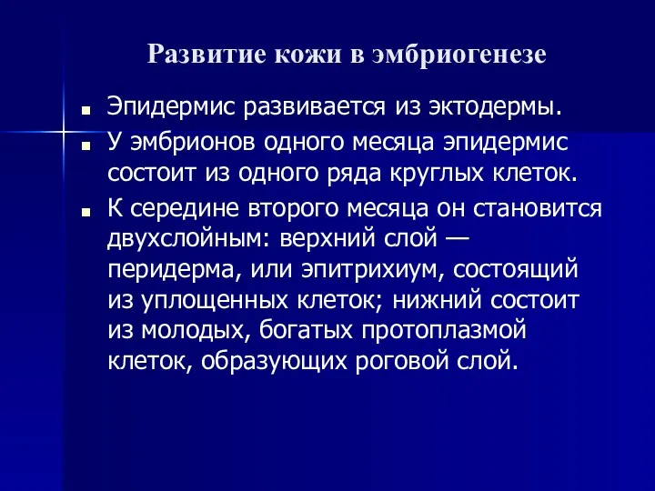 Развитие кожи в эмбриогенезе
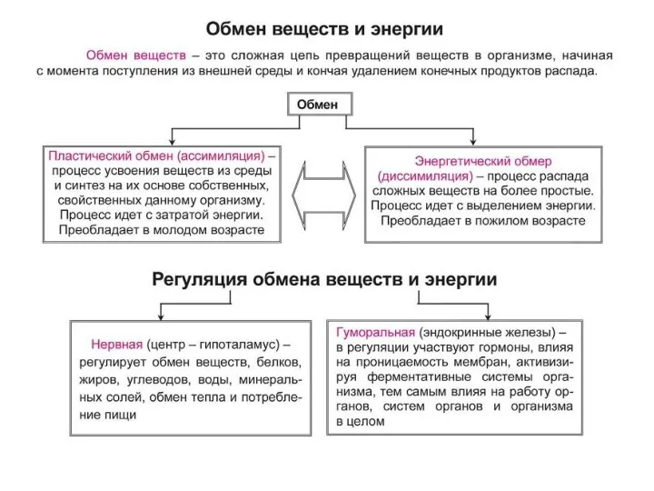 Обмен веществ и энергии. Фотосинтез
 Тип Губки
 Птичий переполох (игра)
 Самые опасные насекомые в мире