- Enterobacteriaceae

Содержание
- 2. The species are subdivided into epidemiologically significant serovars based on O, H, and K antigens. The
- 3. Together with the families Vibrionaceae and others, the Enterobacteriaceae form the group of Gram-negative, facultatively anaerobic
- 4. The taxonomy of the Enterobacteriaceae has seen repeated changes in recent decades and has doubtless not
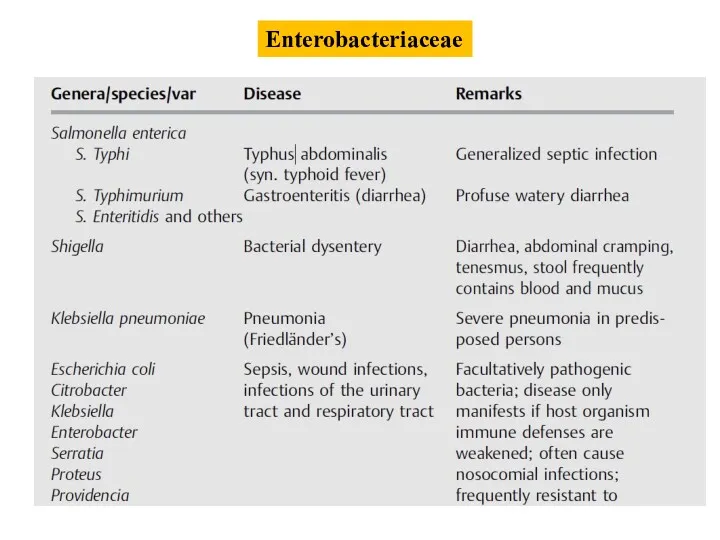
- 5. Enterobacteriaceae
- 7. Salmonella (Gastroenteritis, Typhoid Fever, Paratyphoid Fever) All salmonellae are classified in the species Salmonella enterica with
- 8. Typhoid salmonelloses are caused by the serovars typhi and paratyphi A, B, and C. The salmonellae
- 9. Enteric salmonelloses develop when pathogens are taken up with food. The primary infection source is usually
- 10. Excerpt from the Kauffmann–White Scheme which Covers Over 2000 Serovars
- 12. Overview of the Most Important Differences between Typhoid and Enteric Salmonellae and Salmonelloses
- 14. The cases of typhoid salmonelloses seen in northern and central Europe are imported by travelers. Cases
- 15. Shigella (Bacterial Dysentery) Shigella is the causative pathogen in bacterial dysentery. The genus comprises the species
- 16. Shigellae are only pathogenic in humans. The pathogens are ingested orally. Only a few hundred bacteria
- 17. The invasion is facilitated by outer membrane polypeptides, the invasins, which are coded by inv genes
- 18. Anti-infective agents are the first line of treatment (aminopenicillins, 4-quinolones, cephalosporins). Losses of water and electrolytes
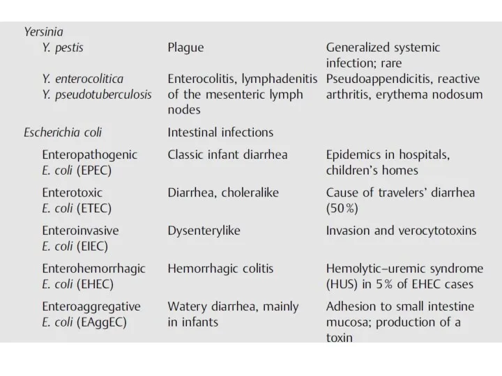
- 19. Yersinia (Plague, Enteritis) Y. pestis is the causative pathogen of plague (black death, bubonic plague). Plague
- 20. Yersinia pestis Y. pestis is a nonflagellated, short, encapsulated, Gram-negative rod bacteria that often shows bipolar
- 21. Diagnosis. The pathogen must be identified in bubo punctate, sputum, or blood by means of microscopy
- 22. Yersinia enterocolitica and Yersinia pseudotuberculosis Y. enterocolitica and Y. pseudotuberculosis cause generalized infections in domestic and
- 23. Escherichia coli The natural habitat of E. coli is the intestinal tract of humans and animals.
- 24. General characteristics The natural habitat of E. coli is the intestines of animals and humans. This
- 25. The Gram-negative, straight rods are peritrichously flagellated. Lactose is broken down rapidly. The complex antigen structure
- 26. Vibrio cholerae (Cholera) Morphology and culture. Cholera vibrios are Gram-negative rod bacteria, usually slightly bent (comma-shaped),
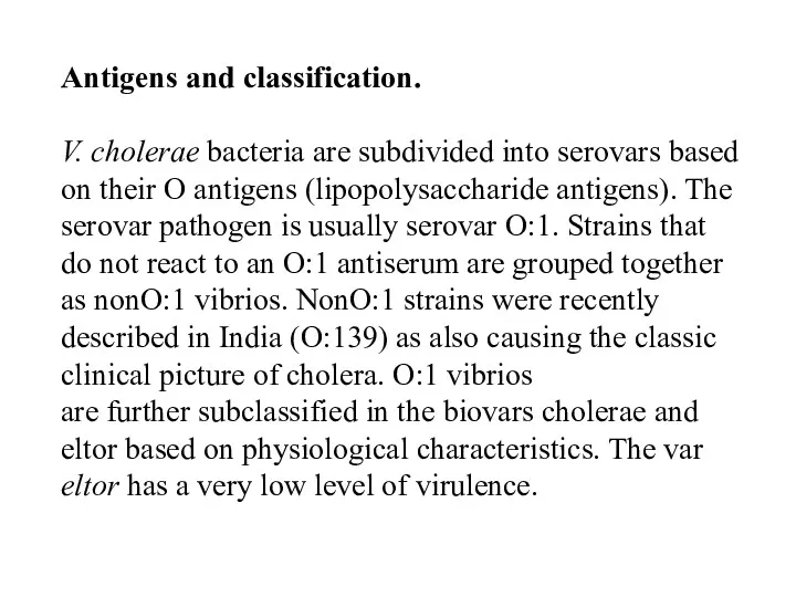
- 27. Antigens and classification. V. cholerae bacteria are subdivided into serovars based on their O antigens (lipopolysaccharide
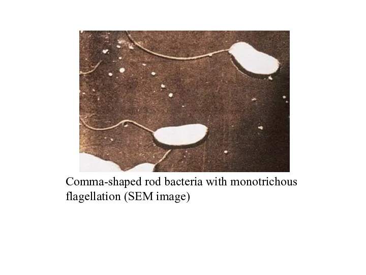
- 28. Comma-shaped rod bacteria with monotrichous flagellation (SEM image)
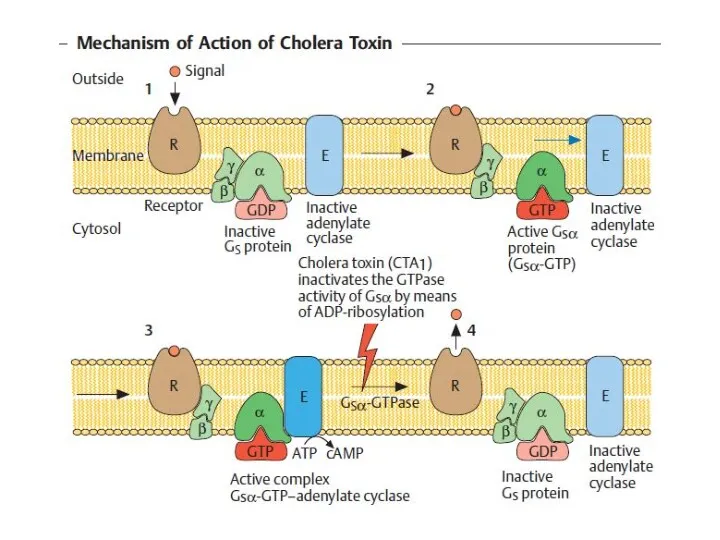
- 30. Pathogenesis and clinical picture Infection results from oral ingestion of the pathogen. The infective dose must
- 31. Diagnosis requires identification of the pathogen in stool or vomit. Sometimes a rapid microscopical diagnosis succeeds
- 32. The most important measure is restoration of the disturbed waternand electrolyte balance in the body. Secondly,
- 33. Epidemiology and prevention Nineteenth-century Europe experienced several cholera pandemics, all of which were caused by the
- 34. Humans are the only source of infection. Infected persons in particular eliminate large numbers of pathogens.
- 35. Protection from exposure to the pathogen is the main thrust of the relevant preventive measures. In
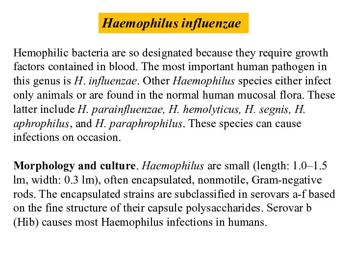
- 36. Haemophilus influenzae Hemophilic bacteria are so designated because they require growth factors contained in blood. The
- 37. a Gram-stained cerebrospinal fluid sediment preparation. Fine, Gram-negative rods surrounded by a capsule (serovar b). Clinical
- 38. H. influenzae is a mucosal parasite of the upper respiratory tract present in 30–50% of healthy
- 39. Haemophilus infections in adults are usually secondary complications of severe primary illnesses or the result of
- 40. Diagnosis The method of choice is identification of the pathogen in cerebrospinal fluid, blood, pus, or
- 41. H. influenzae is found only in humans. The incidence of severe invasive infections (meningitis, sepsis, epiglottitis)
- 42. Campylobacter, Helicobacter, and Spirillum belong to the group of spiral, motile, Gram-negative, microaerophilic bacteria. C. jejuni
- 43. Campylobacter Campylobacter (meaning "curved bacteria") is a genus of Gram-negative bacteria. Campylobacter typically appear comma or
- 44. Helicobacter pylori Morphology and culture H. pylori are spirally shaped, Gram-negative rods with lophotrichous flagellation. Cultures
- 45. H. pylori occurs only in humans and is transmitted by the fecal-oral pathway. The pathogen colonizes
- 46. Once the pathogen has infected the stomach tissues an acute gastritis results, the course of which
- 47. Diagnosis. Histopathological, cultural and, molecular identification of the bacteria in stomach lining biopsies. Antigen detection in
- 48. Legionella is the only genus in the family Legionellaceae. The species Legionella pneumophila is responsible for
- 49. Legionella bacteria were discovered in 1976, occasioned by an epidemic among those attending a conference of
- 50. The persons most likely to contract legionnaire’s disease are those with a primary cardiopulmonary disease and
- 51. Morphology and culture L. pneumophila is a rod bacterium 0.3–1 lm wide and 2–20 lm long.
- 52. Pathogenesis and clinical picture The pathomechanisms employed by legionellae are not yet fully clarified. These organisms
- 53. Diagnosis. Specific antibodies marked with fluorescein are used to detect the pathogens in material from the
- 54. Treponema pallidum Borrelia (Relapsing Fever, Lyme Disease) Leptospira (Leptospirosis, Weil Disease) Rickettsia Chlamydia Mycoplasma
- 56. Скачать презентацию
The species are subdivided into epidemiologically significant serovars based on O,
The species are subdivided into epidemiologically significant serovars based on O,
Together with the families Vibrionaceae and others, the Enterobacteriaceae form the
Together with the families Vibrionaceae and others, the Enterobacteriaceae form the
The taxonomy of the Enterobacteriaceae has seen repeated changes in recent
The taxonomy of the Enterobacteriaceae has seen repeated changes in recent
Enterobacteriaceae
Enterobacteriaceae
Salmonella (Gastroenteritis, Typhoid Fever, Paratyphoid Fever)
All salmonellae are classified in the
Salmonella (Gastroenteritis, Typhoid Fever, Paratyphoid Fever)
All salmonellae are classified in the
Typhoid salmonelloses are caused by the serovars typhi and paratyphi A,
Typhoid salmonelloses are caused by the serovars typhi and paratyphi A,
Enteric salmonelloses develop when pathogens are taken up with food. The
Enteric salmonelloses develop when pathogens are taken up with food. The
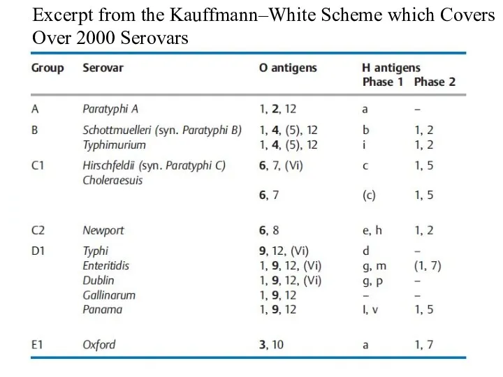
Excerpt from the Kauffmann–White Scheme which Covers Over 2000 Serovars
Excerpt from the Kauffmann–White Scheme which Covers Over 2000 Serovars
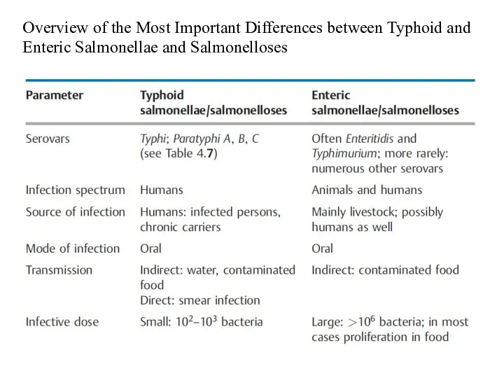
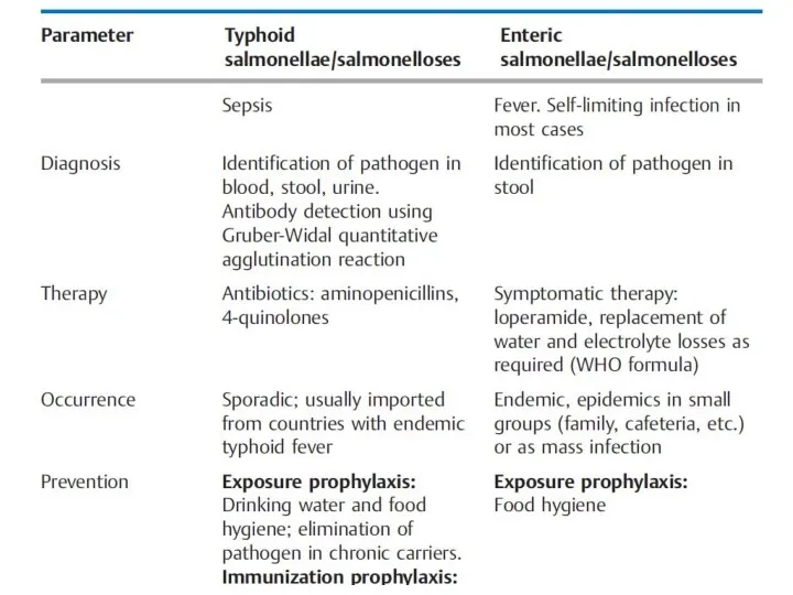
Overview of the Most Important Differences between Typhoid and
Enteric Salmonellae and
Overview of the Most Important Differences between Typhoid and
Enteric Salmonellae and
The cases of typhoid salmonelloses seen in northern and central
Europe are
The cases of typhoid salmonelloses seen in northern and central
Europe are
Shigella (Bacterial Dysentery)
Shigella is the causative pathogen in bacterial dysentery. The
Shigella (Bacterial Dysentery)
Shigella is the causative pathogen in bacterial dysentery. The
Shigellae are only pathogenic in humans. The pathogens are ingested orally.
Shigellae are only pathogenic in humans. The pathogens are ingested orally.
The invasion is facilitated by outer membrane polypeptides, the invasins, which
The invasion is facilitated by outer membrane polypeptides, the invasins, which
Anti-infective agents are the first line of treatment (aminopenicillins, 4-quinolones, cephalosporins).
Anti-infective agents are the first line of treatment (aminopenicillins, 4-quinolones, cephalosporins).
Yersinia (Plague, Enteritis)
Y. pestis is the causative pathogen of plague (black
Yersinia (Plague, Enteritis)
Y. pestis is the causative pathogen of plague (black
Yersinia pestis
Y. pestis is a nonflagellated, short, encapsulated, Gram-negative rod bacteria
Yersinia pestis
Y. pestis is a nonflagellated, short, encapsulated, Gram-negative rod bacteria
Diagnosis. The pathogen must be identified in bubo punctate, sputum, or
Diagnosis. The pathogen must be identified in bubo punctate, sputum, or
Yersinia enterocolitica and Yersinia pseudotuberculosis
Y. enterocolitica and Y. pseudotuberculosis cause
generalized infections
Yersinia enterocolitica and Yersinia pseudotuberculosis
Y. enterocolitica and Y. pseudotuberculosis cause
generalized infections
Escherichia coli
The natural habitat of E. coli is the intestinal tract
Escherichia coli
The natural habitat of E. coli is the intestinal tract
General characteristics
The natural habitat of E. coli is the intestines of
General characteristics
The natural habitat of E. coli is the intestines of
The Gram-negative, straight rods are peritrichously flagellated. Lactose is broken down
The Gram-negative, straight rods are peritrichously flagellated. Lactose is broken down
Vibrio cholerae (Cholera)
Morphology and culture. Cholera vibrios are Gram-negative rod bacteria,
Vibrio cholerae (Cholera)
Morphology and culture. Cholera vibrios are Gram-negative rod bacteria,
Antigens and classification.
V. cholerae bacteria are subdivided into serovars based
Antigens and classification.
V. cholerae bacteria are subdivided into serovars based
Comma-shaped rod bacteria with monotrichous flagellation (SEM image)
Comma-shaped rod bacteria with monotrichous flagellation (SEM image)
Pathogenesis and clinical picture
Infection results from oral ingestion of the pathogen.
Pathogenesis and clinical picture
Infection results from oral ingestion of the pathogen.
Diagnosis requires identification of the pathogen in stool or vomit. Sometimes
Diagnosis requires identification of the pathogen in stool or vomit. Sometimes
The most important measure is restoration of the disturbed waternand electrolyte
The most important measure is restoration of the disturbed waternand electrolyte
Epidemiology and prevention
Nineteenth-century Europe experienced several cholera pandemics, all of which
Epidemiology and prevention
Nineteenth-century Europe experienced several cholera pandemics, all of which
Humans are the only source of infection. Infected persons in particular
Protection from exposure to the pathogen is the main thrust of
Protection from exposure to the pathogen is the main thrust of
Haemophilus influenzae
Hemophilic bacteria are so designated because they require growth factors
Haemophilus influenzae
Hemophilic bacteria are so designated because they require growth factors
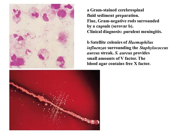
a Gram-stained cerebrospinal
fluid sediment preparation.
Fine, Gram-negative rods surrounded
by a capsule (serovar
a Gram-stained cerebrospinal
fluid sediment preparation.
Fine, Gram-negative rods surrounded
by a capsule (serovar
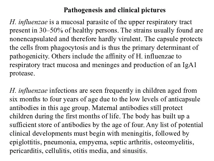
H. influenzae is a mucosal parasite of the upper respiratory tract
H. influenzae is a mucosal parasite of the upper respiratory tract
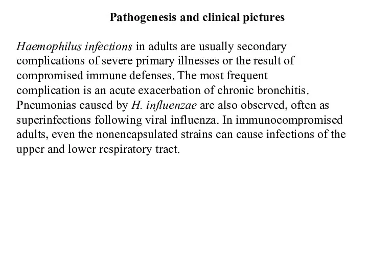
Haemophilus infections in adults are usually secondary complications of severe primary
Haemophilus infections in adults are usually secondary complications of severe primary
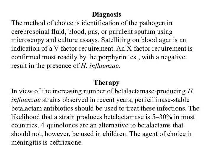
Diagnosis
The method of choice is identification of the pathogen in cerebrospinal
Diagnosis
The method of choice is identification of the pathogen in cerebrospinal
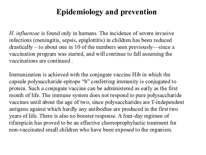
H. influenzae is found only in humans. The incidence of severe
H. influenzae is found only in humans. The incidence of severe
Campylobacter, Helicobacter, and Spirillum belong to the group of spiral, motile,
Campylobacter, Helicobacter, and Spirillum belong to the group of spiral, motile,
Campylobacter
Campylobacter (meaning "curved bacteria") is a genus of Gram-negative bacteria. Campylobacter typically appear comma or s-shaped and motile.
Campylobacter
Campylobacter (meaning "curved bacteria") is a genus of Gram-negative bacteria. Campylobacter typically appear comma or s-shaped and motile.
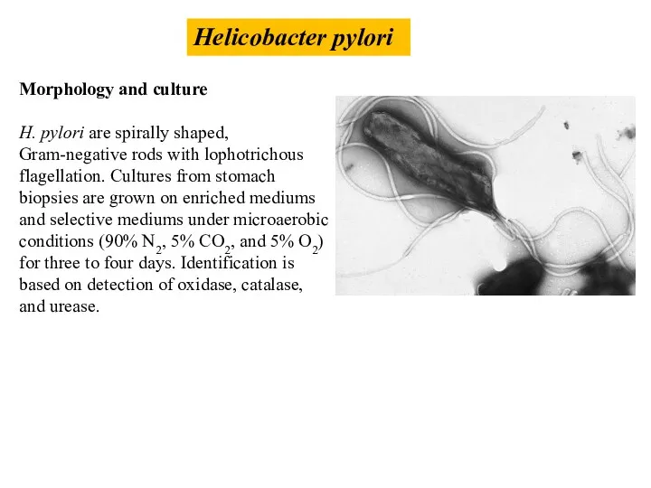
Helicobacter pylori
Morphology and culture
H. pylori are spirally shaped, Gram-negative rods with
Helicobacter pylori
Morphology and culture
H. pylori are spirally shaped, Gram-negative rods with
H. pylori occurs only in humans and is transmitted by the
H. pylori occurs only in humans and is transmitted by the
Once the pathogen has infected the stomach tissues an acute gastritis
Once the pathogen has infected the stomach tissues an acute gastritis
Diagnosis. Histopathological, cultural and, molecular identification of the bacteria in stomach
Diagnosis. Histopathological, cultural and, molecular identification of the bacteria in stomach
Legionella is the only genus in the family Legionellaceae. The species
Legionella is the only genus in the family Legionellaceae. The species
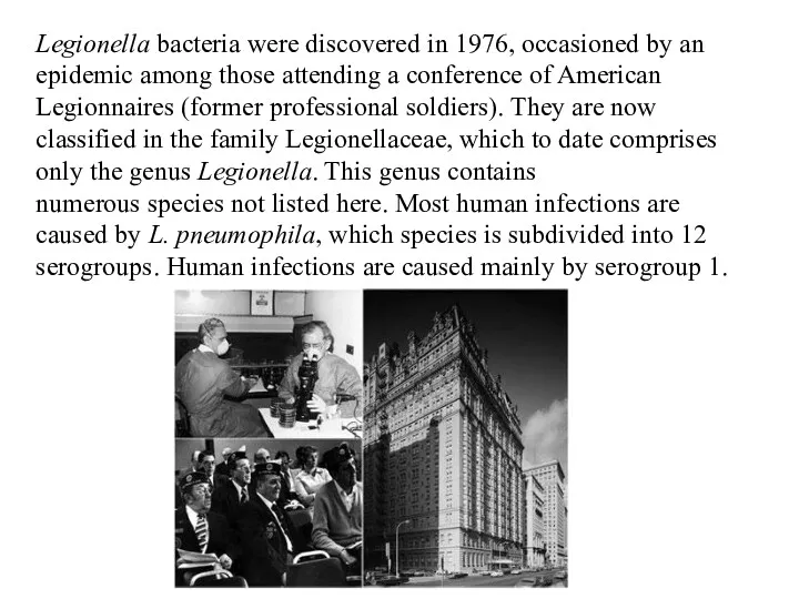
Legionella bacteria were discovered in 1976, occasioned by an
epidemic among those
Legionella bacteria were discovered in 1976, occasioned by an
epidemic among those
The persons most likely to contract legionnaire’s disease are those with
The persons most likely to contract legionnaire’s disease are those with
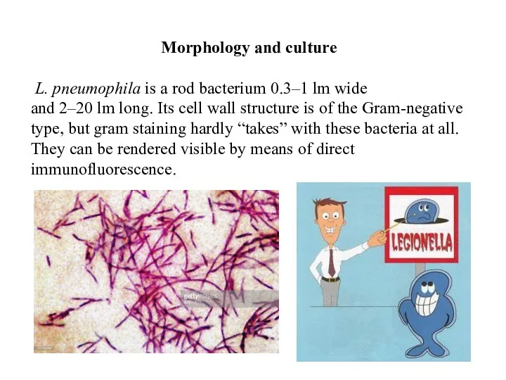
Morphology and culture
L. pneumophila is a rod bacterium 0.3–1 lm
Morphology and culture
L. pneumophila is a rod bacterium 0.3–1 lm
Pathogenesis and clinical picture
The pathomechanisms employed by legionellae are not yet
Pathogenesis and clinical picture
The pathomechanisms employed by legionellae are not yet
Diagnosis. Specific antibodies marked with fluorescein are used to detect the
pathogens
Diagnosis. Specific antibodies marked with fluorescein are used to detect the
pathogens
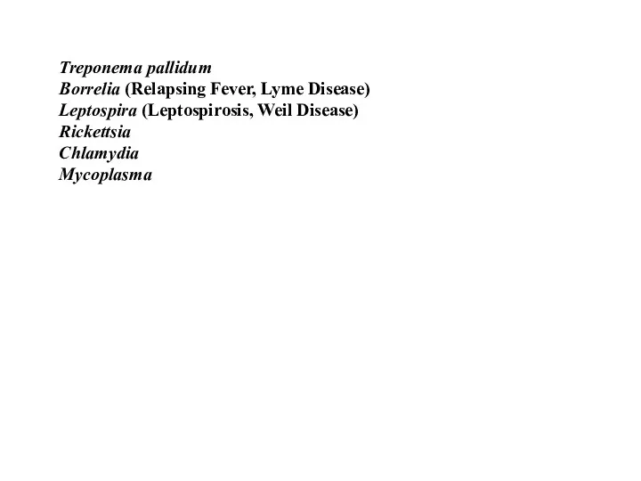
Treponema pallidum
Borrelia (Relapsing Fever, Lyme Disease)
Leptospira (Leptospirosis, Weil Disease)
Rickettsia
Chlamydia
Mycoplasma
Treponema pallidum
Borrelia (Relapsing Fever, Lyme Disease)
Leptospira (Leptospirosis, Weil Disease)
Rickettsia
Chlamydia
Mycoplasma
 Презентация к уроку 5 класса ФГОС
 Жасуша мембранасының биофизикасы
 Фізіологія скелетних та гладких м‘язів
 Вторичный метаболизм растений
 Биологиялық мембраналардың өткізгіштік механизмі. Иондық каналдардың және тасымалдаушылардың құрылысы мен функциясы
 Физиология ретикулярной формации, промежуточного и заднего мозга. Лекция 9
 Як спілкуються тварини. 7 клас
 Приспособленность организмов к определенной среде обитания
 Разнообразие ракообразных и их роли в природе
 Систематические группы надкласса рыб
 сердце
 Методическая разработка раздела программы по темеОпора и движение(8 класс)
 Иммунная система человека. Органы кроветворения
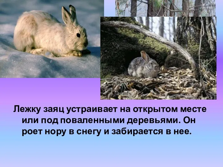 Заяц. Враги зайца
 Птицы синицы. Своя игра
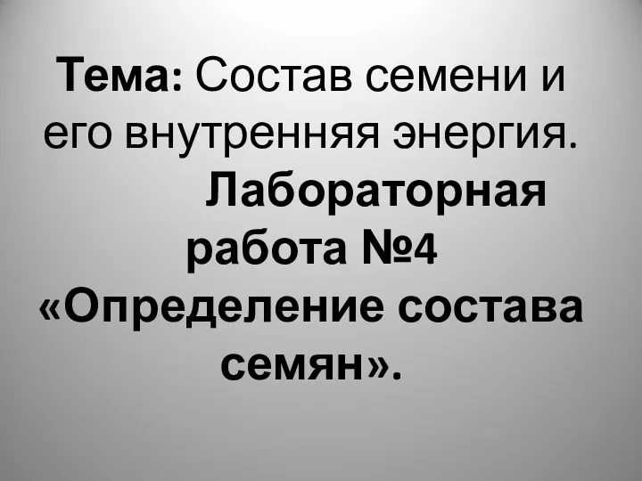 Состав семени и его внутренняя энергия. Лабораторная работа №4 Определение состава семян
 Строение цветка. Соцветие. Значение оплодотворения
 Animal life of the UK
 Своя игра про животных
 Углеводы. 9 класс
 Этапы эволюции человека
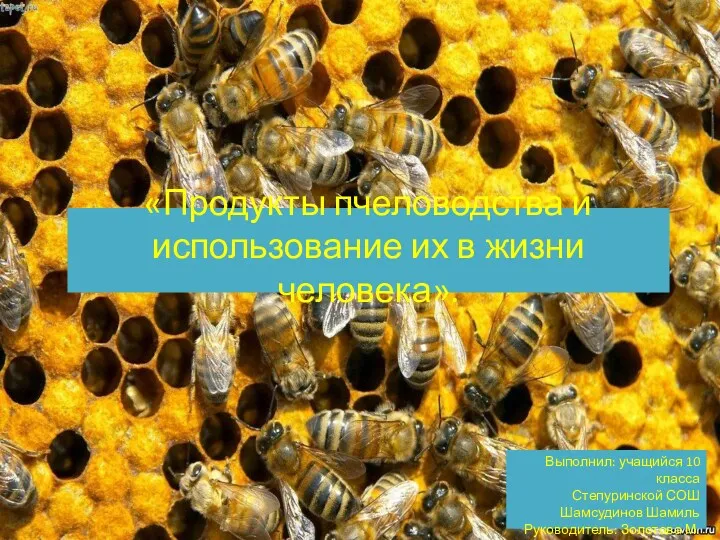 Продукты пчеловодства и использование их в жизни человека
 ПРЕЗЕНТАЦИИ 5 КЛАСС 1 ЧЕТВЕРТЬ
 Cattle breeds
 Приспособленность – результат действия факторов эволюции
 Разработка презентации по биологии по теме Размножение живых организмов.
 Птичьи посиделки
 Метод классификации организмов, применение двойных названий организмов (5 класс)