- Morphology & structure of microorganisms

Содержание
- 2. Morphology & structure of Microorganisms Lecture 1
- 3. Microbiology Microbiology is the study of organisms that too small to be seen without magnification. Microbiology
- 4. History of Microbiology 1. Robert Koch was notable for his discovery of the bacterium Bacillus anthracis
- 5. 2. Robert Hooke: Known for his discovery of the first ever compound microscope, Robert Hooke is
- 6. Microorganisms Are Ubiquitous Microorganisms include: Bacteria Viruses Fungi Parasites (protozoa & helminthes) Algae
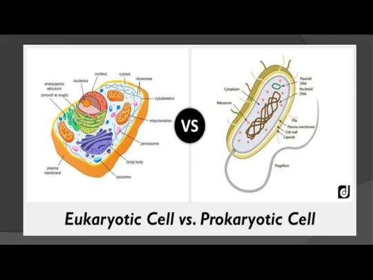
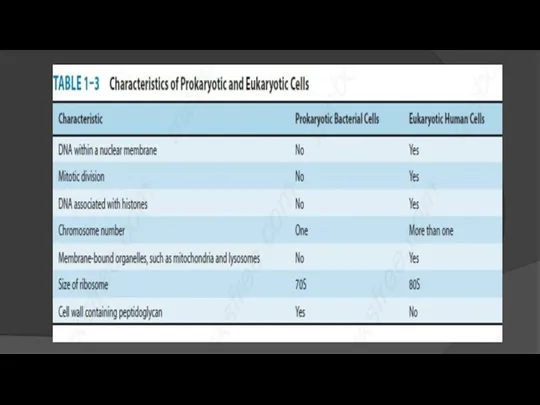
- 7. Introduction There are several classes of living organisms. Based on the organization of their cellular structures,
- 10. Prokaryotic Cells Much smaller (microns) and more simple than eukaryotes. Prokaryotes are molecules surrounding by a
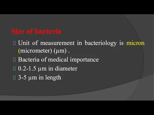
- 12. Size of bacteria Unit of measurement in bacteriology is micron (micrometer) (µm) . Bacteria of medical
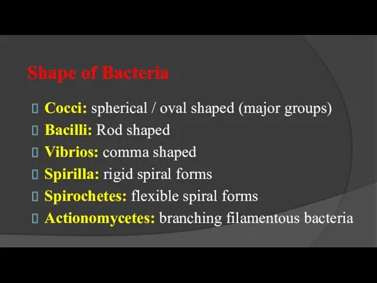
- 13. Shape of Bacteria Cocci: spherical / oval shaped (major groups) Bacilli: Rod shaped Vibrios: comma shaped
- 14. Shape of Bacteria
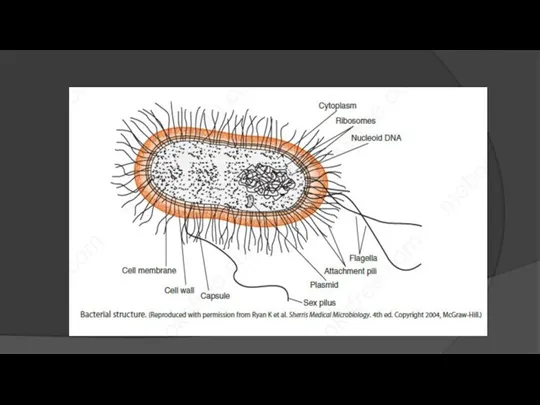
- 15. Outer layer: consist of two components 1- Rigid cell wall. 2-Cytoplasmic (cell or plasma membrane) present
- 16. Structure & Function of Cell Components
- 17. 1- Cell wall: Outermost layer, encloses cytoplasmic membrane. 1- Confers shape and rigidity. 2- Peptidoglycans is
- 18. 4- Cell wall cannot be seen by direct light microscope and do not stain with simple
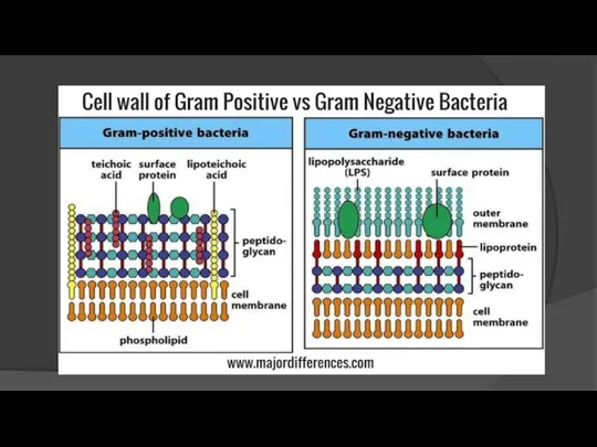
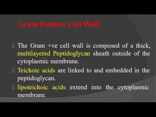
- 20. Gram Positive Cell Wall The Gram +ve cell wall is composed of a thick, multilayered Peptidoglycan
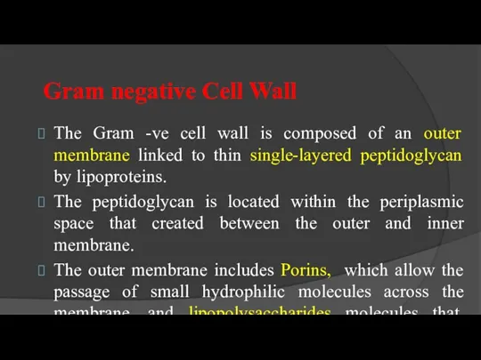
- 21. Gram negative Cell Wall The Gram -ve cell wall is composed of an outer membrane linked
- 22. Comparison
- 23. 2- Cytoplasmic (Plasma) Membrane Thin layer 5-10 nm, separates cell wall from cytoplasm Acts as a
- 24. 3- Cytoplasm Colloidal system of variety of organic & inorganic solutes in viscous watery solution. -
- 25. 2- Mesosomes 1- Multi-laminated structures formed as invaginations of plasma membrane. 2- Principle sites of respiratory
- 26. 4- Nucleus No nucleolus No nuclear membrane Oval or elongated bodies generally 1 per cell Genome-
- 27. Additional Organelles 1-Plasmid: Extra-nuclear genetic elements consisting of DNA. Transmitted to daughter cells during binary fission
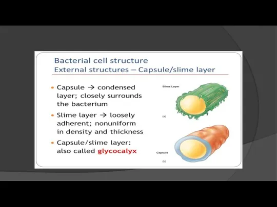
- 28. 2-Capsule & Slime Layer Viscous layer secreted around the cell wall. Polysaccharide or polypeptide in nature..
- 29. B- Slime layer: Is loosely associated with bacterium and can be easily washed off, whereas a
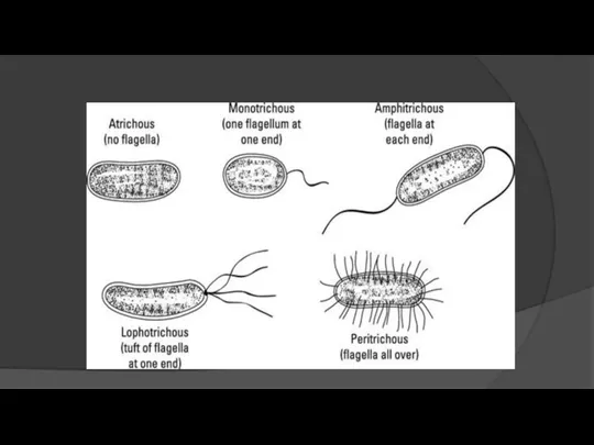
- 31. 3- Flagella Long (3-12µm) filamentous surface appendages. Organs of locomotion. Composed of protein called flagellins The
- 32. Presence shown by motility e.g. hanging drop preparation test. Each flagellum consist of 3 part: 1-
- 34. 4- Fimbriae (Pili) Thin, hair like appendages on the surface of many Gram –ve bacteria 10-20µ
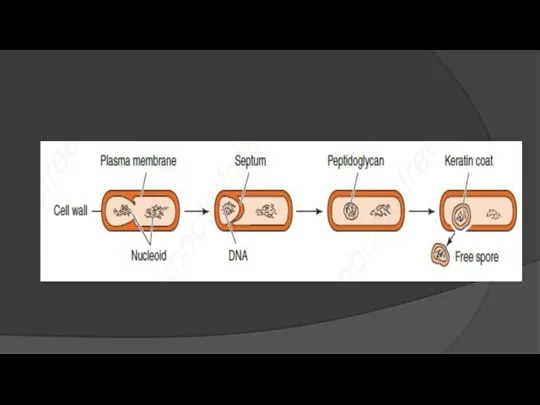
- 35. 5- Spores Highly resistant resting stages formed during adverse environmental (depletion of nutrients). Formed inside the
- 37. Cell Division : most bacteria divide by binary fission into two equal cells. In a growing
- 38. Growth is the orderly increase in the sum of all the components of an organism. The
- 40. Скачать презентацию
Morphology & structure of Microorganisms
Lecture 1
Morphology & structure of Microorganisms
Lecture 1
Microbiology
Microbiology is the study of organisms that too small to be
Microbiology
Microbiology is the study of organisms that too small to be
History of Microbiology
1. Robert Koch was notable for his discovery of
History of Microbiology
1. Robert Koch was notable for his discovery of
2. Robert Hooke: Known for his discovery of the first ever
2. Robert Hooke: Known for his discovery of the first ever
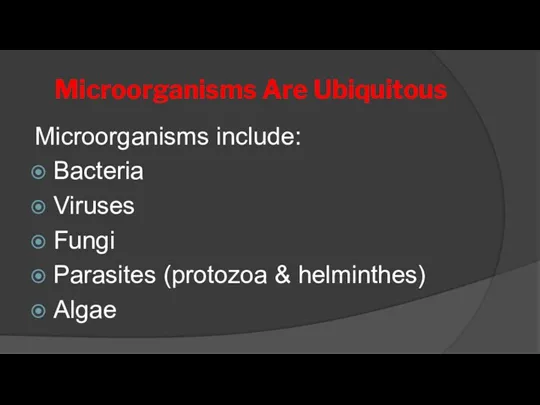
Microorganisms Are Ubiquitous
Microorganisms include:
Bacteria
Viruses
Fungi
Parasites (protozoa & helminthes)
Algae
Microorganisms Are Ubiquitous
Microorganisms include:
Bacteria
Viruses
Fungi
Parasites (protozoa & helminthes)
Algae
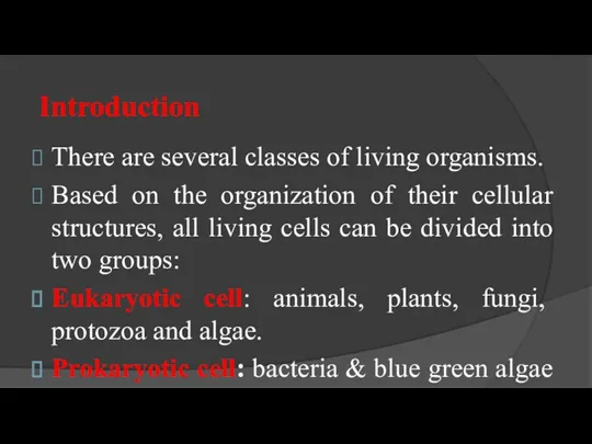
Introduction
There are several classes of living organisms.
Based on the organization
Introduction
There are several classes of living organisms.
Based on the organization
Prokaryotic Cells
Much smaller (microns) and more simple than eukaryotes.
Prokaryotes are molecules
Prokaryotic Cells
Much smaller (microns) and more simple than eukaryotes.
Prokaryotes are molecules
Size of bacteria
Unit of measurement in bacteriology is micron (micrometer) (µm)
Size of bacteria
Unit of measurement in bacteriology is micron (micrometer) (µm)
Shape of Bacteria
Cocci: spherical / oval shaped (major groups)
Bacilli: Rod shaped
Vibrios:
Shape of Bacteria
Cocci: spherical / oval shaped (major groups)
Bacilli: Rod shaped
Vibrios:
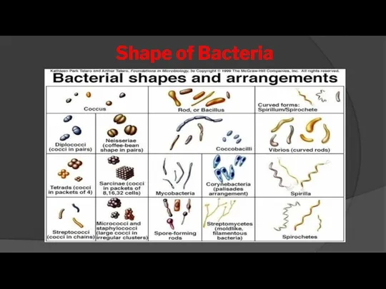
Shape of Bacteria
Shape of Bacteria
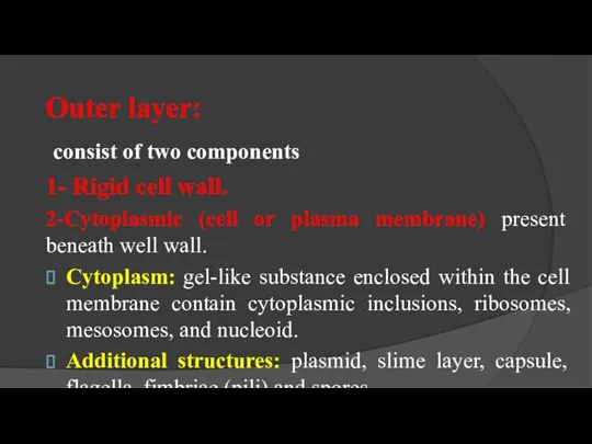
Outer layer:
consist of two components
1- Rigid cell wall.
2-Cytoplasmic (cell or
Outer layer:
consist of two components
1- Rigid cell wall.
2-Cytoplasmic (cell or
Structure & Function
of Cell Components
Structure & Function
of Cell Components
1- Cell wall:
Outermost layer, encloses cytoplasmic membrane.
1- Confers shape and rigidity.
2-
Outermost layer, encloses cytoplasmic membrane.
1- Confers shape and rigidity.
2-
4- Cell wall cannot be seen by direct light microscope and
4- Cell wall cannot be seen by direct light microscope and
Gram Positive Cell Wall
The Gram +ve cell wall is composed of
Gram Positive Cell Wall
The Gram +ve cell wall is composed of
Gram negative Cell Wall
The Gram -ve cell wall is composed of
Gram negative Cell Wall
The Gram -ve cell wall is composed of
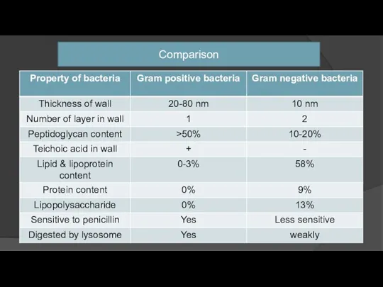
Comparison
Comparison
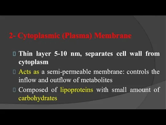
2- Cytoplasmic (Plasma) Membrane
Thin layer 5-10 nm, separates cell wall from
2- Cytoplasmic (Plasma) Membrane
Thin layer 5-10 nm, separates cell wall from
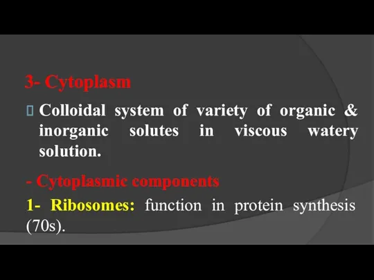
3- Cytoplasm
Colloidal system of variety of organic & inorganic solutes in
3- Cytoplasm
Colloidal system of variety of organic & inorganic solutes in
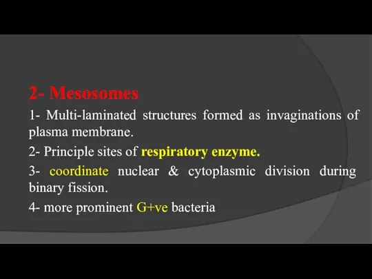
2- Mesosomes
1- Multi-laminated structures formed as invaginations of plasma membrane.
2- Principle
2- Mesosomes
1- Multi-laminated structures formed as invaginations of plasma membrane.
2- Principle
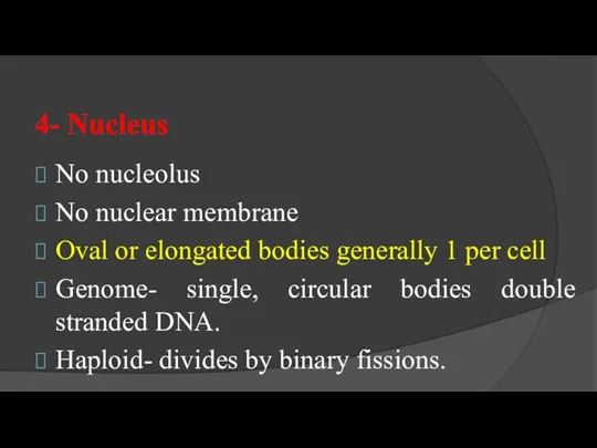
4- Nucleus
No nucleolus
No nuclear membrane
Oval or elongated bodies generally 1 per
4- Nucleus
No nucleolus
No nuclear membrane
Oval or elongated bodies generally 1 per
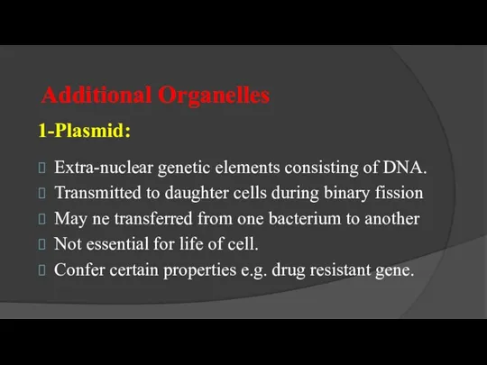
Additional Organelles
1-Plasmid:
Extra-nuclear genetic elements consisting of DNA.
Transmitted to daughter cells during
Additional Organelles
1-Plasmid:
Extra-nuclear genetic elements consisting of DNA.
Transmitted to daughter cells during
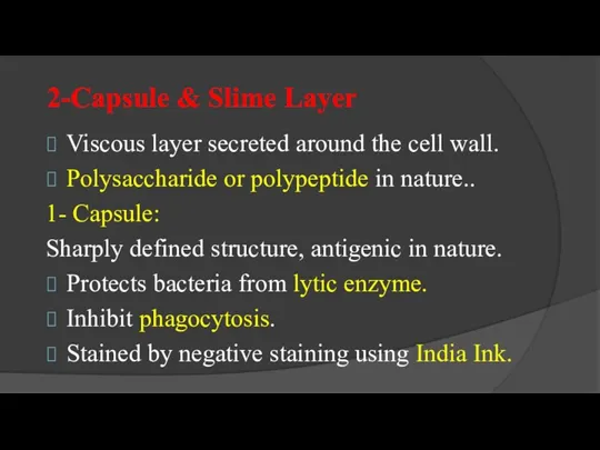
2-Capsule & Slime Layer
Viscous layer secreted around the cell wall.
Polysaccharide or
2-Capsule & Slime Layer
Viscous layer secreted around the cell wall.
Polysaccharide or
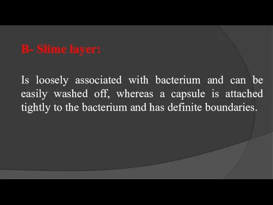
B- Slime layer:
Is loosely associated with bacterium and can be easily
B- Slime layer:
Is loosely associated with bacterium and can be easily
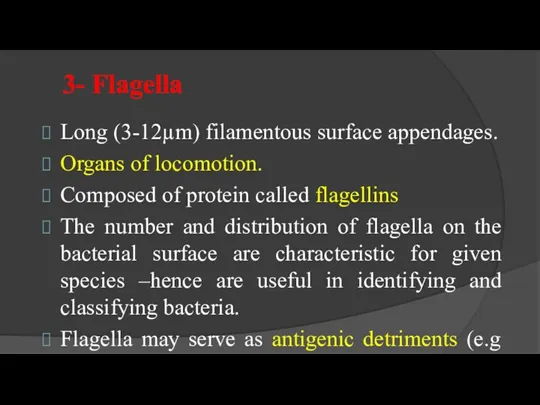
3- Flagella
Long (3-12µm) filamentous surface appendages.
Organs of locomotion.
Composed of protein called
3- Flagella
Long (3-12µm) filamentous surface appendages.
Organs of locomotion.
Composed of protein called
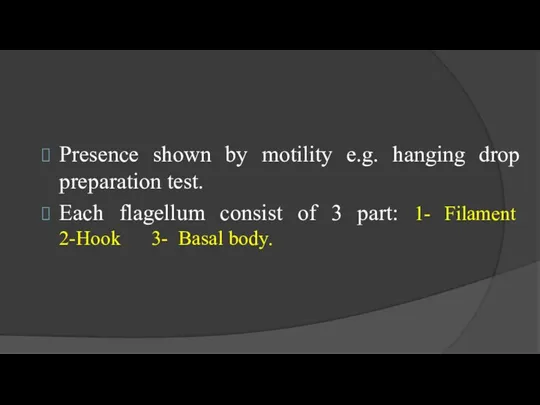
Presence shown by motility e.g. hanging drop preparation test.
Each flagellum consist
Presence shown by motility e.g. hanging drop preparation test.
Each flagellum consist
4- Fimbriae (Pili)
Thin, hair like appendages on the surface of many
4- Fimbriae (Pili)
Thin, hair like appendages on the surface of many
5- Spores
Highly resistant resting stages formed during adverse environmental (depletion of
5- Spores
Highly resistant resting stages formed during adverse environmental (depletion of
Cell Division : most bacteria divide by binary fission into two
Cell Division : most bacteria divide by binary fission into two
Growth is the orderly increase in the sum of all the
Growth is the orderly increase in the sum of all the
 Клеточное строение стебля
 Эволюция прокариот. Лекция 3
 Тесты по биологии
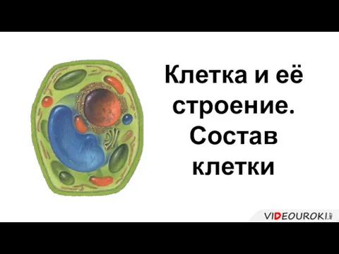 Клетка и её строение. Состав клетки
 презентация Цветок
 Мозжечок (cerebellum). Сагиттальный разрез головного мозга
 Компания Хелатные БиоТехнологии. Органический биостимулятор для открытого и защищённого грунтов
 Влияние жидких синтетических моющих средств на рост и развитие растений на примере прорастания семян фасоли посевной
 Семейство Бобовые - Fabaceae
 4-СӨЖ: Классикалық ПТР. Реал-тайм ПТР
 Урок биологии в 9 классе по теме Антропогенез
 Митоз. Апоптоз. Канцерогенез
 Отдел Лишайники. Lichenophyta. Лихенизированные грибы
 Копытные. Анатомическая особенность пальцев овец
 Деление клеток. Митоз
 Однодольные растения. Liliopsida, Monocotyledones, Monocotyledoneae
 Biochemistry of Blood
 Клеточный цикл, митоз и апоптоз
 Логические задачи по основам экологии
 Этнические процессы
 Естественный отбор
 Отличие палеонтологии от других биологических наук
 Кольчатые черви
 Презентация к уроку Тип Кольчатые черви.Класс Малощетинковые
 Қазақстанның оңтүстік және солтүстік облыс аудандарының флораларының ерекшеліктері
 Лев - король звірів
 5 весняних квіток
 Методы изучения генетики человека