- Общая гистология. Ткань

Содержание
- 2. DEVELOPMENT of TISSUES - HISTOGENESIS It is a formation of an embryonal tissue rudiment and its
- 3. ПЕРИОДЫ ДИФФЕРЕНЦИРОВКИ ЗАРОДЫШЕВОГО МАТЕРИАЛА Periods of differentiation of embryonal material Оотипическая дифференцировка в зиготе – образование
- 4. Сперматозоид акросома ядро жгутик Яйцеклетка курицы (резко телолецитальная) Гранулы желтка Ядро Яйцеклетка млекопитающих (вторично изолецитальная) Ядро
- 5. Оплодотворение и образование зиготы Fertilization and a zygote formation Сближение половых клеток Проникновение сперматозоида в цитоплазму
- 6. Зигота Zygote (couple cell) Ядро Цитоплазма с желтком Оболочка оплодотворения Оотипическая дифференцировка зиготы (перед дроблением): –
- 7. Бластомерная дифференцировка в процессе дробления Blastomere differentiation during cleavage Differences of cleavage from mitosis: daughter cells
- 8. 1. Blastomeres form the dense cellular ball – MORULA. 2. Cells of morula allocate a liquid
- 9. Морула (Morula) Бластула (Blastula) Стадия двух бластомеров Стадия 4-х бластомеров Стадия 8 бластомеров Полость cavity Бластодерма
- 10. Дробление зародыша курицы - частичное дискоидальное Crushing of bird’s embryon – partial diskoidal Образование зародышевого диска
- 11. Зачатковая дифференцировка Germinal differentiation Gastrulation happens to 2 stages: 1 stage – formation of germ layers
- 12. 1 этап гаструляции – деляминация 1 stage of gastrulation - lamination Дискобластула Diskoblastula Гаструла Gastrula Стенка
- 13. 2nd stage of gastrulation – immigration, germ differentiation Blastomeres installate between ectoderm and entoderm and build
- 14. Закладка хордомезодермального зачатка Laying of chorda-mesoderm germ 2 этап гаструляции 2nd stage of gastrulation primary streak
- 15. A chord and mesoderm are pawned from a primary strip. A head chord shoot is pawned
- 16. Образование хорды и мезодермы Formation of chorda and mesoderm chord mesoderm nervous tube
- 17. Neurulation Above a chord an ectoderm cells become high and form a nervous plate. Then it
- 18. Нейруляция Neurulation Нервная пластинка Кожная эктодерма Нервный желобок Энтодерма Хорда Мезодерма Нервная трубка Закладка спинного мозга
- 19. Закладка мозговых пузырей Germ of Cerebral bladders Дифференцировка мезодермы Differentiation of mesoderm on dorsal and ventral
- 20. Тотальный препарат зародыша курицы на сомитной стадии Germ of bird on somite stage Мозговые пузыри (Cerebral
- 21. Dorsal mesoderm is segmented on somites. Ventral mesoderm is split on 2 layers - parietal (near
- 22. Поперечный срез зародыша на сомитной стадии Germ of bird on somite stage (diametrical cut) Кожная эктодерма
- 23. Закладка туловищной складки и тканевая дифференцировка зачатков Туловищная складка trunk fold Амниотическая складка amnion fold Нервная
- 24. Группы тканей (groups of tissues) Эпителиальные ткани Epithelial tissues Соединительные ткани Connective tissues Кровь и лимфа
- 25. Влияние одних частей зародыша (индукторов) на другие части (реагирующие системы) называется ЭМБРИОНАЛЬНАЯ ИНДУКЦИЯ (Шпеман, 1901) Индуктор
- 26. ЭМБРИОНАЛЬНАЯ ИНДУКЦИЯ EMBRYONAL INDUCTION Гомотипическая (развитие по типу индуктора) При формировании зачатка ткани Гетеротипическая (развитие по
- 27. Этапы дифференцировки клетки 1 этап – ДЕТЕРМИНАЦИЯ – латентный (скрытый), обратимый. Клетка не меняет строение, но
- 28. Период покоя или выполнения специфических функций G1 (период роста клетки) (period of cell growth) G2 (подготовка
- 30. Скачать презентацию
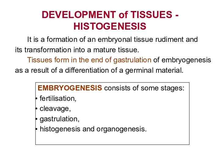
DEVELOPMENT of TISSUES - HISTOGENESIS
It is a formation of an
DEVELOPMENT of TISSUES - HISTOGENESIS
It is a formation of an
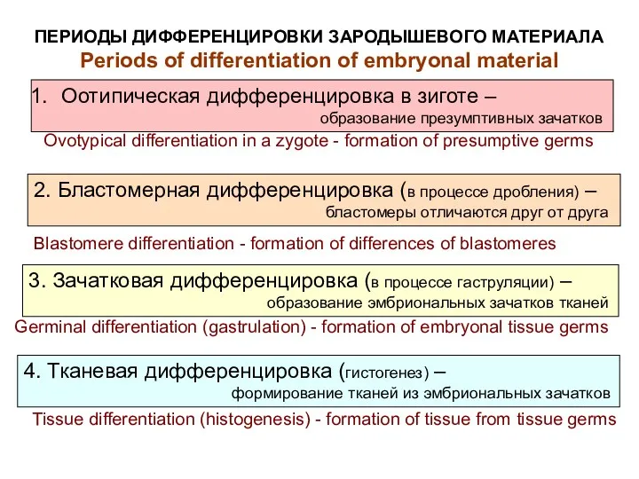
ПЕРИОДЫ ДИФФЕРЕНЦИРОВКИ ЗАРОДЫШЕВОГО МАТЕРИАЛА
Periods of differentiation of embryonal material
Оотипическая дифференцировка
ПЕРИОДЫ ДИФФЕРЕНЦИРОВКИ ЗАРОДЫШЕВОГО МАТЕРИАЛА
Periods of differentiation of embryonal material
Оотипическая дифференцировка
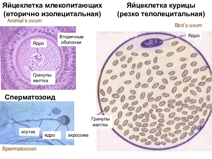
Сперматозоид
акросома
ядро
жгутик
Яйцеклетка курицы
(резко телолецитальная)
Гранулы
желтка
Ядро
Яйцеклетка млекопитающих
(вторично изолецитальная)
Ядро
Гранулы
желтка
Вторичные
оболочки
Animal’s ovum
Bird’s ovum
Spermatozoon
Сперматозоид
акросома
ядро
жгутик
Яйцеклетка курицы
(резко телолецитальная)
Гранулы
желтка
Ядро
Яйцеклетка млекопитающих
(вторично изолецитальная)
Ядро
Гранулы
желтка
Вторичные
оболочки
Animal’s ovum
Bird’s ovum
Spermatozoon
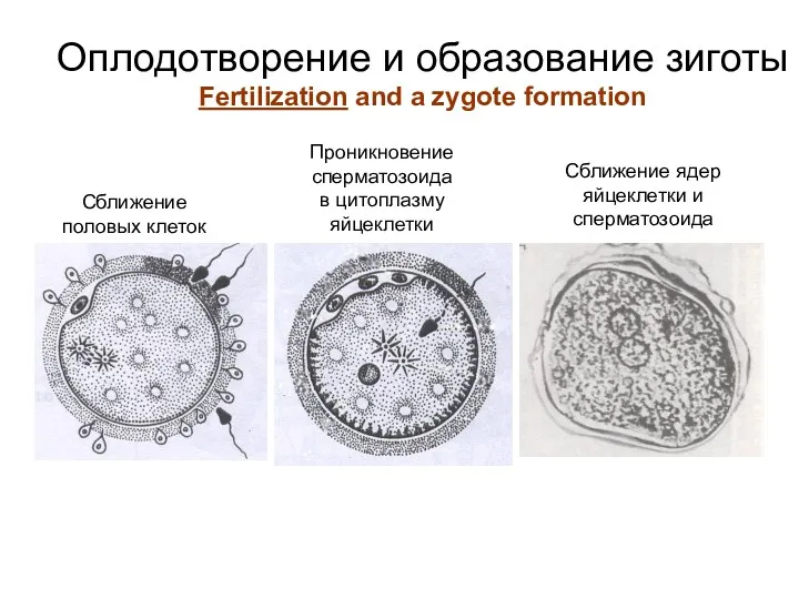
Оплодотворение и образование зиготы
Fertilization and a zygote formation
Сближение
половых клеток
Проникновение
сперматозоида
в
Оплодотворение и образование зиготы
Fertilization and a zygote formation
Сближение
половых клеток
Проникновение
сперматозоида
в
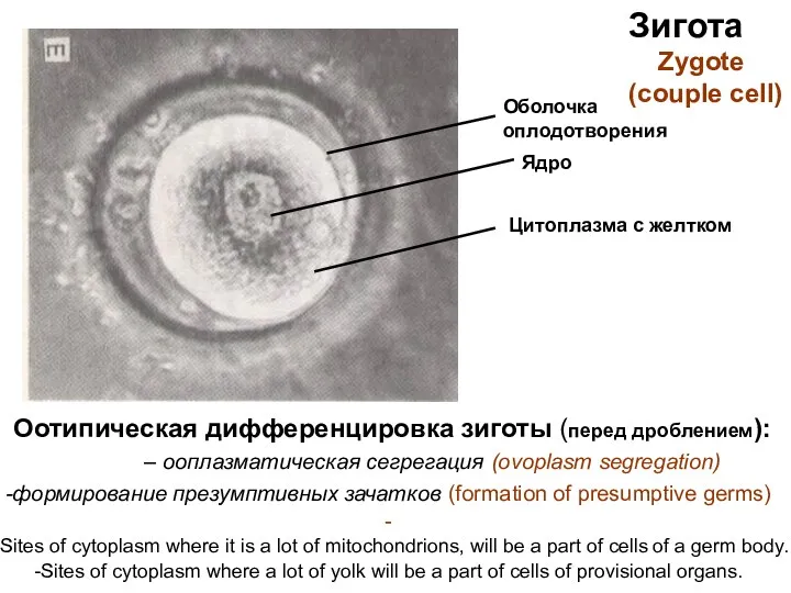
Зигота
Zygote
(couple cell)
Ядро
Цитоплазма с желтком
Оболочка
оплодотворения
Оотипическая дифференцировка зиготы (перед дроблением):
Зигота
Zygote
(couple cell)
Ядро
Цитоплазма с желтком
Оболочка
оплодотворения
Оотипическая дифференцировка зиготы (перед дроблением):
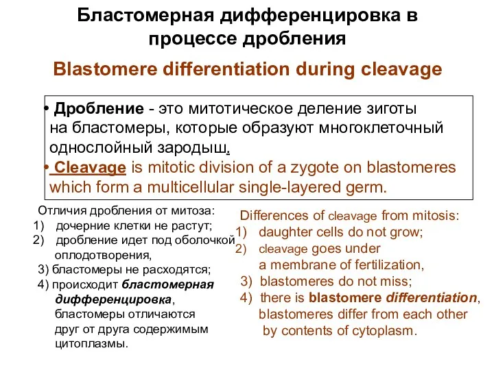
Бластомерная дифференцировка в процессе дробления
Blastomere differentiation during cleavage
Differences of cleavage
Бластомерная дифференцировка в процессе дробления
Blastomere differentiation during cleavage
Differences of cleavage
1. Blastomeres form the dense cellular ball – MORULA.
2. Cells
1. Blastomeres form the dense cellular ball – MORULA.
2. Cells
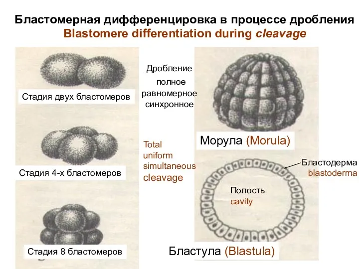
Морула (Morula)
Бластула (Blastula)
Стадия двух бластомеров
Стадия 4-х бластомеров
Стадия 8 бластомеров
Полость
cavity
Бластодерма
blastoderma
Бластомерная дифференцировка в
Морула (Morula)
Бластула (Blastula)
Стадия двух бластомеров
Стадия 4-х бластомеров
Стадия 8 бластомеров
Полость
cavity
Бластодерма
blastoderma
Бластомерная дифференцировка в
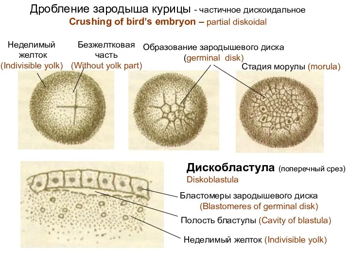
Дробление зародыша курицы - частичное дискоидальное
Crushing of bird’s embryon – partial
Дробление зародыша курицы - частичное дискоидальное
Crushing of bird’s embryon – partial
Зачатковая дифференцировка
Germinal differentiation
Gastrulation happens to 2 stages:
1 stage – formation
Зачатковая дифференцировка
Germinal differentiation
Gastrulation happens to 2 stages:
1 stage – formation
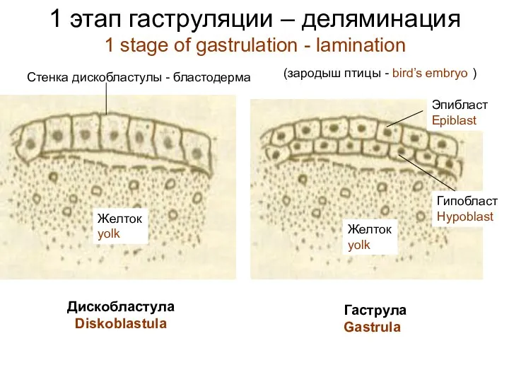
1 этап гаструляции – деляминация
1 stage of gastrulation - lamination
Дискобластула
Diskoblastula
Гаструла
Gastrula
Стенка дискобластулы
1 этап гаструляции – деляминация
1 stage of gastrulation - lamination
Дискобластула
Diskoblastula
Гаструла
Gastrula
Стенка дискобластулы
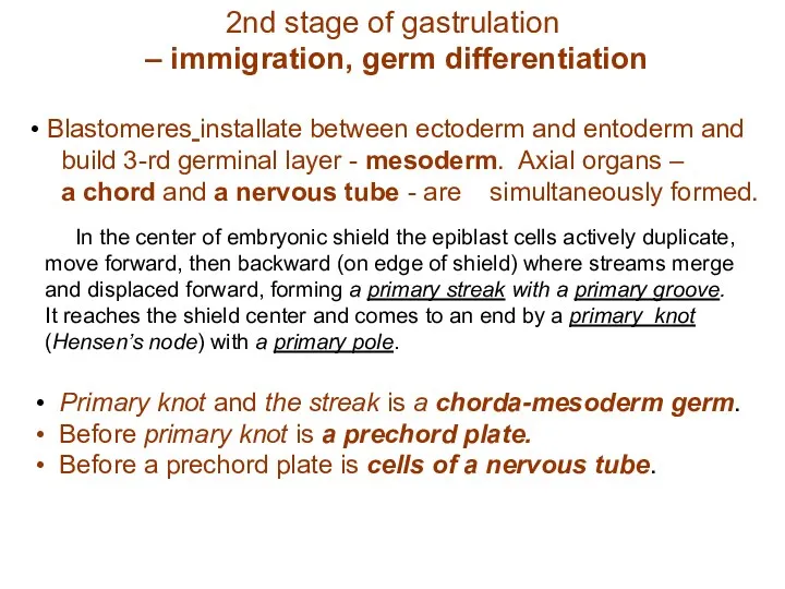
2nd stage of gastrulation
– immigration, germ differentiation
Blastomeres installate
2nd stage of gastrulation
– immigration, germ differentiation
Blastomeres installate
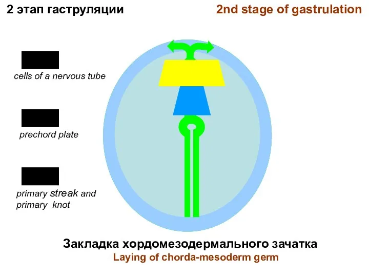
Закладка хордомезодермального зачатка
Laying of chorda-mesoderm germ
2 этап гаструляции 2nd stage of
Закладка хордомезодермального зачатка
Laying of chorda-mesoderm germ
2 этап гаструляции 2nd stage of
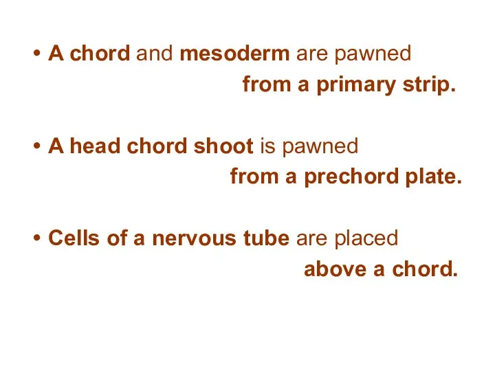
A chord and mesoderm are pawned
from a primary strip.
A chord and mesoderm are pawned
from a primary strip.
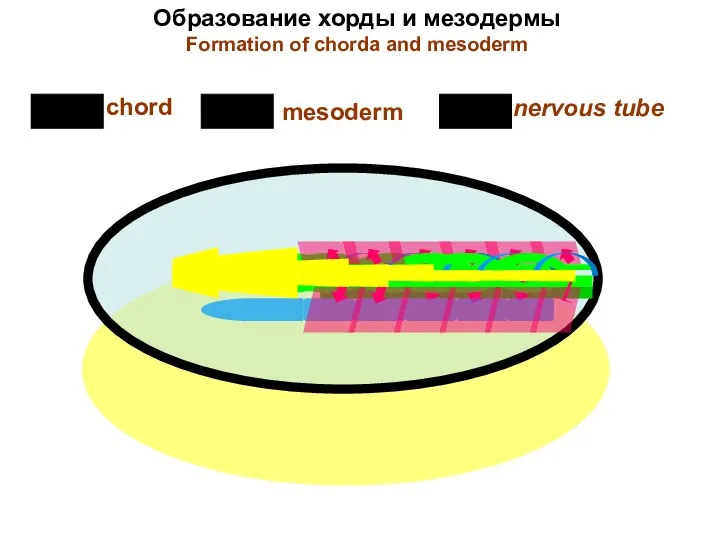
Образование хорды и мезодермы
Formation of chorda and mesoderm
chord
mesoderm
nervous tube
Образование хорды и мезодермы
Formation of chorda and mesoderm
chord
mesoderm
nervous tube
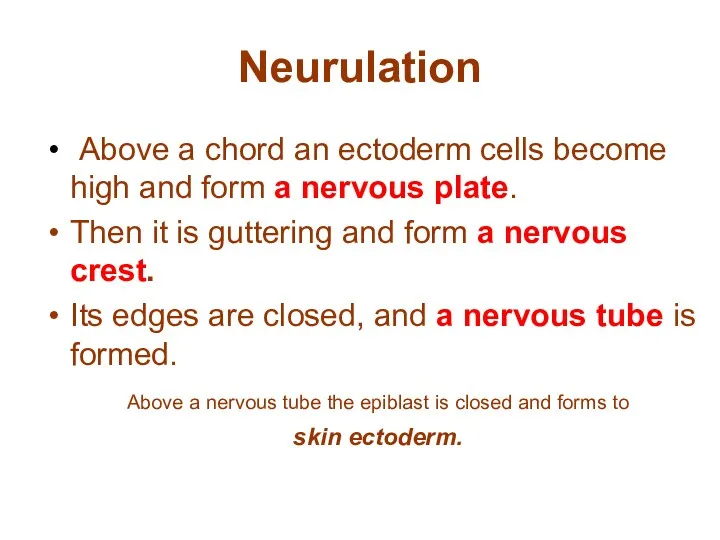
Neurulation
Above a chord an ectoderm cells become high and
Neurulation
Above a chord an ectoderm cells become high and
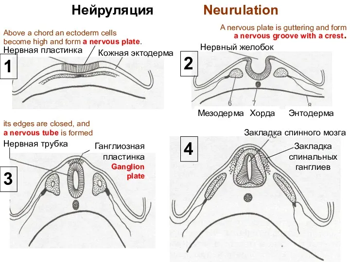
Нейруляция Neurulation
Нервная пластинка
Кожная эктодерма
Нервный желобок
Энтодерма
Хорда
Мезодерма
Нервная трубка
Закладка спинного мозга
Закладка
спинальных
ганглиев
1
2
3
4
Above a chord an
Нейруляция Neurulation
Нервная пластинка
Кожная эктодерма
Нервный желобок
Энтодерма
Хорда
Мезодерма
Нервная трубка
Закладка спинного мозга
Закладка
спинальных
ганглиев
1
2
3
4
Above a chord an
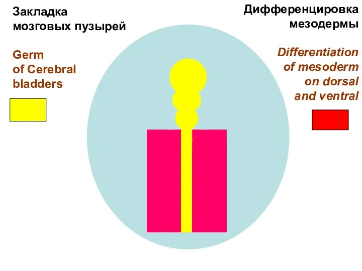
Закладка
мозговых пузырей
Germ
of Cerebral
bladders
Дифференцировка
мезодермы
Differentiation
of mesoderm
on dorsal
and ventral
Закладка
мозговых пузырей
Germ
of Cerebral
bladders
Дифференцировка
мезодермы
Differentiation
of mesoderm
on dorsal
and ventral
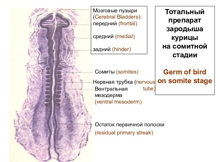
Тотальный
препарат
зародыша
курицы
на сомитной
стадии
Germ of bird
on somite stage
Мозговые пузыри
(Cerebral
Тотальный
препарат
зародыша
курицы
на сомитной
стадии
Germ of bird
on somite stage
Мозговые пузыри
(Cerebral
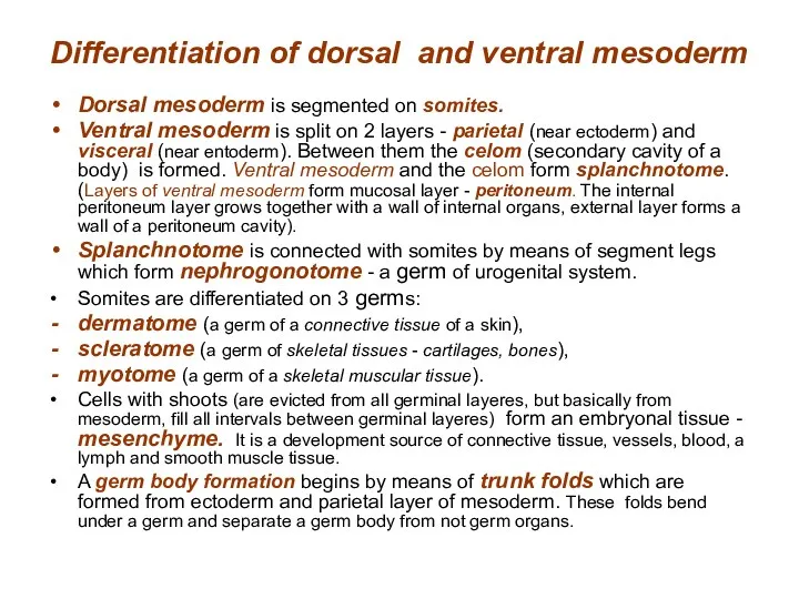
Dorsal mesoderm is segmented on somites.
Ventral mesoderm is split on
Dorsal mesoderm is segmented on somites.
Ventral mesoderm is split on
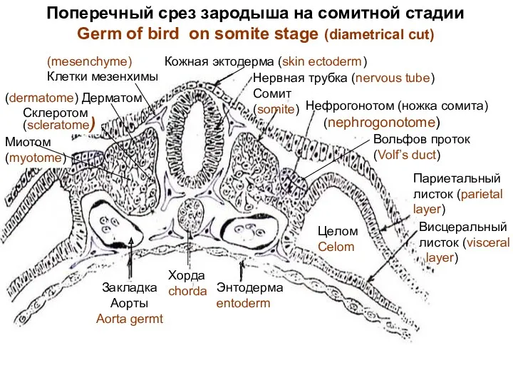
Поперечный срез зародыша на сомитной стадии
Germ of bird on somite
Поперечный срез зародыша на сомитной стадии
Germ of bird on somite
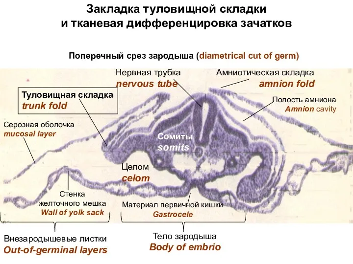
Закладка туловищной складки
и тканевая дифференцировка зачатков
Туловищная складка
trunk fold
Амниотическая складка
amnion fold
Нервная трубка
nervous
Закладка туловищной складки
и тканевая дифференцировка зачатков
Туловищная складка
trunk fold
Амниотическая складка
amnion fold
Нервная трубка
nervous
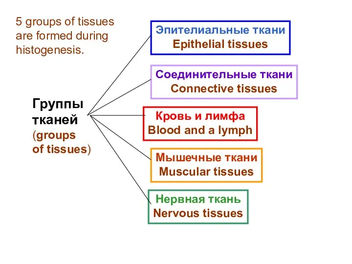
Группы
тканей
(groups
of tissues)
Эпителиальные ткани
Epithelial tissues
Соединительные ткани
Connective tissues
Кровь и лимфа
Blood and
Группы
тканей
(groups
of tissues)
Эпителиальные ткани
Epithelial tissues
Соединительные ткани
Connective tissues
Кровь и лимфа
Blood and
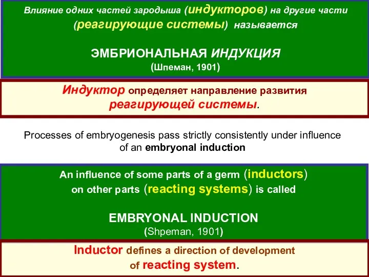
Влияние одних частей зародыша (индукторов) на другие части (реагирующие системы) называется
Влияние одних частей зародыша (индукторов) на другие части (реагирующие системы) называется
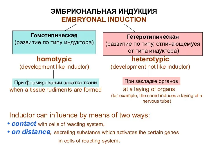
ЭМБРИОНАЛЬНАЯ ИНДУКЦИЯ
EMBRYONAL INDUCTION
Гомотипическая
(развитие по типу индуктора)
При формировании зачатка ткани
Гетеротипическая
(развитие по типу,
ЭМБРИОНАЛЬНАЯ ИНДУКЦИЯ
EMBRYONAL INDUCTION
Гомотипическая
(развитие по типу индуктора)
При формировании зачатка ткани
Гетеротипическая
(развитие по типу,
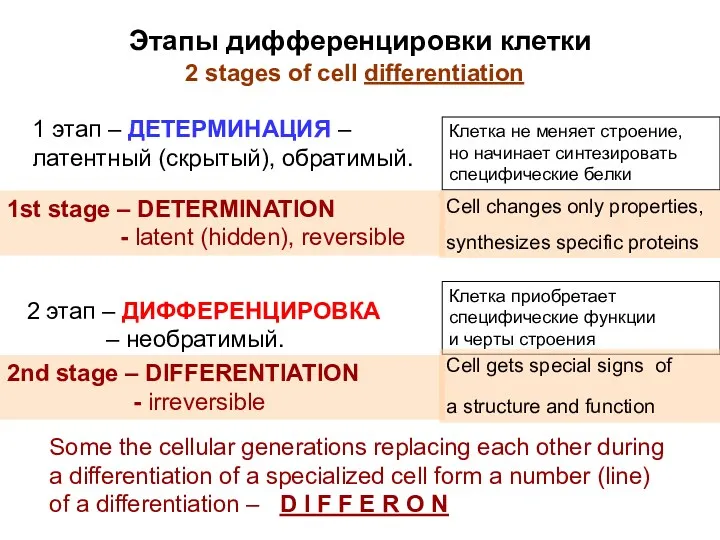
Этапы дифференцировки клетки
1 этап – ДЕТЕРМИНАЦИЯ – латентный (скрытый), обратимый.
Клетка
Этапы дифференцировки клетки
1 этап – ДЕТЕРМИНАЦИЯ – латентный (скрытый), обратимый.
Клетка
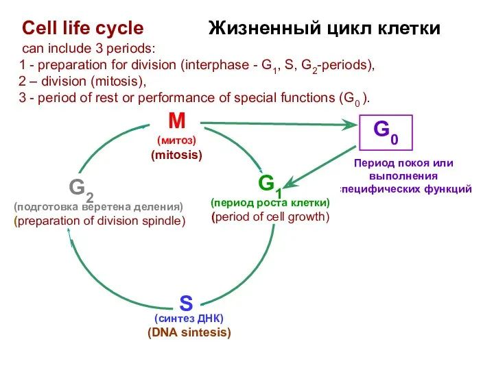
Период покоя или
выполнения специфических функций
G1
(период роста клетки)
(period of cell growth)
G2
(подготовка
Период покоя или
выполнения специфических функций
G1
(период роста клетки)
(period of cell growth)
G2
(подготовка
 Общая экология. Экология популяций
 Головной мозг - строение
 Презентация Глобальный экологический кризис
 Презентация по биологии Гуморальная регуляция для 8 класса
 Экзотические животные Африки
 Приспособленность живых организмов к среде обитания
 Вид. Критерии вида. 9 класс
 Презентация Растения хищники
 Строение бактериальной клетки
 Микробиологическая лаборатория, устройства оснащения, правила работы, изучение морфологии бактерий
 Окружающая среда
 Життя підводних мешканців
 Сухоцветы. Составление зимних букетов
 Вирусология и открытие вирусов
 Витамин А
 Матричный принцип, как основа современной эволюционной теории
 Презентация к уроку биологии в 6 классе
 Функциональная структура биосферы
 Вторично чувствующие органы чувств: орган слуха, орган равновесия, орган вкуса
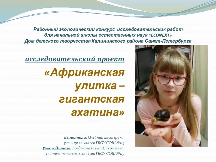 Африканская улитка - гигантская ахатина
 Кейс-метод
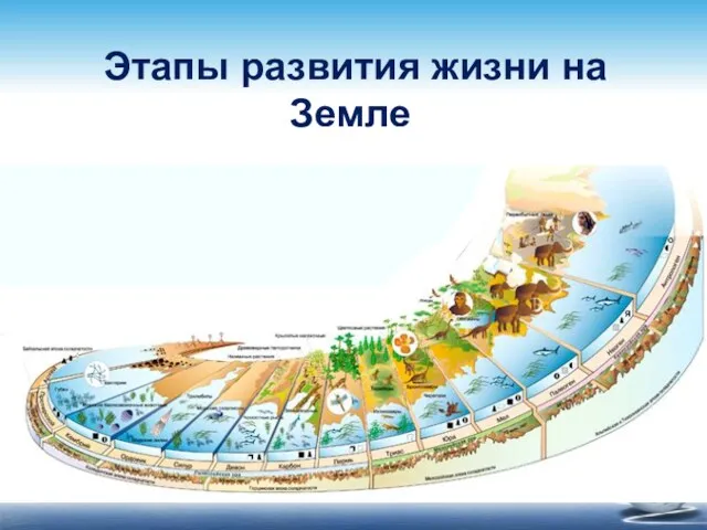 Этапы развития жизни на Земли
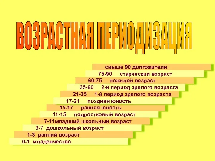 Возрастная периодизация
 Наследование групп крови человека
 Червонокнижні види рослин України
 Нуклеиновые кислоты: состав, строение, функции. Биология. 10 класс
 Ткани животных и растений
 Биохимия липидов. Классификация, биологические функции. Переваривание и всасывание. Обмен липопротеидов