- Respiratory system

Содержание
- 2. Introduction The Respiratory System is mainly concerned with gaseous exchange which occurs in the lungs at
- 3. Objectives: Description of the main functional units of the respiratory system and its division into upper
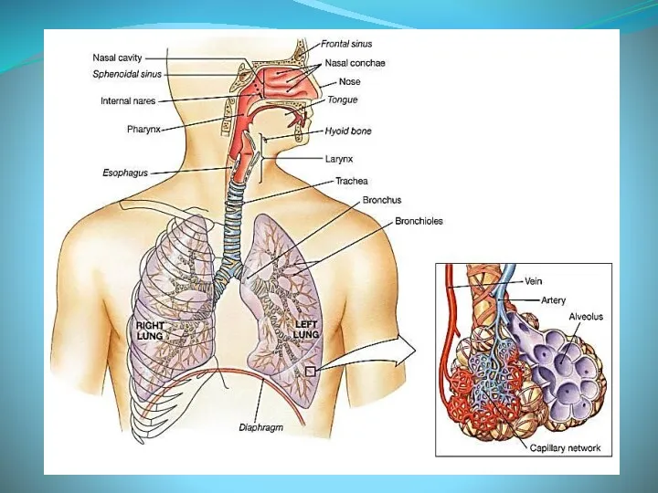
- 5. Organization of the Respiratory System The upper respiratory tract consists of: (Nose and nasal cavity, Pharynx
- 6. Function of the Respiratory System Conducting portion transports air. - includes the nose, nasal cavity, pharynx,
- 7. Upper Respiratory Tract
- 8. Nose It consists of external nose and nasal cavity. The external nose extends the nasal cavities
- 9. Nasal Cavity Four walled pyramidal space. Each nasal cavity consists of three general regions-the nasal vestibule,
- 10. Nasal Conchae are folds in the mucous membrane that increase air turbulence and ensures that most
- 11. Respiratory mucosa Most of the conducting portion is lined with ciliated pseudostratified columnar epithelium that contains
- 12. Paranasal Sinuses They are closed cavities in the frontal, maxillary, ethmoid, and sphenoid bones. They are
- 13. Communication with nasal cavity The frontal sinus drains through frontonasal duct to the semilunar hiatus of
- 14. Functions of the Paranasal sinuses Decrease skull bone weight. Warm, moisten and filter incoming air. Add
- 15. Paranasal sinuses X-Ray
- 16. Pharynx The pharynx is a musculo-fascial half cylinder that links the oral and nasal cavities in
- 17. Pharyngeal mucosa Superior-most region of the nasopharynx is covered with pseudostratified ciliated columnar epithelium. Posterior nasopharynx
- 18. Larynx It is a cylindrical musculo-ligamentous structure with a cartilaginous framework that caps the lower respiratory
- 19. Laryngeal Cartilages Nine C-rings of cartilage form the framework of the larynx Thyroid cartilage – (1)
- 20. Larynx Muscular walls aid in voice production and the swallowing reflex. Glottis – the superior opening
- 21. Sound Production The cavity of larynx has two folds (ligaments): Upper Vestibular folds are false vocal
- 22. Sound Production Intermittent release of exhaled air through the vocal folds Loudness – depends on the
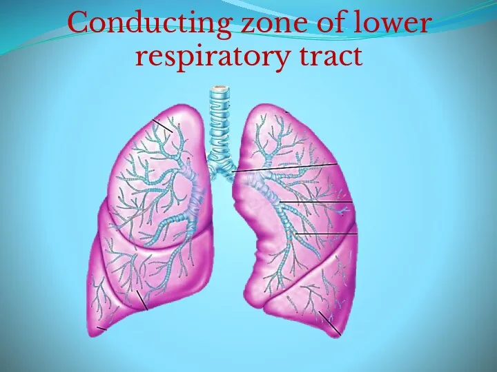
- 23. Conducting zone of lower respiratory tract
- 24. Trachea A flexible tube also called windpipe. Extends through the mediastinum and lies anterior to the
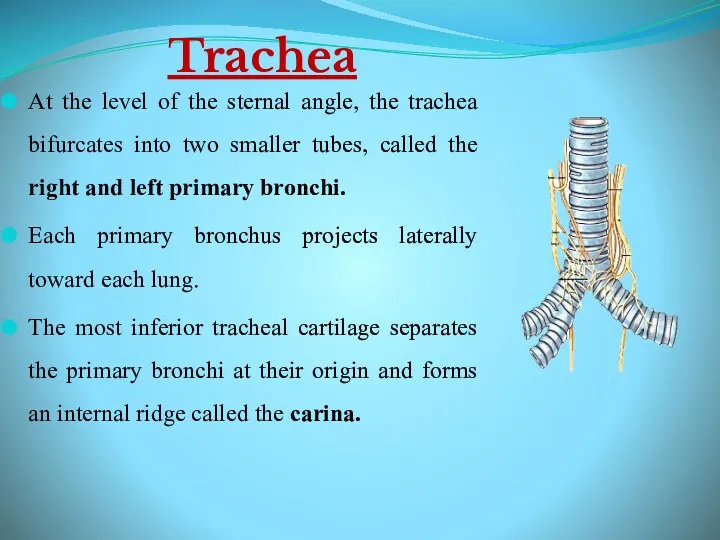
- 25. Trachea At the level of the sternal angle, the trachea bifurcates into two smaller tubes, called
- 26. Bronchial Tree A highly branched system of air-conducting passages that originate from the left and right
- 27. Bronchial Tree The primary bronchi enter the hilus of each lung together with the pulmonary vessels,
- 28. Bronchial Tree Secondary bronchi? Tertiary bronchi? Bronchioles? Terminal bronchioles. With successive branching amount of cartilage decreases
- 29. Respiratory Zone of Lower Respiratory Tract
- 30. Conduction vs. Respiratory zones Most of the tubing in the lungs makes up conduction zone. Consists
- 31. Respiratory Bronchioles, Alveolar Ducts, and Alveoli Lungs contain small saccular outpocketings called alveoli. They have a
- 32. Respiratory Membrane Squamous cells of alveoli . Basement membrane of alveoli. Basement membrane of capillaries Simple
- 33. Cells in the Alveolus Type I cells : simple squamous cells forming lining. Type II cells
- 34. Gross Anatomy of the Lungs Each lung has a conical shape. Its superior region called the
- 35. Lungs Left lung Divided into 2 lobes by oblique fissure. Smaller than the right lung. Cardiac
- 37. Скачать презентацию
Introduction
The Respiratory System is mainly concerned with gaseous exchange which occurs
Introduction
The Respiratory System is mainly concerned with gaseous exchange which occurs
Objectives:
Description of the main functional units of the respiratory system and
Objectives:
Description of the main functional units of the respiratory system and
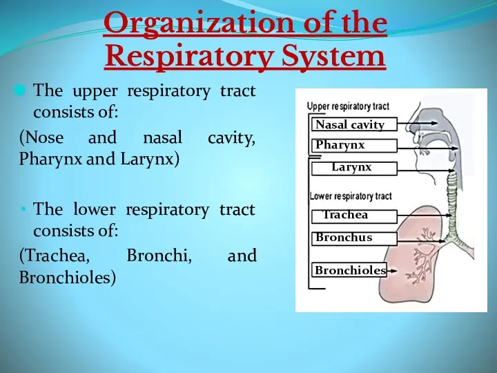
Organization of the Respiratory System
The upper respiratory tract consists of:
(Nose and
Organization of the Respiratory System
The upper respiratory tract consists of:
(Nose and
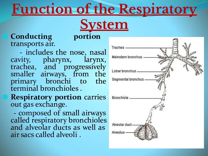
Function of the Respiratory System
Conducting portion transports air.
- includes
Function of the Respiratory System
Conducting portion transports air.
- includes
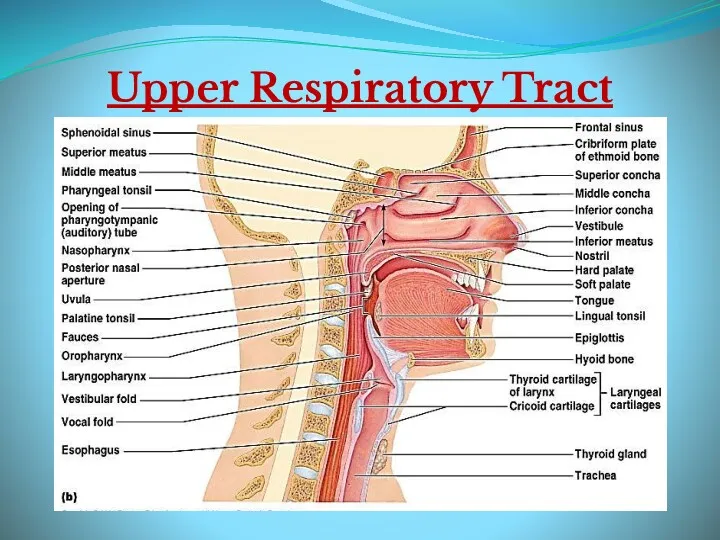
Upper Respiratory Tract
Upper Respiratory Tract
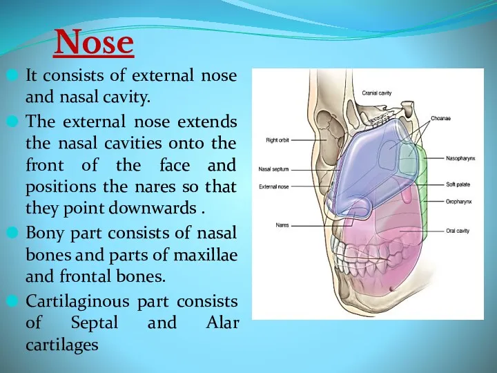
Nose
It consists of external nose and nasal cavity.
The external nose extends
Nose
It consists of external nose and nasal cavity.
The external nose extends
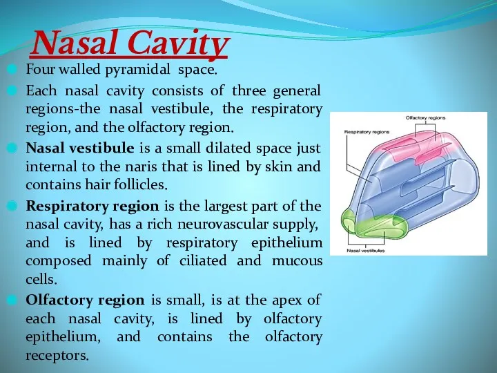
Nasal Cavity
Four walled pyramidal space.
Each nasal cavity consists of three general
Nasal Cavity
Four walled pyramidal space.
Each nasal cavity consists of three general
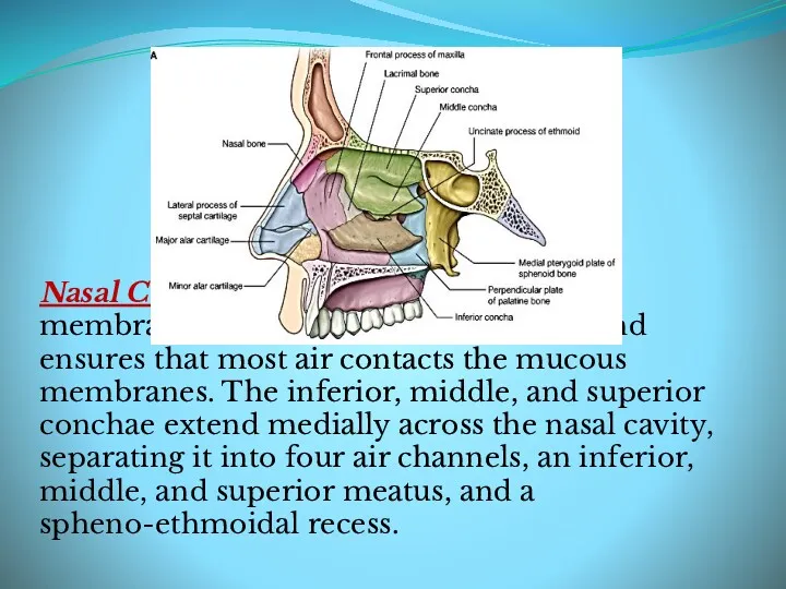
Nasal Conchae are folds in the mucous membrane that increase air
Nasal Conchae are folds in the mucous membrane that increase air
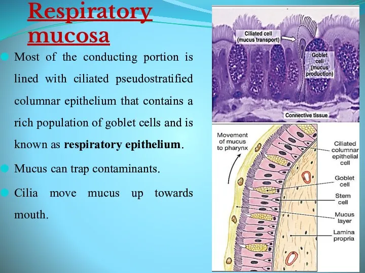
Respiratory mucosa
Most of the conducting portion is lined with ciliated pseudostratified
Respiratory mucosa
Most of the conducting portion is lined with ciliated pseudostratified
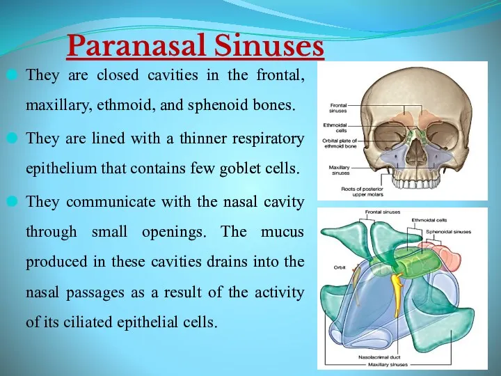
Paranasal Sinuses
They are closed cavities in the frontal, maxillary, ethmoid, and
Paranasal Sinuses
They are closed cavities in the frontal, maxillary, ethmoid, and
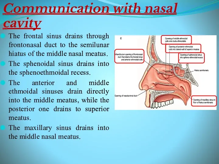
Communication with nasal cavity
The frontal sinus drains through frontonasal duct to
Communication with nasal cavity
The frontal sinus drains through frontonasal duct to
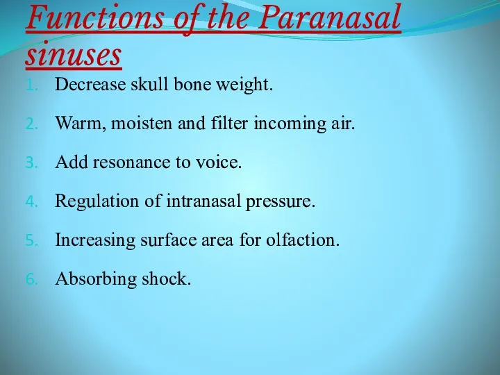
Functions of the Paranasal sinuses
Decrease skull bone weight.
Warm, moisten and filter
Functions of the Paranasal sinuses
Decrease skull bone weight.
Warm, moisten and filter
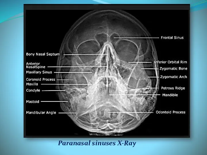
Paranasal sinuses X-Ray
Paranasal sinuses X-Ray
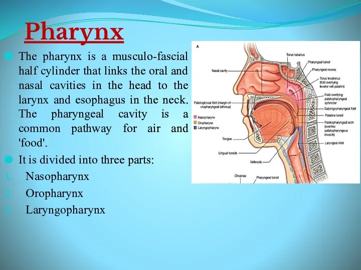
Pharynx
The pharynx is a musculo-fascial half cylinder that links the oral
Pharynx
The pharynx is a musculo-fascial half cylinder that links the oral
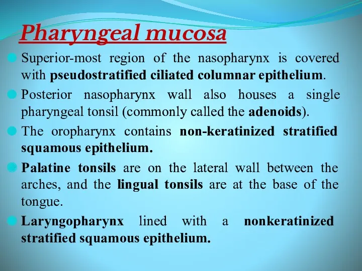
Pharyngeal mucosa
Superior-most region of the nasopharynx is covered with pseudostratified ciliated
Pharyngeal mucosa
Superior-most region of the nasopharynx is covered with pseudostratified ciliated
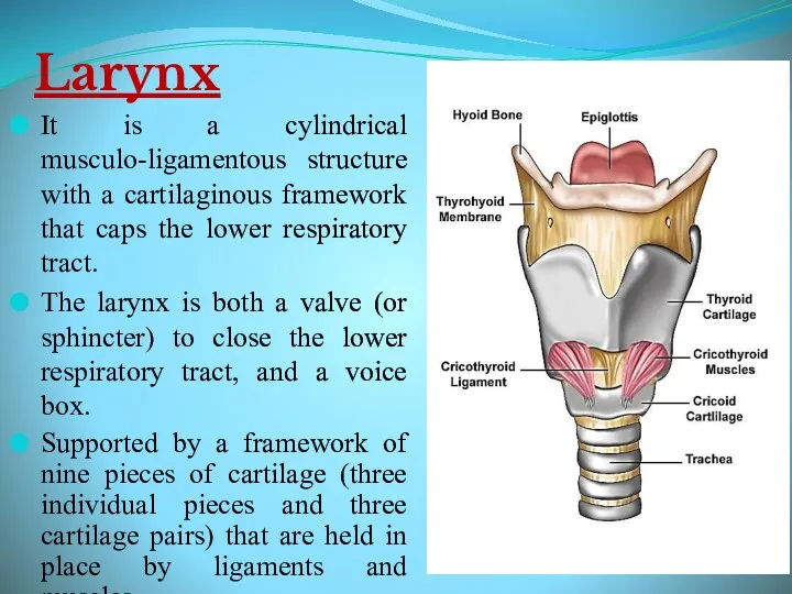
Larynx
It is a cylindrical musculo-ligamentous structure with a cartilaginous framework that
Larynx
It is a cylindrical musculo-ligamentous structure with a cartilaginous framework that
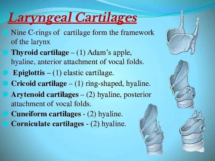
Laryngeal Cartilages
Nine C-rings of cartilage form the framework of the larynx
Thyroid
Laryngeal Cartilages
Nine C-rings of cartilage form the framework of the larynx
Thyroid
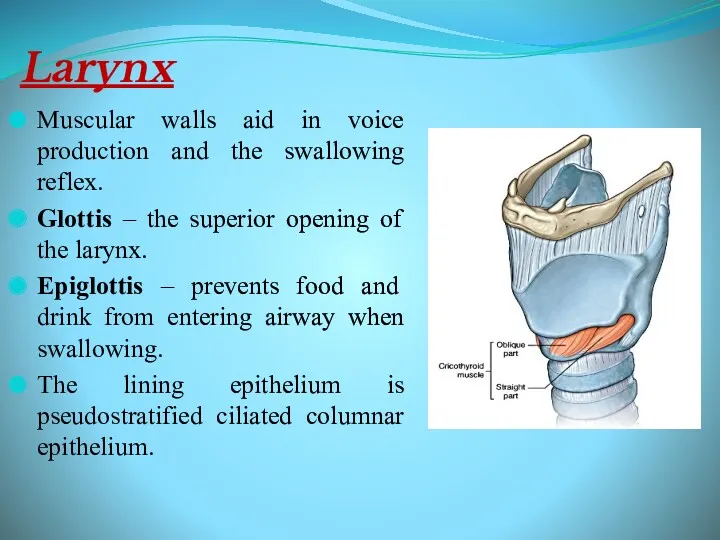
Larynx
Muscular walls aid in voice production and the swallowing reflex.
Glottis
Larynx
Muscular walls aid in voice production and the swallowing reflex.
Glottis
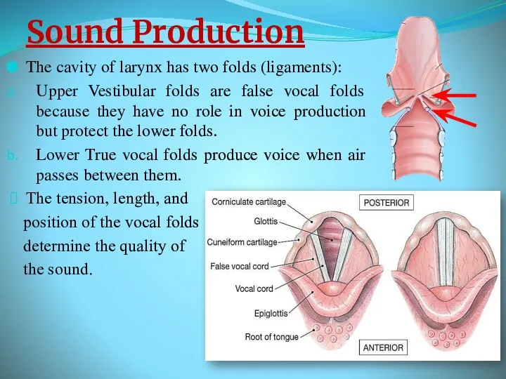
Sound Production
The cavity of larynx has two folds (ligaments):
Upper Vestibular folds
Sound Production
The cavity of larynx has two folds (ligaments):
Upper Vestibular folds
Sound Production
Intermittent release of exhaled air through the vocal folds
Loudness –
Sound Production
Intermittent release of exhaled air through the vocal folds
Loudness –
Conducting zone of lower respiratory tract
Conducting zone of lower respiratory tract
Trachea
A flexible tube also called windpipe.
Extends through the mediastinum and lies
Trachea
A flexible tube also called windpipe.
Extends through the mediastinum and lies
Trachea
At the level of the sternal angle, the trachea bifurcates into
Trachea
At the level of the sternal angle, the trachea bifurcates into
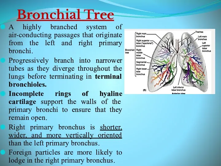
Bronchial Tree
A highly branched system of air-conducting passages that originate from
Bronchial Tree
A highly branched system of air-conducting passages that originate from
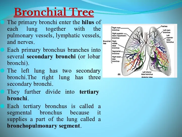
Bronchial Tree
The primary bronchi enter the hilus of each lung together
Bronchial Tree
The primary bronchi enter the hilus of each lung together
Bronchial Tree
Secondary bronchi? Tertiary bronchi? Bronchioles? Terminal bronchioles.
With successive branching
Bronchial Tree
Secondary bronchi? Tertiary bronchi? Bronchioles? Terminal bronchioles.
With successive branching
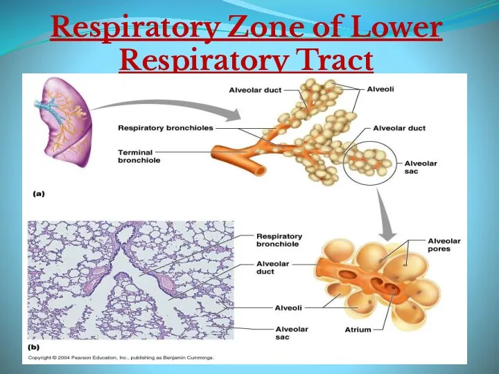
Respiratory Zone of Lower Respiratory Tract
Respiratory Zone of Lower Respiratory Tract
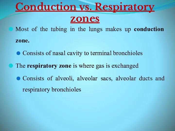
Conduction vs. Respiratory zones
Most of the tubing in the lungs makes
Conduction vs. Respiratory zones
Most of the tubing in the lungs makes
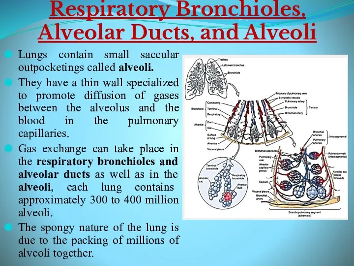
Respiratory Bronchioles, Alveolar Ducts, and Alveoli
Lungs contain small saccular outpocketings
Respiratory Bronchioles, Alveolar Ducts, and Alveoli
Lungs contain small saccular outpocketings
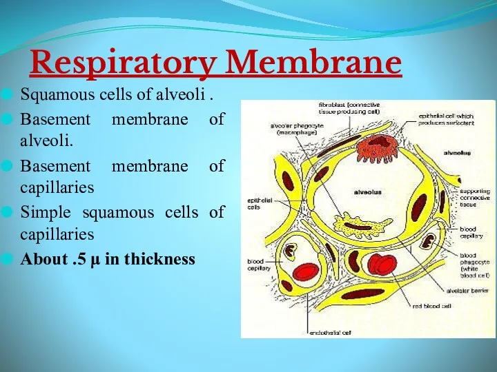
Respiratory Membrane
Squamous cells of alveoli .
Basement membrane of alveoli.
Basement membrane of
Respiratory Membrane
Squamous cells of alveoli .
Basement membrane of alveoli.
Basement membrane of
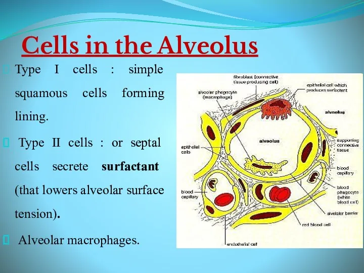
Cells in the Alveolus
Type I cells : simple squamous cells forming
Cells in the Alveolus
Type I cells : simple squamous cells forming
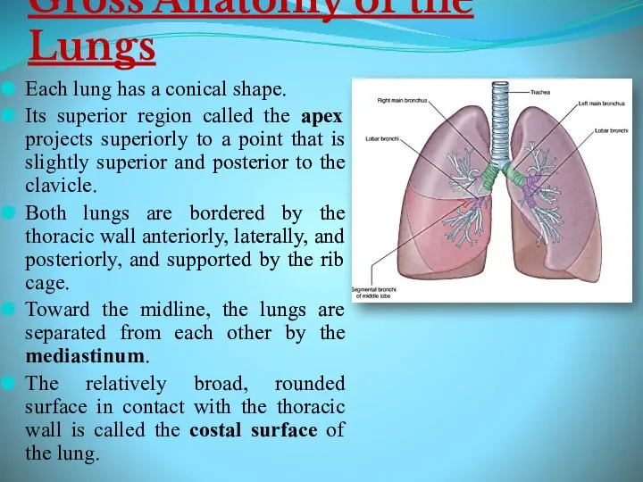
Gross Anatomy of the Lungs
Each lung has a conical shape.
Its
Gross Anatomy of the Lungs
Each lung has a conical shape.
Its
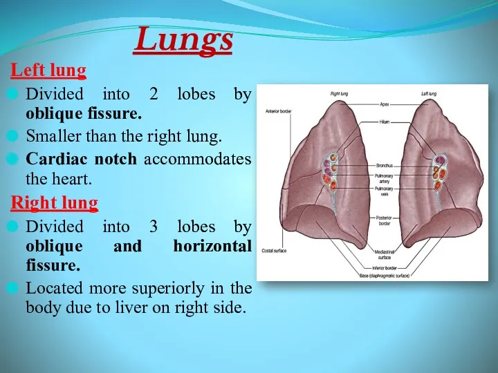
Lungs
Left lung
Divided into 2 lobes by oblique fissure.
Smaller than the right
Lungs
Left lung
Divided into 2 lobes by oblique fissure.
Smaller than the right
 Кровоносні судини (урок)
 Из опыта работы Проектно – исследовательская деятельность на уроках биологии
 Биологические свойства воды
 Цитогенетика
 Законы наследственности
 Пищеварение в ротовой полости
 Питание и пищеварение
 Конспект урока биологии 7 класса Тип Кишечнополостные, УМК Н.И.Сонина
 Эмбриональное развитие организма
 Птахи, які народжують взимку
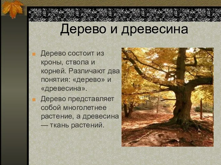 Дерево и древесина
 Царство Животные Подцарство Простейшие (Одноклеточные)
 Химия и здоровье
 Цитология. Клеточная теория
 Анатомия и физиология нервной системы
 Анализ типичных ошибок участников ЕГЭ по биологии 2019 года
 Влияние факторов внешней среды на микроорганизмы. Взаимоотношения микробов друг с другом и макроорганизмом
 Ткани растений
 Тип членистоногие. Подтип жабродышащие, подтип хелицеровые
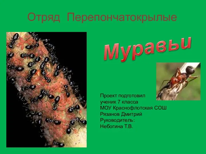 Презентация Общественные насекомые. Муравьи.
 Мутации. Классификация мутаций
 Особенности обмена веществ у детей дошкольного возраста
 WDC - Whale and Dolphin Conservation
 Вегетативная нервная система
 Белки. Содержание белков в различных тканях человека
 Интеллектуально - развлекательная игра В здоровом теле здоровый дух
 Царство растения
 Определение рефлекса. Принципы рефлекторной деятельности