Слайд 2UPPER RESPIRATORY TRACT
Figure 13.2
Слайд 3ANATOMY OF THE NASAL CAVITY
Olfactory receptors are located in the mucosa on the
superior surface
The rest of the cavity is lined with respiratory mucosa
Moistens air
Traps incoming foreign particles
Слайд 4ANATOMY OF THE NASAL CAVITY
Lateral walls have projections called conchae
Increases surface area
Increases air
turbulence within the nasal cavity
The nasal cavity is separated from the oral cavity by the palate
Anterior hard palate (bone)
Posterior soft palate (muscle)
Слайд 5PARANASAL SINUSES
Cavities within bones surrounding the nasal cavity
Frontal bone
Sphenoid bone
Ethmoid bone
Maxillary bone
Слайд 6PARANASAL SINUSES
Function of the sinuses
Lighten the skull
Act as resonance chambers for speech
Produce mucus
that drains into the nasal cavity Produce mucus that drains into the nasal cavity
Слайд 7PHARYNX (THROAT)
Muscular passage from nasal cavity to larynx
Three regions of the pharynx
Nasopharynx –
superior region behind nasal cavity
Oropharynx – middle region behind mouth
Laryngopharynx – inferior region attached to larynx
The oropharynx and laryngopharynx are common passageways for air and food
Слайд 8STRUCTURES OF THE PHARYNX
Auditory tubes enter the nasopharynx
Tonsils of the pharynx
Pharyngeal tonsil (adenoids)
in the nasopharynx
Palatine tonsils in the oropharynx
Lingual tonsils at the base of the tongue
Слайд 9LARYNX (VOICE BOX)
Routes air and food into proper channels
Plays a role in speech
Made
of eight rigid hyaline cartilages and a spoon-shaped flap of elastic cartilage (epiglottis)
Vocal cords - vibrate with expelled air to create sound (speech)
Слайд 10STRUCTURES OF THE LARYNX
Thyroid cartilage
Largest hyaline cartilage
Protrudes anteriorly (Adam’s apple)
Epiglottis
Superior opening of the
larynx
Routes food to the larynx and air toward the trachea
Glottis – opening between vocal cords
Слайд 11TRACHEA (WINDPIPE)
Connects larynx with bronchi
Lined with ciliated mucosa
Beat continuously in the opposite direction
of incoming air
Expel mucus loaded with dust and other debris away from lungs
Walls are reinforced with C-shaped hyaline cartilage
Слайд 12PRIMARY BRONCHI
Formed by division of the trachea
Enters the lung at the hilus
(medial
depression)
Right bronchus is wider, shorter,
and straighter than left
Bronchi subdivide into smaller
and smaller branches
Слайд 13LUNGS
Ocupy most of the thoracic cavity
Apex is near the clavicle (superior portion)
Each lung
is divided into lobes by fissures
Left lung – two lobes
Right lung – three lobes
Слайд 15COVERINGS OF THE LUNGS
Pulmonary (visceral) pleura covers the lung surface
Parietal pleura lines the
walls of the thoracic cavity
Pleural fluid fills the area between layers of pleura to allow gliding
Слайд 16RESPIRATORY TREE DIVISIONS
Primary bronchi
Secondary bronchi
Tertiary bronchi
Bronchioli
Terminal bronchioli
Слайд 17BRONCHIOLES
Smallest branches of the bronchi
All but the smallest branches have reinforcing cartilage
Terminal bronchioles
end in alveoli
Figure 13.5a
Слайд 18ALVEOLI
Structure of alveoli
Alveolar duct
Alveolar sac
Alveolus
Gas exchange takes place within the alveoli in the
respiratory membrane
Squamous epithelial lining alveolar walls
Covered with pulmonary capillaries on external surfaces
Слайд 20COVERINGS OF THE LUNGS
Pulmonary (visceral) pleura covers the lung surface
Parietal pleura lines the
walls of the thoracic cavity
Pleural fluid fills the area between layers of pleura to allow gliding
Слайд 21RESPIRATORY TREE DIVISIONS
Primary bronchi
Secondary bronchi
Tertiary bronchi
Bronchioli
Terminal bronchioli
Слайд 22BRONCHIOLES
Smallest branches of the bronchi
All but the smallest branches have reinforcing cartilage
Terminal bronchioles
end in alveoli
Figure 13.5a
Слайд 23ALVEOLI
Structure of alveoli
Alveolar duct
Alveolar sac
Alveolus
Gas exchange takes place within the alveoli in the
respiratory membrane
Squamous epithelial lining alveolar walls
Covered with pulmonary capillaries on external surfaces
Слайд 24Figure 17-2b
MUSCLES USED FOR VENTILATION
Слайд 25Figure 17-3
THE RESPIRATORY SYSTEM
The relationship between the pleural sac and the lung
Слайд 26Figure 17-2e
BRANCHING OF AIRWAYS
Слайд 27MECHANICS OF BREATHING
(PULMONARY VENTILATION)
Mechanical process
Depends on volume changes in the thoracic cavity
Volume
changes lead to pressure changes, which lead to equalize pressure of flow of gases
2 phases
Inspiration – flow of air into lung
Expiration – air leaving lung
Слайд 28INSPIRATION
Diaphragm and intercostal muscles contract
The size of the thoracic cavity increases
External air
is pulled into the lungs due to an increase in intrapulmonary volume
Слайд 29EXPIRATION
Passive process dependent up on natural lung elasticity
As muscles relax, air is pushed
out of the lungs
Forced expiration can occur mostly by contracting internal intercostal muscles to depress the rib cage
Слайд 31PRESSURE DIFFERENCES IN THE THORACIC CAVITY
Normal pressure within the pleural space is always
negative (intrapleural pressure)
Differences in lung and pleural space pressures keep lungs from collapsing
Слайд 32NONRESPIRATORY AIR MOVEMENTS
Caused by reflexes or voluntary actions
Examples
Cough and sneeze – clears lungs
of debris
Laughing
Crying
Yawn
Hiccup
Слайд 33RESPIRATORY VOLUMES AND CAPACITIES
Normal breathing moves about 500 ml of air with each
breath - tidal volume (TV)
Many factors that affect respiratory capacity
A person’s size
Sex
Age
Physical condition
Residual volume of air – after exhalation, about 1200 ml of air remains in the lungs
Слайд 34RESPIRATORY VOLUMES AND CAPACITIES
Inspiratory reserve volume (IRV)
Amount of air that can be taken
in forcibly over the tidal volume
Usually between 2100 and 3200 ml
Expiratory reserve volume (ERV)
Amount of air that can be forcibly exhaled
Approximately 1200 ml
Residual volume
Air remaining in lung after expiration
About 1200 ml
Слайд 35RESPIRATORY VOLUMES AND CAPACITIES
Functional volume
Air that actually reaches the respiratory zone
Usually about 350
ml
Respiratory capacities are measured with a spirometer
Слайд 37GAS LAWS
Pgas = Patm × % of gas in atmosphere
Слайд 38Figure 17-7
LUNGS VOLUMES AND CAPACITIES
Слайд 39Figure 17-8
CILIATED RESPIRATORY EPITHELIUM
Слайд 40RESPIRATORY SOUNDS
Sounds are monitored with a stethoscope
Bronchial sounds – produced by air rushing
through trachea and bronchi
Vesicular breathing sounds – soft sounds of air filling alveoli
Слайд 41EXTERNAL RESPIRATION
Oxygen movement into the blood
The alveoli always has more oxygen than the
blood
Oxygen moves by diffusion towards the area of lower concentration
Pulmonary capillary blood gains oxygen
Слайд 42VENTILATION
Auscultation = diagnostic technique
Obstructive lung diseases
Asthma
Emphysema
Chronic bronchitis
Слайд 43SUMMARY
Respiratory system
Cellular respiration, external respiration, respiratory system, upper respiratory tract, pharynx, and larynx
Lower
respiratory tract, trachea, bronchi, bronchioles, alveoli, Type I and Type II alveolar cells
Diaphragm, intercostal muscles, lung, pleural sac, and plural fluid
Gas Laws: Dalton’s law and Boyle’s law
Слайд 44SUMMARY
Ventilation
Tidal volume, vital capacity, residual volume, and respiratory cycle
Alveolar pressure, active expiration, intrapleural
pressures, compliance, elastance, surfactant, bronchoconstriction, and bronchodilation
Total pulmonary ventilation, alveolar ventilation, hyperventilation, and hypoventilation

 Действие физических факторов на микроорганизмы. (Лекция 10)
 ОГЭ по обществознанию 9 класс Тема 1. Человек
 Строение, свойства, биологическая роль нуклеотидов и нуклеиновых кислот. Катаболизм нуклеиновых кислот
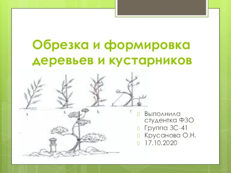 Обрезка и формировка деревьев и кустарников
 Самые интересные факты о птицах
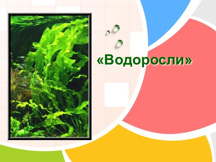 Водоросли. 5 класс
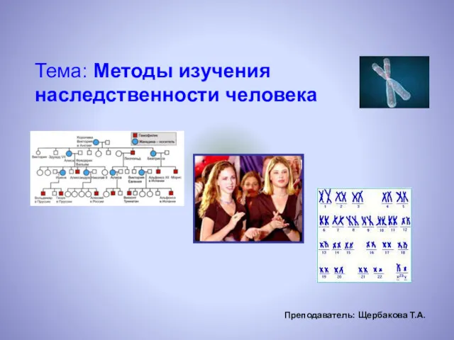 Методы изучения наследственности человека
 Биологический чемпионат
 Алоэ.Что мы о нем не знали
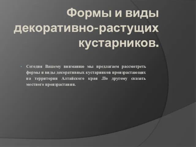 Формы и виды декоративно-растущих кустарников
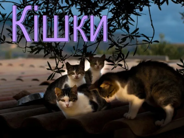 Породи кішок
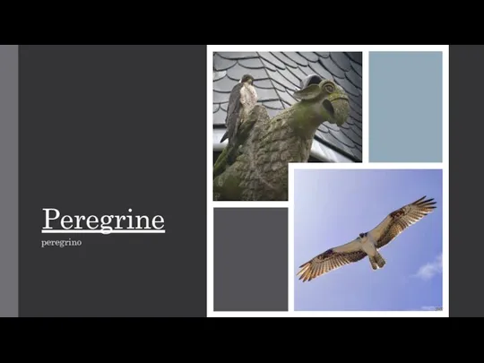 Peregrine falcon is the fastest bird
 Семейство лютиковые
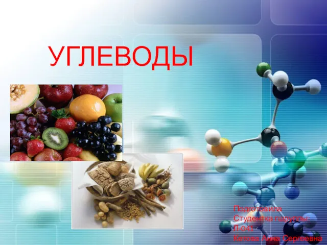 Полифункциональные соединения углеводы
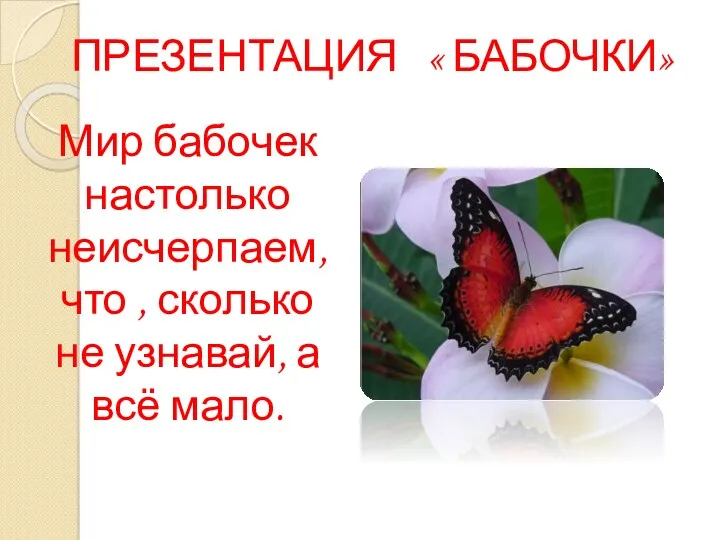 Мир бабочек. Бабочка из бумаги
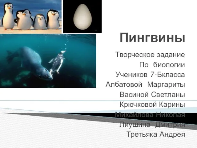 Пингвины
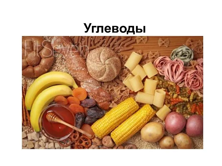 Углеводы. Функции углеводов
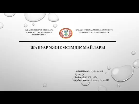 Жануар және өсімдік майлары
 Контрольная работа по материалам темы Органы растений
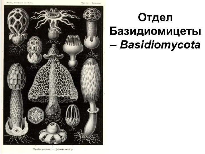 Отдел базидиомицеты (basidiomycota)
 Многообразие и происхождение культурных растений. 6 класс
 Нервная ткань
 Царство Бактерии
 Комнатные растения
 Метаболизм белков и аминокислот. Образование и обезвреживание аммиака. Тема 13.а
 Презентация Интегративное обучение в изучении предметов географии и биологии
 Видообразование как результат эволюции
 Насекомые луга