- The Cytoskeleton: Intermediate Filaments and Microtubules

Содержание
- 2. The Cytoskeleton Includes Dynamic Networks Of Microfilaments And Microfilaments
- 3. The cytoskeleton consists of three major types of filaments plus many filament-associated proteins including molecular motors
- 4. YPET – MAP R – MT PLUS END KERITAN – INTERMEDIATE FIL. ACTIN – STRESS FIBERS
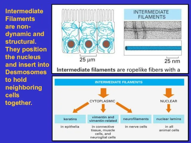
- 5. Intermediate Filaments are non- dynamic and structural. They position the nucleus and insert into Desmosomes to
- 6. Intermediate Filaments polymerize to form strong rope-like fibers. The basic structural unit is a coiled-coil dimer.
- 7. The inner side of the nuclear envelope is lined by a network of intermediate filaments called
- 8. Intermediate filament networks flare out from the nucleus and insert into plasma membrane junctions called desmosomes.
- 9. Microtubules Make Up Dynamic Networks
- 10. Microtubules serve four functions: To give shape to the cell. Example: nerve axons contain numerous micro-
- 11. Microtubules Are Made Of Tubulin Protofilaments
- 12. Microtubules as seen by Electron Microscopy 1) thin section 2) freeze dried And platinum Shadowed
- 13. Microtubules are stabilized by capping at their Plus and minus ends. Centrosomes and Microtubule organizing centers
- 14. The centrosome consists of centrioles surrounded by a “protein cloud”. Minus ends of microtubules are capped
- 15. Microtubule assembly at plus end is governed by GTP hydrolysis; GTP- tubulin is required for polymerization;
- 16. Catastrophic Disassembly can occur if growth at the plus end stops or is slow; but the
- 17. DYNAMIC INSTABILITY IN A MICROTUBULE ASTER
- 18. MICROTUBULE DYNAMICS SEEN WITH FLUORESCENT PLUS END PROTEINS
- 19. MICROTUBULE DYNAMICS SEEN WITH FLUORESCENT PLUS END PROTEINS
- 20. Microtubule associated proteins also stabilize microtubules. Acetylation and tyrosylation do too.
- 21. Drugs can stabilize or destabilize microtubules; Taxol stabilizes existing mts; cholchicine destabilizes microtubules by monomer binding
- 22. Motor proteins “walk” on microtubules and microfilaments via their heads acting as “motors”
- 23. Kinesin, like myosin, hydrolyzes ATP as it walks During this process chemical energy is transformed into
- 24. MOTOR PROTEINS MOVE VESICLES ON MICROTUBULE TRACKS – A CONFORMATIONAL CYCLE THAT HYDROLYZES ATP
- 25. MOTOR PROTEINS MOVE VESICLES ON MICROTUBULE TRACKS
- 26. Direction of vesicle Transport on microtubules FIBROBLAST NEURON Movement of pigment granules on MTs
- 27. Cilia And Flagella: A Different Form Of Motility
- 29. Dynein provides Motive force to move one MT doublet relative to a neighboring MT doublet
- 31. Скачать презентацию
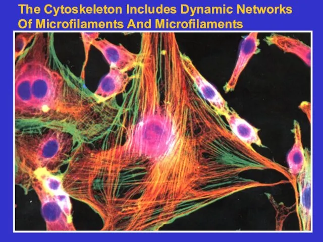
The Cytoskeleton Includes Dynamic Networks
Of Microfilaments And Microfilaments
The Cytoskeleton Includes Dynamic Networks
Of Microfilaments And Microfilaments
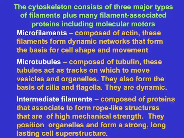
The cytoskeleton consists of three major types
of filaments plus many
The cytoskeleton consists of three major types
of filaments plus many
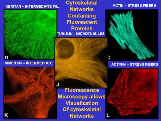
YPET – MAP R – MT PLUS END
KERITAN – INTERMEDIATE FIL.
ACTIN
YPET – MAP R – MT PLUS END
KERITAN – INTERMEDIATE FIL.
ACTIN
Intermediate
Filaments
are non-
dynamic and
structural.
They position
the nucleus
and insert into
Desmosomes
to hold
neighboring
cells
together.
Intermediate
Filaments
are non-
dynamic and
structural.
They position
the nucleus
and insert into
Desmosomes
to hold
neighboring
cells
together.
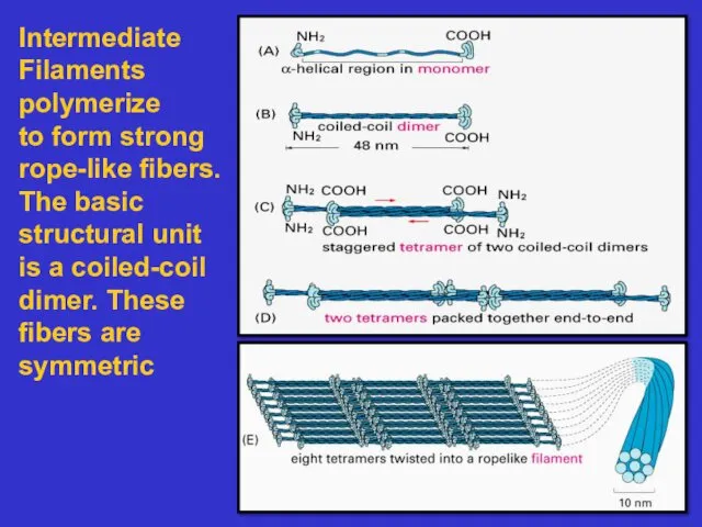
Intermediate
Filaments
polymerize
to form strong
rope-like fibers. The basic
structural unit is a coiled-coil
dimer.
Intermediate
Filaments
polymerize
to form strong
rope-like fibers. The basic
structural unit is a coiled-coil
dimer.
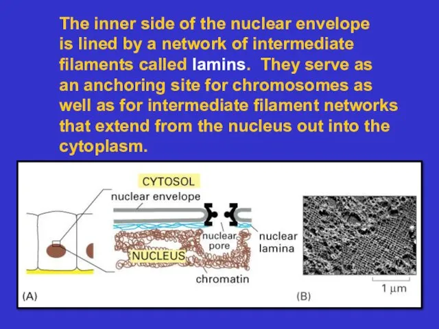
The inner side of the nuclear envelope
is lined by a network
The inner side of the nuclear envelope
is lined by a network
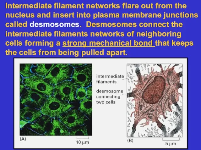
Intermediate filament networks flare out from the nucleus and insert into
Intermediate filament networks flare out from the nucleus and insert into
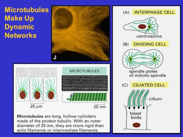
Microtubules
Make Up
Dynamic
Networks
Microtubules
Make Up
Dynamic
Networks
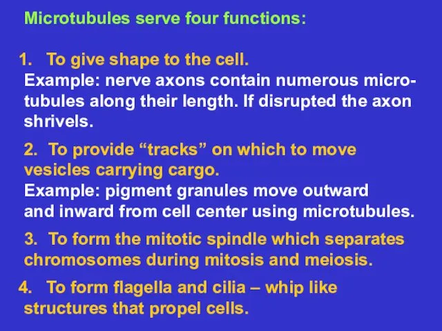
Microtubules serve four functions:
To give shape to the cell.
Example: nerve axons
Microtubules serve four functions:
To give shape to the cell.
Example: nerve axons
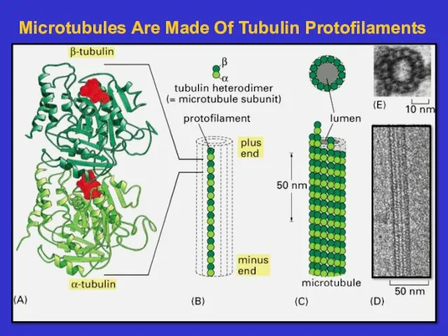
Microtubules Are Made Of Tubulin Protofilaments
Microtubules Are Made Of Tubulin Protofilaments
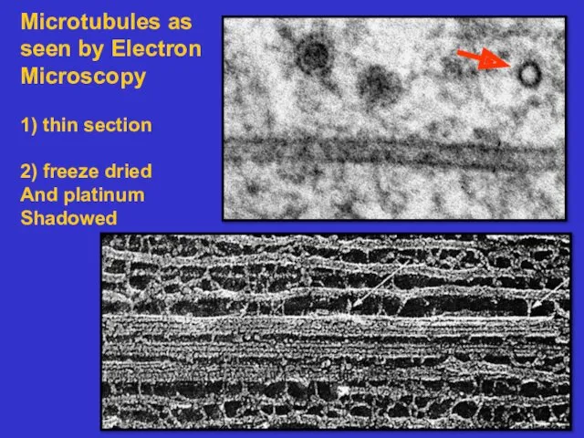
Microtubules as seen by Electron
Microscopy
1) thin section
2) freeze dried
And platinum
Shadowed
Microtubules as seen by Electron
Microscopy
1) thin section
2) freeze dried
And platinum
Shadowed
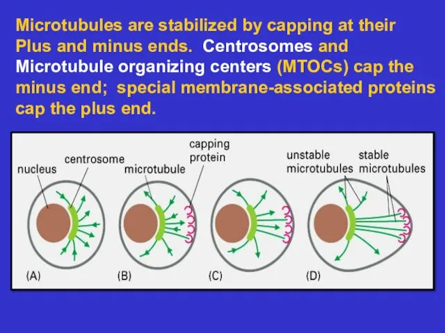
Microtubules are stabilized by capping at their
Plus and minus ends. Centrosomes
Microtubules are stabilized by capping at their
Plus and minus ends. Centrosomes
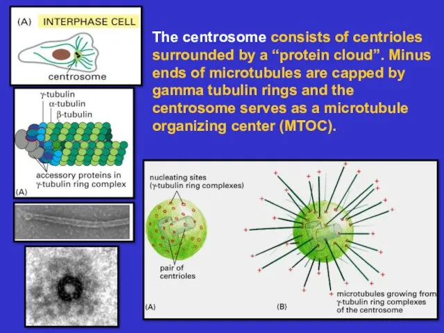
The centrosome consists of centrioles surrounded by a “protein cloud”. Minus
The centrosome consists of centrioles surrounded by a “protein cloud”. Minus
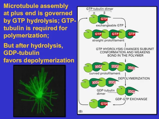
Microtubule assembly
at plus end is governed
by GTP hydrolysis; GTP-
tubulin is
Microtubule assembly
at plus end is governed
by GTP hydrolysis; GTP-
tubulin is
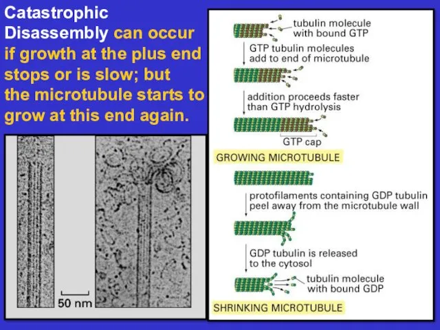
Catastrophic
Disassembly can occur if growth at the plus end stops
Catastrophic
Disassembly can occur if growth at the plus end stops
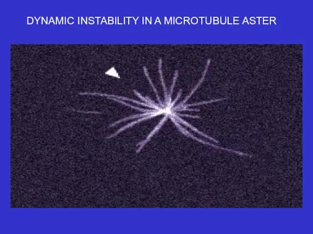
DYNAMIC INSTABILITY IN A MICROTUBULE ASTER
DYNAMIC INSTABILITY IN A MICROTUBULE ASTER
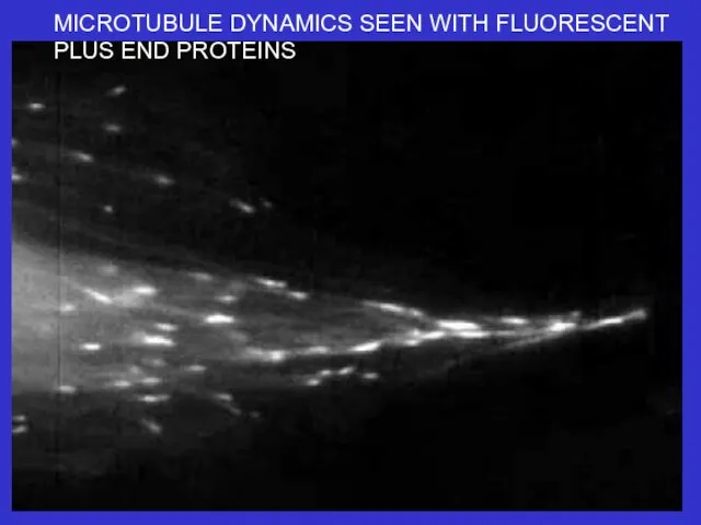
MICROTUBULE DYNAMICS SEEN WITH FLUORESCENT PLUS END PROTEINS
MICROTUBULE DYNAMICS SEEN WITH FLUORESCENT PLUS END PROTEINS
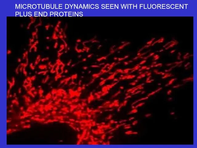
MICROTUBULE DYNAMICS SEEN WITH FLUORESCENT PLUS END PROTEINS
MICROTUBULE DYNAMICS SEEN WITH FLUORESCENT PLUS END PROTEINS
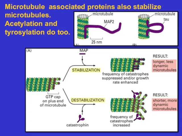
Microtubule associated proteins also stabilize microtubules.
Acetylation and
tyrosylation do too.
Microtubule associated proteins also stabilize microtubules.
Acetylation and
tyrosylation do too.
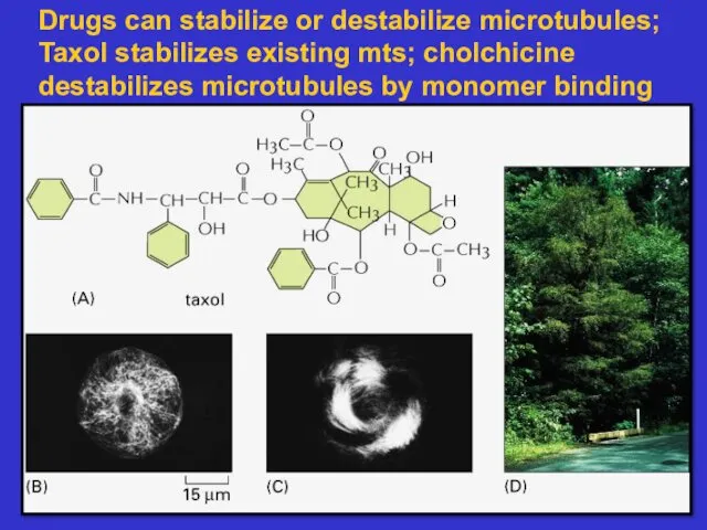
Drugs can stabilize or destabilize microtubules;
Taxol stabilizes existing mts; cholchicine
Drugs can stabilize or destabilize microtubules;
Taxol stabilizes existing mts; cholchicine
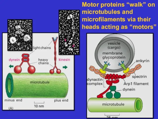
Motor proteins “walk” on microtubules and microfilaments via their heads acting
Motor proteins “walk” on microtubules and microfilaments via their heads acting

Kinesin, like myosin, hydrolyzes ATP as it walks
During this process chemical
Kinesin, like myosin, hydrolyzes ATP as it walks
During this process chemical
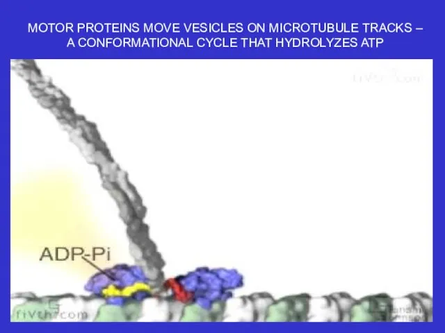
MOTOR PROTEINS MOVE VESICLES ON MICROTUBULE TRACKS –
A CONFORMATIONAL CYCLE
MOTOR PROTEINS MOVE VESICLES ON MICROTUBULE TRACKS –
A CONFORMATIONAL CYCLE
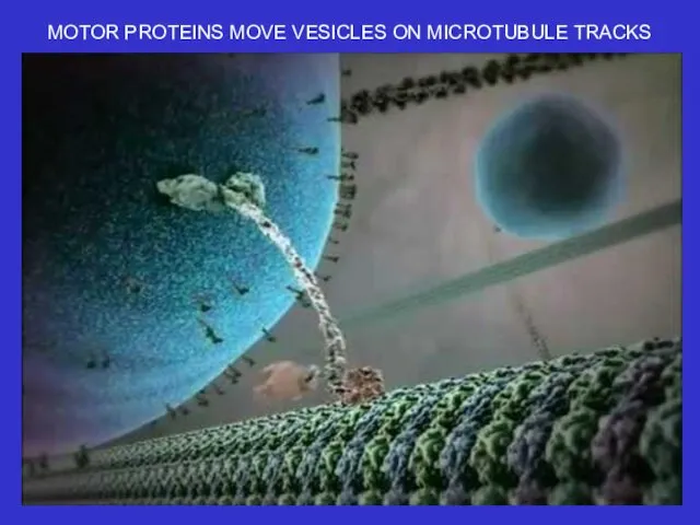
MOTOR PROTEINS MOVE VESICLES ON MICROTUBULE TRACKS
MOTOR PROTEINS MOVE VESICLES ON MICROTUBULE TRACKS
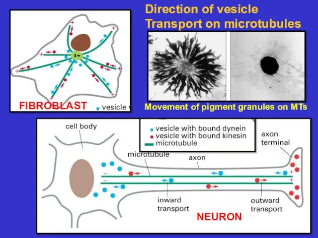
Direction of vesicle
Transport on microtubules
FIBROBLAST
NEURON
Movement of pigment granules on MTs
Direction of vesicle
Transport on microtubules
FIBROBLAST
NEURON
Movement of pigment granules on MTs
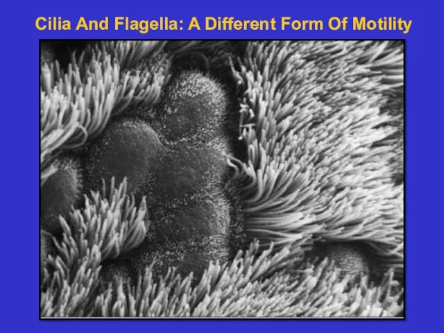
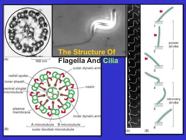
Cilia And Flagella: A Different Form Of Motility
Cilia And Flagella: A Different Form Of Motility
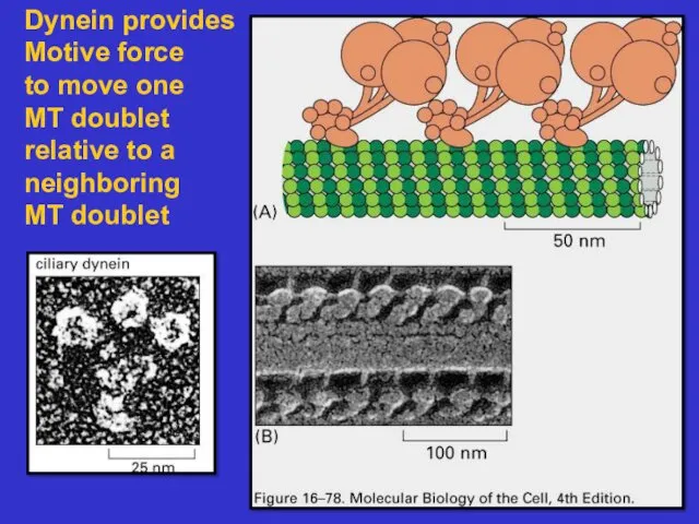
Dynein provides Motive force
to move one
MT doublet
relative to a
neighboring
MT doublet
Dynein provides Motive force
to move one
MT doublet
relative to a
neighboring
MT doublet
 Рефлекторная регуляция
 Ленинградский зоопарк
 Изменчивость: наследственная и ненаследственная. Мутации
 Орієнтування тварин
 Витамины. Какие манипуляции с продуктами позволяют сохранить в них витамины
 Репликация. Синтез ДНК по матрице ДНК
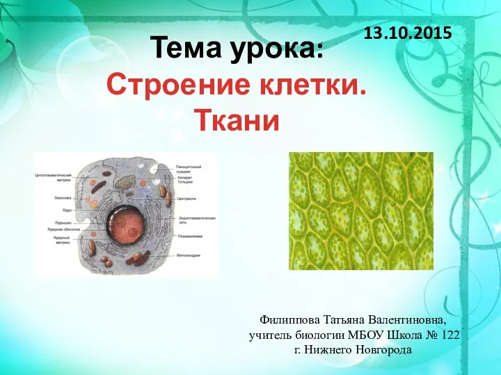 Строение клетки ткани
 Живое вещество биосферы, его функции
 Репликация. Прокариоты. Репликация фагов
 Zoológico de Madrid
 Собачий клещ (Ixodes ricinus)
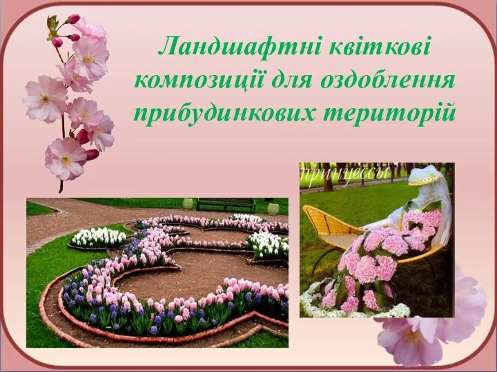 Ландшафтні квіткові композиції для оздоблення прибудинкових територій
 ПРЕЗЕНТАЦИЯ ДЛЯ ИНТЕРАКТИВНОЙ ДОСКИ. Тест Рыбы 7 класс.
 Презентация по биологии на тему _Плауны,хвощи,папоротники_(5 класс) (1)
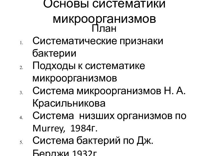 Основы систематики микроорганизмов
 Хрящевые рыбы
 Основы сравнительной эмбриологии
 Клетка и методы цитологии
 Орган слуха и равновесия
 Функциональная анатомия вегетативной нервной системы. Симпатическая часть ВНС (лекция № 23)
 Комнатные растения в интерьере помещения
 Хронобиология и биоритмы человека
 Окапи
Окапи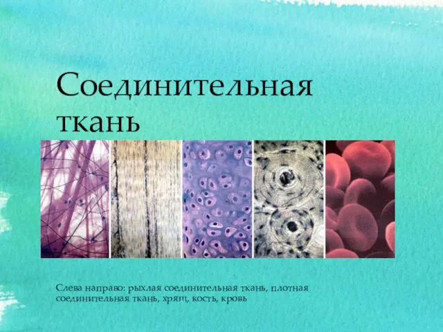 Соединительная ткань
 Овчарки со всего мира. Фотографии
 Общие признаки для всех живых организмов
 Изоляция
 Соцветия. Строение соцветия