- The nervous system and nervous tissue

Содержание
- 2. MAJOR CHAPTER OBJECTIVES Name the major divisions of the nervous system, both anatomical and functional Describe
- 3. 12.1 BASIC STRUCTURE AND FUNCTION OF THE NERVOUS SYSTEM MAJOR SECTION OBJECTIVES Identify the anatomical and
- 4. FIGURE 12.2 Central and Peripheral Nervous System The structures of the PNS are referred to as
- 5. FIGURE 12.3 Gray Matter and White Matter A brain removed during an autopsy, with a partial
- 6. FIGURE 12.4 What Is a Nucleus? The nucleus of an atom contains its protons and neutrons.
- 7. FIGURE 12.5 Optic Nerve Versus Optic Tract This drawing of the connections of the eye to
- 8. TABLE 12.1 Structures of the CNS and PNS
- 9. FIGURE 12.6 Somatic, Autonomic, and Enteric Structures of the Nervous System Somatic structures include the spinal
- 10. Image source: Adapted from Marieb’s Anatomy and Physiology, 9th edition, Pearson. RELATIONSHIPS BETWEEN THE SUBDIVISIONS OF
- 11. 12.2 NERVOUS TISSUE MAJOR SECTION OBJECTIVES Describe the basic structure of a neuron Identify the different
- 12. FIGURE 12.8 Parts of a Neuron The major parts of the neuron are labeled on a
- 13. FIGURE 12.9 Neuron Classification by Shape Unipolar cells have one process that includes both the axon
- 14. FIGURE 12.10 Other Neuron Classifications Three examples of neurons that are classified on the basis of
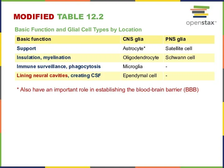
- 15. MODIFIED TABLE 12.2 Basic Function and Glial Cell Types by Location * Also have an important
- 16. FIGURE 12.11 Glial Cells of the CNS The CNS has astrocytes, oligodendrocytes, microglia, and ependymal cells
- 17. THE FOUR MAJOR GLIAL CELL TYPES OF THE CNS Image source: Adapted from Marieb’s Anatomy and
- 18. FIGURE 12.12 Glial Cells of the PNS The PNS has satellite cells and Schwann cells.
- 19. FIGURE 12.13 The Process of Myelination Myelinating glia wrap several layers of cell membrane around the
- 20. 12.3 NERVOUS TISSUE MAJOR SECTION OBJECTIVES Distinguish the major functions of the nervous system: sensation integration
- 21. FIGURE 12.14
- 22. FIGURE 12.15 The Sensory Input Receptors in the skin sense the temperature of the water.
- 23. FIGURE 12.16 The Motor Response On the basis of the sensory input and the integration in
- 24. 12.4 THE ACTION POTENTIAL MAJOR SECTION OBJECTIVES Describe the components of the membrane that establish the
- 25. FIGURE 12.17 Cell Membrane and Transmembrane Proteins The cell membrane is composed of a phospholipid bilayer
- 26. FIGURE 12.18 Ligand-Gated Channels When the ligand, in this case the neurotransmitter acetylcholine, binds to a
- 27. FIGURE 12.19 Mechanically Gated Channels When a mechanical change occurs in the surrounding tissue, such as
- 28. FIGURE 12.20 Voltage-Gated Channels Voltage-gated channels open when the transmembrane voltage changes around them. Amino acids
- 29. FIGURE 12.21 Leakage Channels In certain situations, ions need to move across the membrane randomly. The
- 30. FIGURE 12.22 Measuring Charge across a Membrane with a Voltmeter A recording electrode is inserted into
- 31. FIGURE 12.23 Graph of Action Potential Plotting voltage measured across the cell membrane against time, the
- 32. FIGURE 12.24 Stages of an Action Potential Plotting voltage measured across the cell membrane against time,
- 33. GENERATION OF AN ACTION POTENTIAL Resting state. No ions move through voltage-gated channels. Depolarization is caused
- 34. 12.5 THE GRADED POTENTIALS MAJOR SECTION OBJECTIVES Explain the differences between the types of graded potentials
- 35. FIGURE 12.25 Graded Potentials Graded potentials are temporary changes in the membrane voltage, the characteristics of
- 36. FIGURE 12.26 Postsynaptic Potential Summation The result of summation of postsynaptic potentials is the overall change
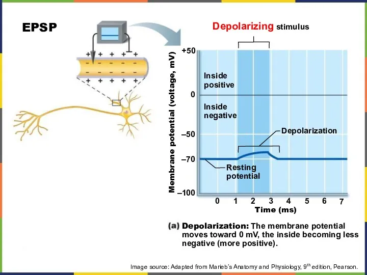
- 37. Depolarizing stimulus Inside positive Inside negative Depolarization Resting potential Membrane potential (voltage, mV) Depolarization: The membrane
- 38. Hyperpolarizing stimulus Membrane potential (voltage, mV) Time (ms) +50 0 –50 –70 –100 0 1 2
- 39. Stimulus Depolarized region Plasma membrane Depolarization: A small patch of the membrane (red area) depolarizes. Image
- 40. Active area (site of initial depolarization) Resting potential Membrane potential (mV) Distance (a few mm) Decay
- 41. SYNAPTIC INTEGRATION: SUMMATION Most neurons receive both excitatory and inhibitory inputs from thousands of other neurons
- 42. EXAMPLE 1: NO SUMMATION (EPSPS) Image source: Adapted from Marieb’s Anatomy and Physiology, 9th edition, Pearson.
- 43. TEMPORAL SUMMATION (EPSPS) Image source: Adapted from Marieb’s Anatomy and Physiology, 9th edition, Pearson.
- 44. SPATIAL SUMMATION (EPSPS) Image source: Adapted from Marieb’s Anatomy and Physiology, 9th edition, Pearson.
- 45. SUMMATION BUT NO AP (EPSPS AND IPSPS) Image source: Adapted from Marieb’s Anatomy and Physiology, 9th
- 46. INTEGRATION: SYNAPTIC POTENTIATION Synaptic potentiation: the repeated use of a given synapse increases ability of presynaptic
- 47. SYNAPSES Electrical Physical connection of pre- and post-synaptic elements Electric signals go through Most abundant in
- 48. FIGURE 12.27 The Chemical Synapse The synapse is a connection between a neuron and its target
- 49. INFORMATION TRANSFER ACROSS CHEMICAL SYNAPSES Image source: Adapted from Marieb’s Anatomy and Physiology, 9th edition, Pearson.
- 50. CHEMICAL SYNAPSE (1/3) 1- Action potential arrives at axon terminal. 2- Voltage-gated Ca2+ channels open and
- 51. CHEMICAL SYNAPSE (2/3) 3- Ca2+ entry (binding to synaptotagmin) causes synaptic vesicles to release neurotransmitter by
- 52. CHEMICAL SYNAPSE (2/3) Graded potential 5- Binding of neuro-transmitter opens ion channels, resulting in graded potentials.
- 53. FIGURE 12.28 Receptor Types An ionotropic receptor is a channel that opens when the neurotransmitter binds
- 54. MODIFIED TABLE 12.3 Characteristics of Selected Neurotransmitters Legend: (I), ionotropic or direct signaling; (M) metabotropic or
- 55. EVERYDAY CONNECTIONS Potassium Concentration and Astrocytes Glial cells, especially astrocytes, are responsible for maintaining the chemical
- 56. DISORDERS & HOMEOSTATIC IMBALANCES Demyelination Disorders Diseases of genetic, infectious or autoimmune origins can cause a
- 57. DISORDERS & HOMEOSTATIC IMBALANCES Proteopathies For proteins to function correctly, their linear sequence of amino acids
- 58. INTERACTIVE LINKS Visit the Nobel Prize web site http://openstaxcollege.org/l/nobel_2 to play an interactive game that demonstrates
- 59. INTERACTIVE LINKS FYI - Visit this site http://openstaxcollege.org/l/neurolab to see a virtual neurophysiology lab, and to
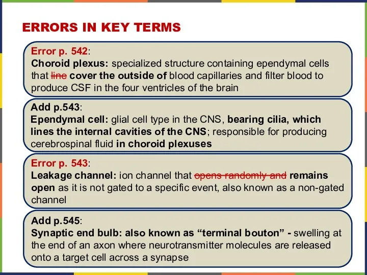
- 60. ERRORS IN KEY TERMS Error p. 542: Choroid plexus: specialized structure containing ependymal cells that line
- 61. This PowerPoint presentation is copyright 2011-2015, Rice University. All Rights Reserved. Last modified: 09/2017 / Dr.
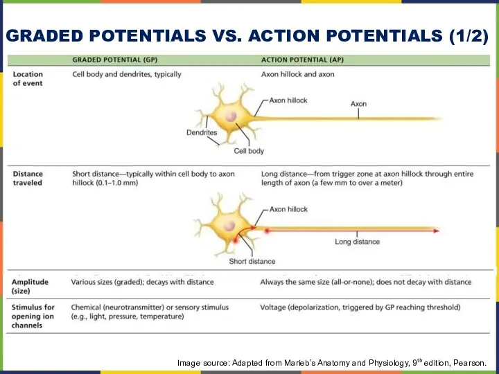
- 62. GRADED POTENTIALS VS. ACTION POTENTIALS (1/2) Image source: Adapted from Marieb’s Anatomy and Physiology, 9th edition,
- 64. Скачать презентацию
MAJOR CHAPTER OBJECTIVES
Name the major divisions of the nervous system, both
MAJOR CHAPTER OBJECTIVES
Name the major divisions of the nervous system, both
12.1 BASIC STRUCTURE AND FUNCTION OF THE NERVOUS SYSTEM
MAJOR SECTION OBJECTIVES
Identify
12.1 BASIC STRUCTURE AND FUNCTION OF THE NERVOUS SYSTEM
MAJOR SECTION OBJECTIVES
Identify
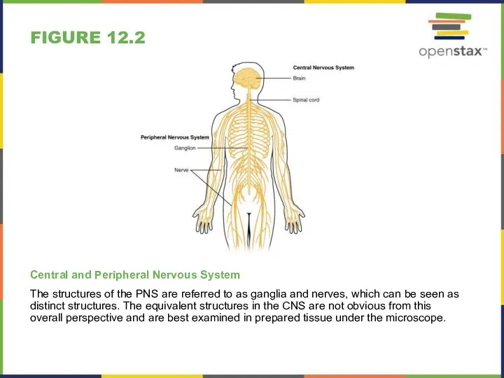
FIGURE 12.2
Central and Peripheral Nervous System
The structures of the PNS are
FIGURE 12.2
Central and Peripheral Nervous System
The structures of the PNS are
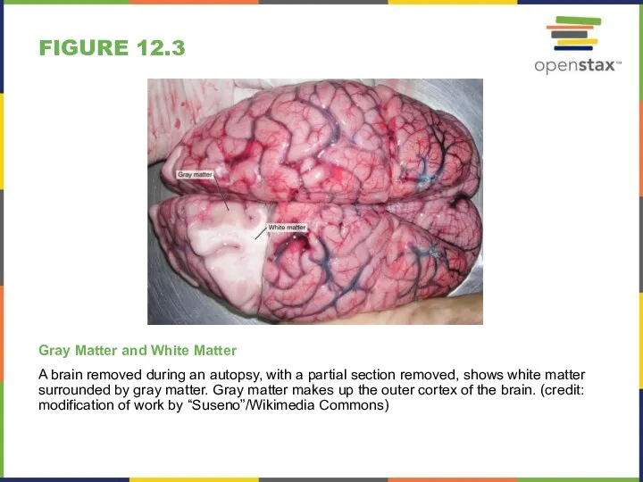
FIGURE 12.3
Gray Matter and White Matter
A brain removed during an autopsy,
FIGURE 12.3
Gray Matter and White Matter
A brain removed during an autopsy,
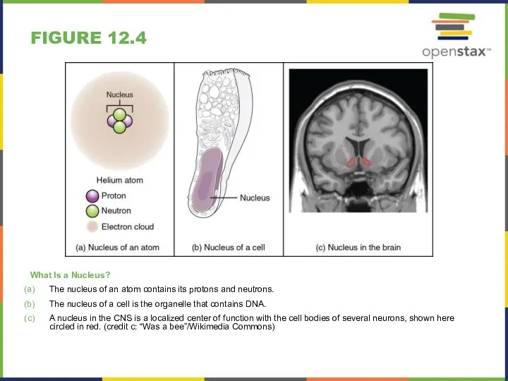
FIGURE 12.4
What Is a Nucleus?
The nucleus of an atom contains its
FIGURE 12.4
What Is a Nucleus?
The nucleus of an atom contains its
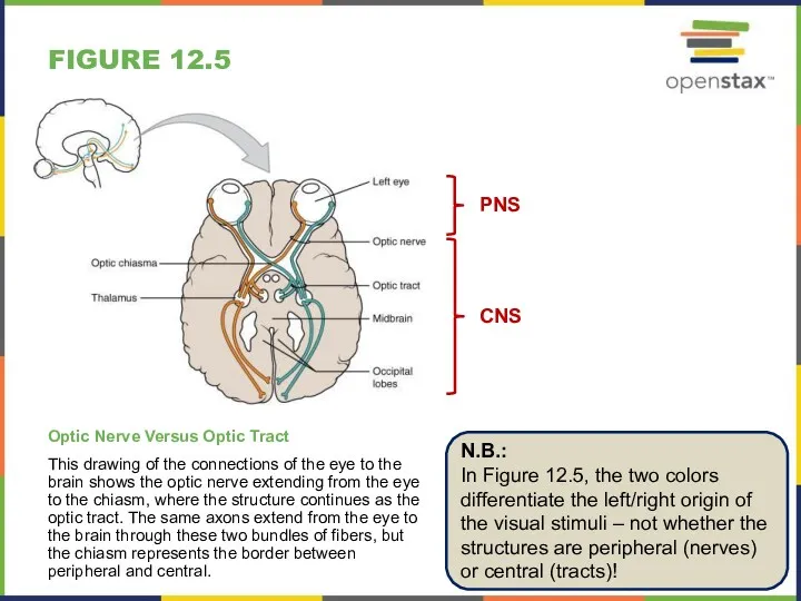
FIGURE 12.5
Optic Nerve Versus Optic Tract
This drawing of the connections of
FIGURE 12.5
Optic Nerve Versus Optic Tract
This drawing of the connections of
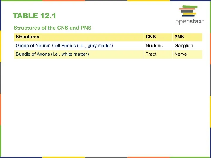
TABLE 12.1
Structures of the CNS and PNS
TABLE 12.1
Structures of the CNS and PNS
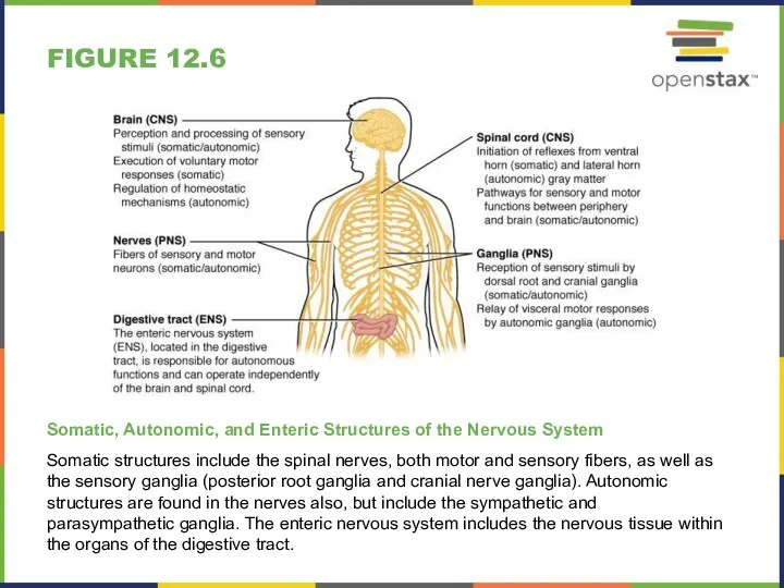
FIGURE 12.6
Somatic, Autonomic, and Enteric Structures of the Nervous System
Somatic structures
FIGURE 12.6
Somatic, Autonomic, and Enteric Structures of the Nervous System
Somatic structures
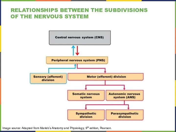
Image source: Adapted from Marieb’s Anatomy and Physiology, 9th edition, Pearson.
RELATIONSHIPS
Image source: Adapted from Marieb’s Anatomy and Physiology, 9th edition, Pearson.
RELATIONSHIPS
12.2 NERVOUS TISSUE
MAJOR SECTION OBJECTIVES
Describe the basic structure of a neuron
Identify
12.2 NERVOUS TISSUE
MAJOR SECTION OBJECTIVES
Describe the basic structure of a neuron
Identify
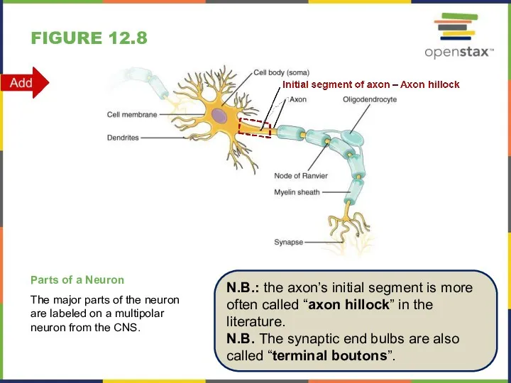
FIGURE 12.8
Parts of a Neuron
The major parts of the neuron are
FIGURE 12.8
Parts of a Neuron
The major parts of the neuron are
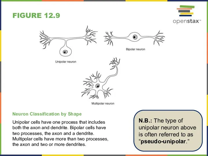
FIGURE 12.9
Neuron Classification by Shape
Unipolar cells have one process that includes
FIGURE 12.9
Neuron Classification by Shape
Unipolar cells have one process that includes
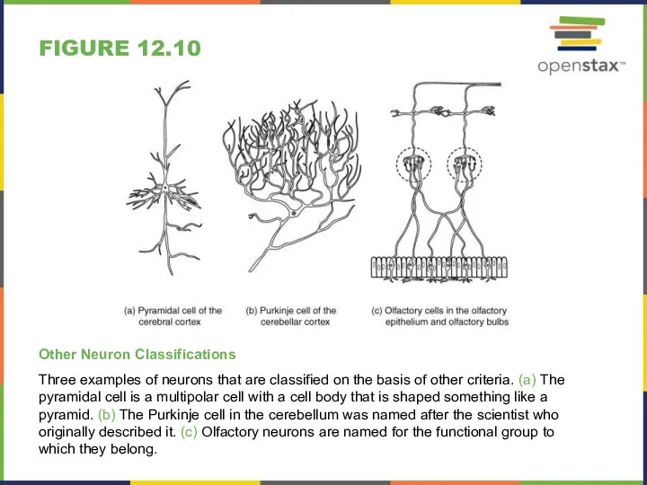
FIGURE 12.10
Other Neuron Classifications
Three examples of neurons that are classified on
FIGURE 12.10
Other Neuron Classifications
Three examples of neurons that are classified on
MODIFIED TABLE 12.2
Basic Function and Glial Cell Types by Location
* Also
MODIFIED TABLE 12.2
Basic Function and Glial Cell Types by Location
* Also
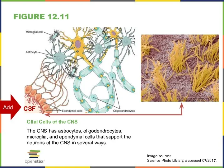
FIGURE 12.11
Glial Cells of the CNS
The CNS has astrocytes, oligodendrocytes, microglia,
FIGURE 12.11
Glial Cells of the CNS
The CNS has astrocytes, oligodendrocytes, microglia,
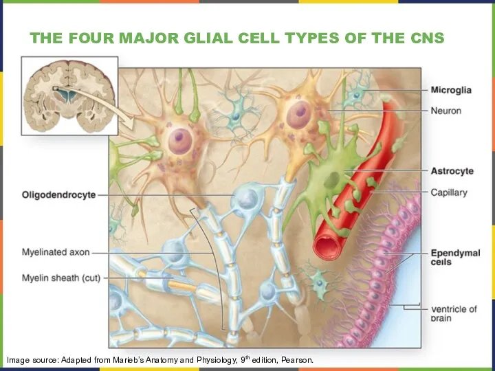
THE FOUR MAJOR GLIAL CELL TYPES OF THE CNS
Image source: Adapted
THE FOUR MAJOR GLIAL CELL TYPES OF THE CNS
Image source: Adapted
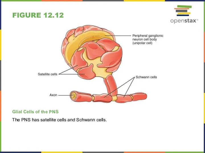
FIGURE 12.12
Glial Cells of the PNS
The PNS has satellite cells and
FIGURE 12.12
Glial Cells of the PNS
The PNS has satellite cells and
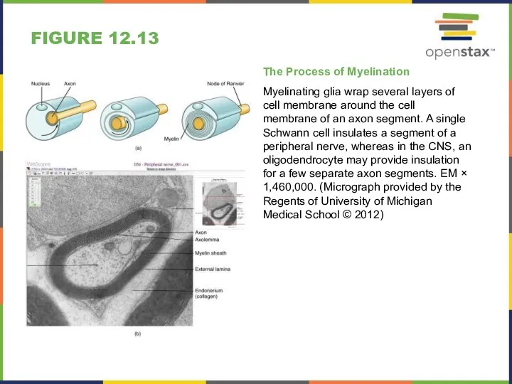
FIGURE 12.13
The Process of Myelination
Myelinating glia wrap several layers of cell
FIGURE 12.13
The Process of Myelination
Myelinating glia wrap several layers of cell
12.3 NERVOUS TISSUE
MAJOR SECTION OBJECTIVES
Distinguish the major functions of the nervous
12.3 NERVOUS TISSUE
MAJOR SECTION OBJECTIVES
Distinguish the major functions of the nervous
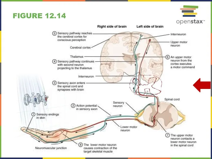
FIGURE 12.14
FIGURE 12.14
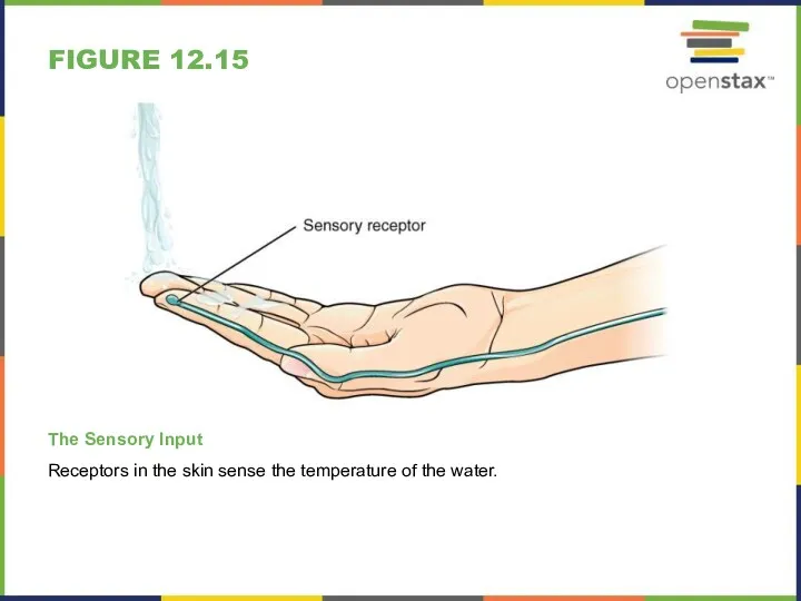
FIGURE 12.15
The Sensory Input
Receptors in the skin sense the temperature of
FIGURE 12.15
The Sensory Input
Receptors in the skin sense the temperature of
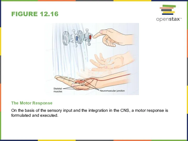
FIGURE 12.16
The Motor Response
On the basis of the sensory input and
FIGURE 12.16
The Motor Response
On the basis of the sensory input and
12.4 THE ACTION POTENTIAL
MAJOR SECTION OBJECTIVES
Describe the components of the membrane
12.4 THE ACTION POTENTIAL
MAJOR SECTION OBJECTIVES
Describe the components of the membrane
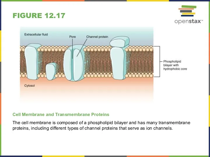
FIGURE 12.17
Cell Membrane and Transmembrane Proteins
The cell membrane is composed of
FIGURE 12.17
Cell Membrane and Transmembrane Proteins
The cell membrane is composed of
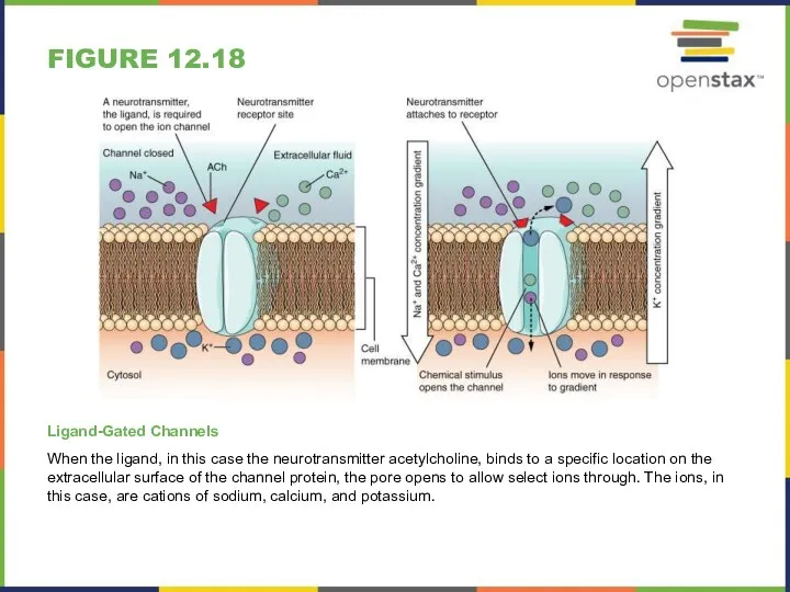
FIGURE 12.18
Ligand-Gated Channels
When the ligand, in this case the neurotransmitter acetylcholine,
FIGURE 12.18
Ligand-Gated Channels
When the ligand, in this case the neurotransmitter acetylcholine,
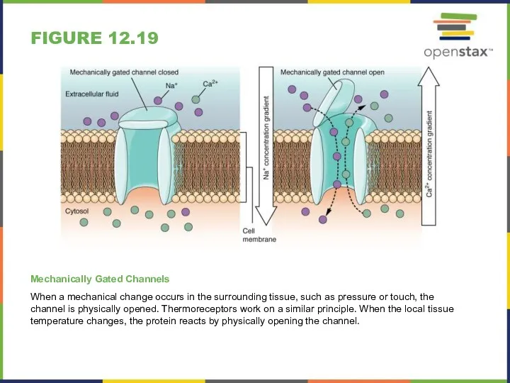
FIGURE 12.19
Mechanically Gated Channels
When a mechanical change occurs in the surrounding
FIGURE 12.19
Mechanically Gated Channels
When a mechanical change occurs in the surrounding
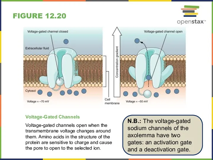
FIGURE 12.20
Voltage-Gated Channels
Voltage-gated channels open when the transmembrane voltage changes around
FIGURE 12.20
Voltage-Gated Channels
Voltage-gated channels open when the transmembrane voltage changes around
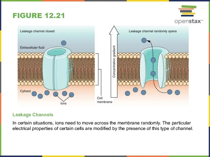
FIGURE 12.21
Leakage Channels
In certain situations, ions need to move across the
FIGURE 12.21
Leakage Channels
In certain situations, ions need to move across the
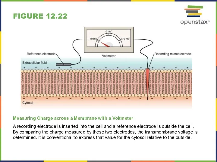
FIGURE 12.22
Measuring Charge across a Membrane with a Voltmeter
A recording electrode
FIGURE 12.22
Measuring Charge across a Membrane with a Voltmeter
A recording electrode
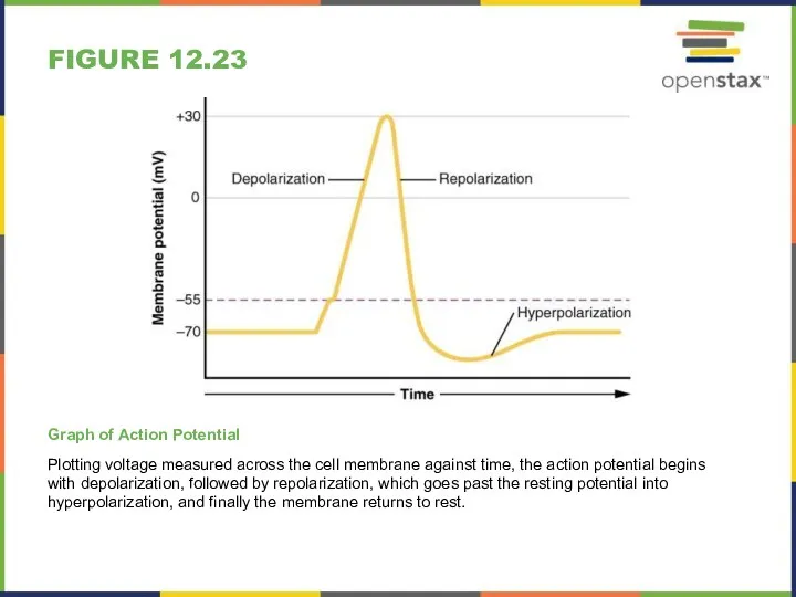
FIGURE 12.23
Graph of Action Potential
Plotting voltage measured across the cell membrane
FIGURE 12.23
Graph of Action Potential
Plotting voltage measured across the cell membrane
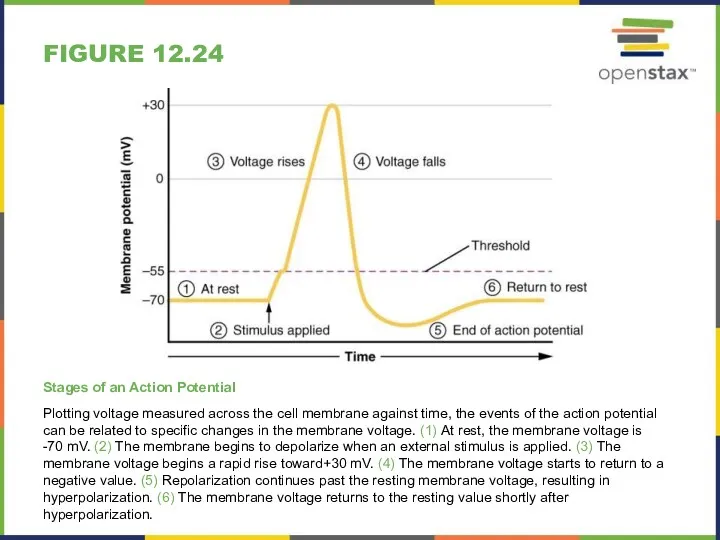
FIGURE 12.24
Stages of an Action Potential
Plotting voltage measured across the cell
FIGURE 12.24
Stages of an Action Potential
Plotting voltage measured across the cell
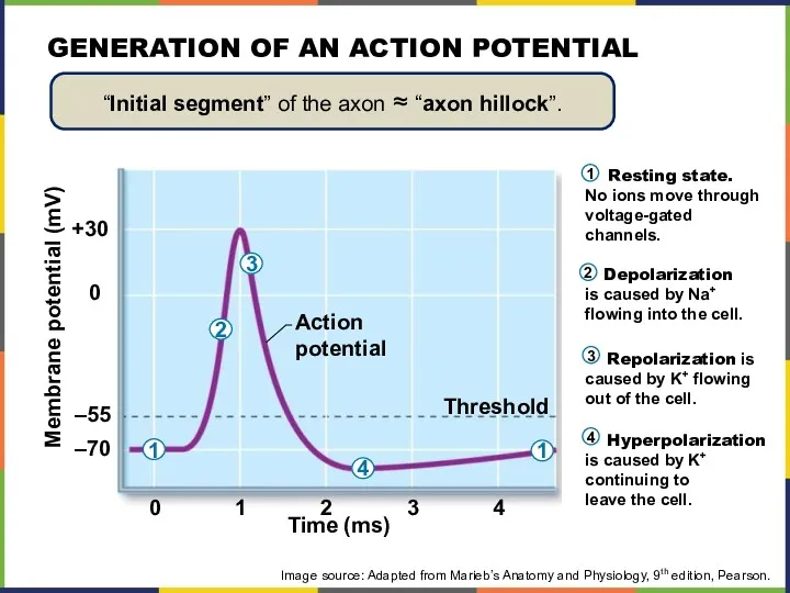
GENERATION OF AN ACTION POTENTIAL
Resting state.
No ions move through
voltage-gated
channels.
GENERATION OF AN ACTION POTENTIAL
Resting state.
No ions move through
voltage-gated
channels.
12.5 THE GRADED POTENTIALS
MAJOR SECTION OBJECTIVES
Explain the differences between the types
12.5 THE GRADED POTENTIALS
MAJOR SECTION OBJECTIVES
Explain the differences between the types
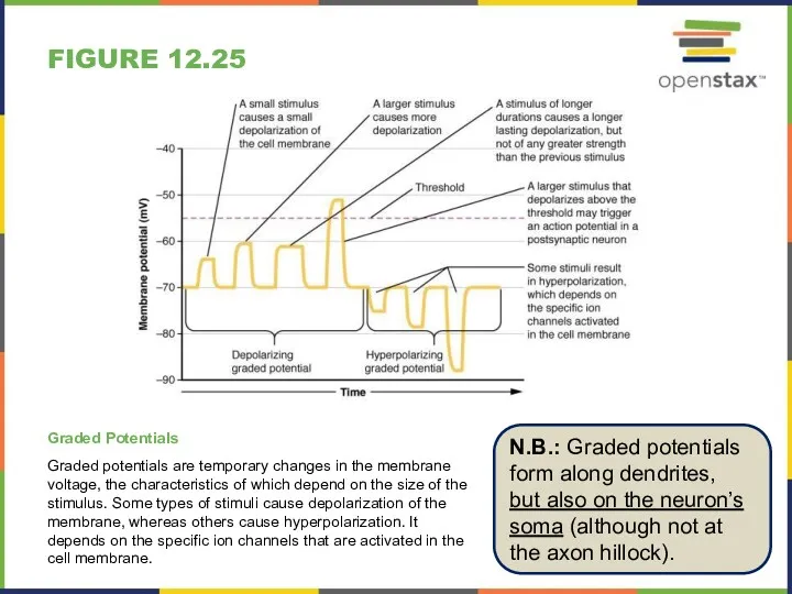
FIGURE 12.25
Graded Potentials
Graded potentials are temporary changes in the membrane voltage,
FIGURE 12.25
Graded Potentials
Graded potentials are temporary changes in the membrane voltage,
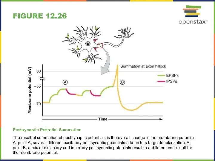
FIGURE 12.26
Postsynaptic Potential Summation
The result of summation of postsynaptic potentials is
FIGURE 12.26
Postsynaptic Potential Summation
The result of summation of postsynaptic potentials is
Depolarizing stimulus
Inside
positive
Inside
negative
Depolarization
Resting
potential
Membrane potential (voltage, mV)
Depolarization: The membrane potential
moves toward 0 mV,
Depolarizing stimulus
Inside
positive
Inside
negative
Depolarization
Resting
potential
Membrane potential (voltage, mV)
Depolarization: The membrane potential
moves toward 0 mV,
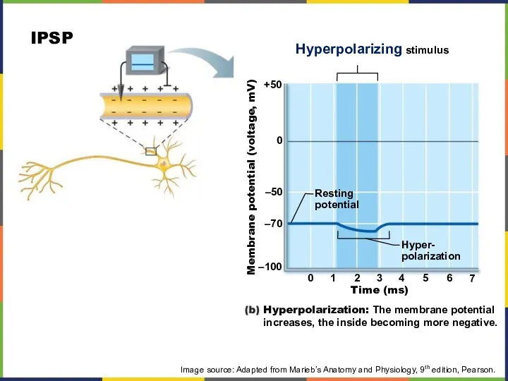
Hyperpolarizing stimulus
Membrane potential (voltage, mV)
Time (ms)
+50
0
–50
–70
–100
0
1
2
3
4
5
6
7
Hyperpolarization: The membrane potential
increases, the inside
Hyperpolarizing stimulus
Membrane potential (voltage, mV)
Time (ms)
+50
0
–50
–70
–100
0
1
2
3
4
5
6
7
Hyperpolarization: The membrane potential
increases, the inside
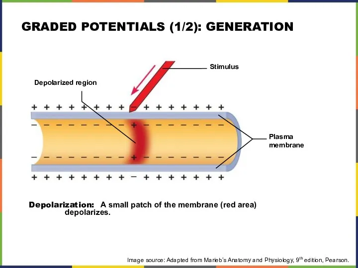
Stimulus
Depolarized region
Plasma
membrane
Depolarization: A small patch of the membrane (red area)
depolarizes.
Image
Stimulus
Depolarized region
Plasma
membrane
Depolarization: A small patch of the membrane (red area)
depolarizes.
Image
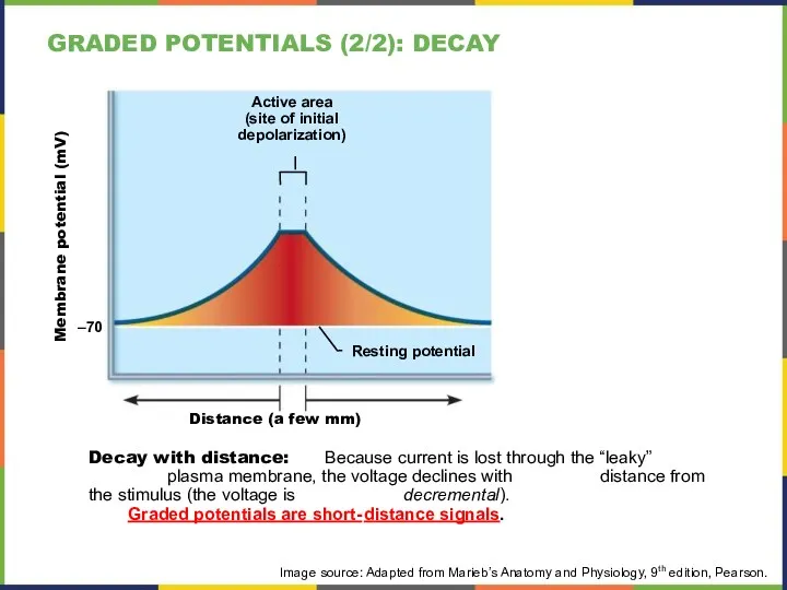
Active area
(site of initial
depolarization)
Resting potential
Membrane potential (mV)
Distance (a few mm)
Decay with
Active area
(site of initial
depolarization)
Resting potential
Membrane potential (mV)
Distance (a few mm)
Decay with
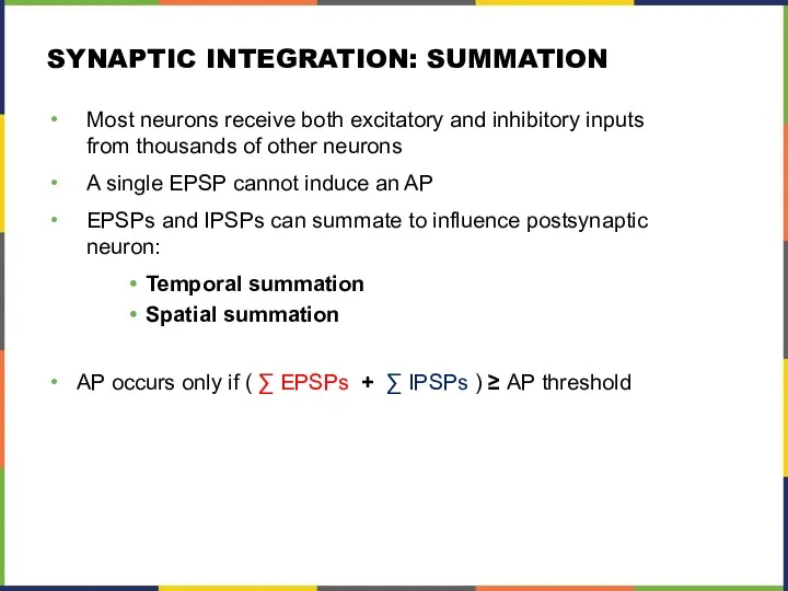
SYNAPTIC INTEGRATION: SUMMATION
Most neurons receive both excitatory and inhibitory inputs from
SYNAPTIC INTEGRATION: SUMMATION
Most neurons receive both excitatory and inhibitory inputs from
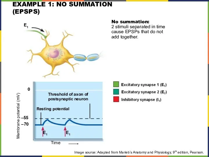
EXAMPLE 1: NO SUMMATION (EPSPS)
Image source: Adapted from Marieb’s Anatomy and
EXAMPLE 1: NO SUMMATION (EPSPS)
Image source: Adapted from Marieb’s Anatomy and
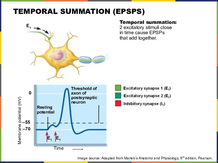
TEMPORAL SUMMATION (EPSPS)
Image source: Adapted from Marieb’s Anatomy and Physiology, 9th
TEMPORAL SUMMATION (EPSPS)
Image source: Adapted from Marieb’s Anatomy and Physiology, 9th
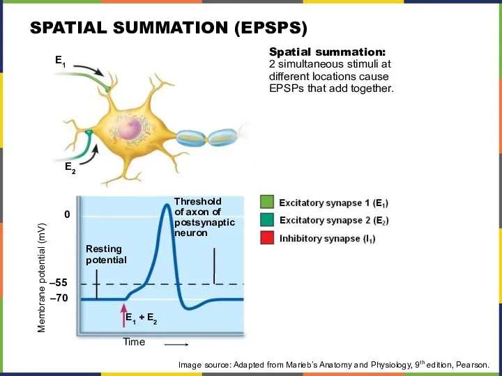
SPATIAL SUMMATION (EPSPS)
Image source: Adapted from Marieb’s Anatomy and Physiology, 9th
SPATIAL SUMMATION (EPSPS)
Image source: Adapted from Marieb’s Anatomy and Physiology, 9th
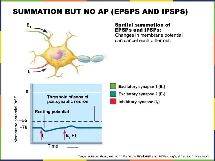
SUMMATION BUT NO AP (EPSPS AND IPSPS)
Image source: Adapted from Marieb’s
SUMMATION BUT NO AP (EPSPS AND IPSPS)
Image source: Adapted from Marieb’s
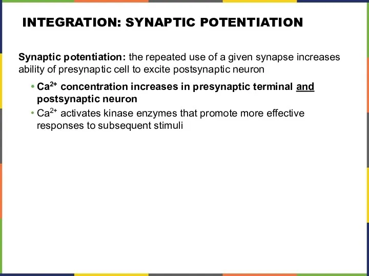
INTEGRATION: SYNAPTIC POTENTIATION
Synaptic potentiation: the repeated use of a given synapse
INTEGRATION: SYNAPTIC POTENTIATION
Synaptic potentiation: the repeated use of a given synapse
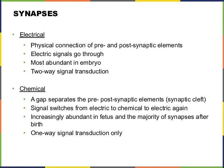
SYNAPSES
Electrical
Physical connection of pre- and post-synaptic elements
Electric signals go through
Most abundant
SYNAPSES
Electrical
Physical connection of pre- and post-synaptic elements
Electric signals go through
Most abundant
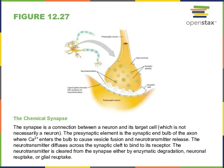
FIGURE 12.27
The Chemical Synapse
The synapse is a connection between a neuron
FIGURE 12.27
The Chemical Synapse
The synapse is a connection between a neuron
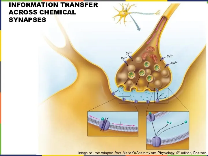
INFORMATION TRANSFER ACROSS CHEMICAL SYNAPSES
Image source: Adapted from Marieb’s Anatomy and
INFORMATION TRANSFER ACROSS CHEMICAL SYNAPSES
Image source: Adapted from Marieb’s Anatomy and
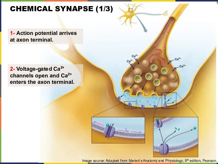
CHEMICAL SYNAPSE (1/3)
1- Action potential arrives
at axon terminal.
2- Voltage-gated
CHEMICAL SYNAPSE (1/3)
1- Action potential arrives
at axon terminal.
2- Voltage-gated
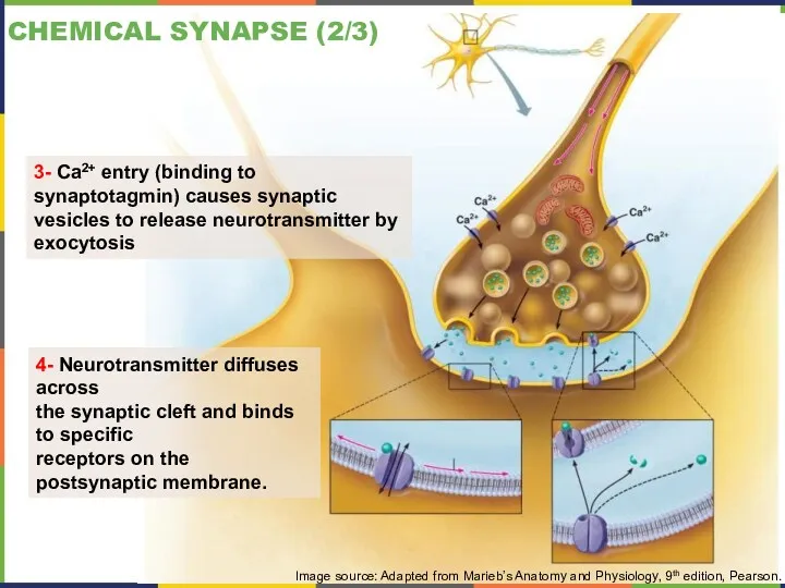
CHEMICAL SYNAPSE (2/3)
3- Ca2+ entry (binding to synaptotagmin) causes synaptic vesicles
CHEMICAL SYNAPSE (2/3)
3- Ca2+ entry (binding to synaptotagmin) causes synaptic vesicles
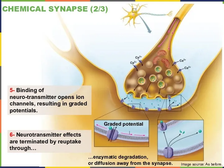
CHEMICAL SYNAPSE (2/3)
Graded potential
5- Binding of neuro-transmitter opens ion channels, resulting
CHEMICAL SYNAPSE (2/3)
Graded potential
5- Binding of neuro-transmitter opens ion channels, resulting
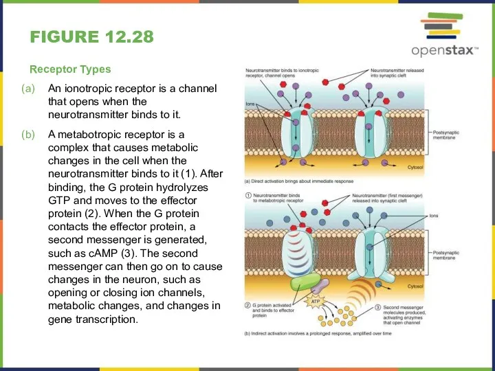
FIGURE 12.28
Receptor Types
An ionotropic receptor is a channel that opens when
FIGURE 12.28
Receptor Types
An ionotropic receptor is a channel that opens when
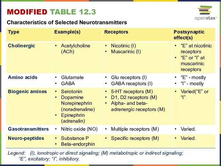
MODIFIED TABLE 12.3
Characteristics of Selected Neurotransmitters
Legend: (I), ionotropic or direct
MODIFIED TABLE 12.3
Characteristics of Selected Neurotransmitters
Legend: (I), ionotropic or direct
EVERYDAY CONNECTIONS
Potassium Concentration and Astrocytes
Glial cells, especially astrocytes, are responsible for
EVERYDAY CONNECTIONS
Potassium Concentration and Astrocytes
Glial cells, especially astrocytes, are responsible for
DISORDERS & HOMEOSTATIC IMBALANCES
Demyelination Disorders
Diseases of genetic, infectious or autoimmune origins
DISORDERS & HOMEOSTATIC IMBALANCES
Demyelination Disorders
Diseases of genetic, infectious or autoimmune origins
DISORDERS & HOMEOSTATIC IMBALANCES
Proteopathies
For proteins to function correctly, their linear sequence
DISORDERS & HOMEOSTATIC IMBALANCES
Proteopathies
For proteins to function correctly, their linear sequence
INTERACTIVE LINKS
Visit the Nobel Prize web site http://openstaxcollege.org/l/nobel_2 to play an
INTERACTIVE LINKS
Visit the Nobel Prize web site http://openstaxcollege.org/l/nobel_2 to play an
INTERACTIVE LINKS
FYI - Visit this site http://openstaxcollege.org/l/neurolab to see a virtual
INTERACTIVE LINKS
FYI - Visit this site http://openstaxcollege.org/l/neurolab to see a virtual
ERRORS IN KEY TERMS
Error p. 542:
Choroid plexus: specialized structure containing
ERRORS IN KEY TERMS
Error p. 542:
Choroid plexus: specialized structure containing

This PowerPoint presentation is copyright 2011-2015, Rice University. All Rights Reserved.
Last
This PowerPoint presentation is copyright 2011-2015, Rice University. All Rights Reserved.
Last
GRADED POTENTIALS VS. ACTION POTENTIALS (1/2)
Image source: Adapted from Marieb’s Anatomy
GRADED POTENTIALS VS. ACTION POTENTIALS (1/2)
Image source: Adapted from Marieb’s Anatomy
 Биоэлектрогенез. Строение и физические свойства биологических мембран
 Пищеварительная система
 Структурная организация микробной клетки
 Биотехнология как источник био-риска
 Фізіологія. Механізми регуляції
 Систематика Покритонасінних рослин. Відділ Магноліофіти, Квіткові або Покритонасінні
 Общая физиология сенсорных систем
 Отряд голенастые
 Белгілерді ң тұқым қуалауының негізгі зандары, т ұқым қуалайтын белгілерді ңберілу ережелері
 Правила техники безопасности в кабинете биологии
 Анатомия нервной системы
 Тип членистоногие. Класс насекомые
 Тип Членистоногие
 Листериоз. Род Listeria
 Интегрированный урок по биологии,химии и физике. Викторина Колосок
 Дыхательная система. Органы дыхания
 Поведение собак
 Основы селекции. Работы Н.И. Вавилова
 Краткая история изучения клетки
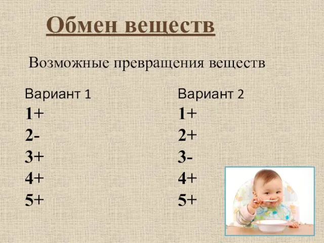 Обмен веществ. Возможные превращения веществ
 Тканевые элементы нервной системы
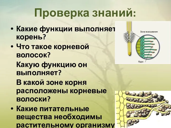 Какие функции выполняет корень?
 Лисиця. Представниця класу ссавці, ряду хижі звірі
 Анатомия крысы
 Зимующие птицы
 ГМО в пищевой промышленности
 Зоология - наука о животных. Подготовка к ОГЭ и ЕГЭ по биологии
 Основные фосфолипиды и гликолипиды тканей человека