- Tissues. The histophysiology of the epithelial tissue

Содержание
- 2. Tissue. The basic types of tissues. The common characteristics of epithelia. Histogenesis of the epithelia. The
- 3. The tissue – is the morphological or morphofisiological system. Tissue is the team of the same
- 4. The tissues are systems of cells and noncellular structures characterized by similar structural, functional properties and
- 5. Differentiation – the morphofunctional exchange of the same organized cells. The main result of the differentiation
- 6. Layers Stratum corneum Stratum lucidum Stratum granulosum Stratum spinosum Stratum basale CONNECTIVE TISSUE Time-differentiation – the
- 7. There are elements of the ciliar epithelium: CILIAR CELLS GOBLET CELLS BASE CELLS INTERMEDIATE CELLS ENDOCRINE
- 8. EXOCRINE AND ENDOCRINE CELLS OF THE INTESTINAL EPITHELIUM Biochemical-differentiation – creation of the cells producing specific
- 9. First of all start to differentiate the steam cells source the differon. Steam cell’s characteristics: They
- 10. Regeneration – the capability of the tissue to recover itself after violation. There are known different
- 11. Physiological regeneration – the recovering of the cell’s population after the death of the some cells.
- 12. The History. 1665 год. Robert Hook was describe the “cell”. 1830 год. Jan Purcinje - cytoplasm.
- 13. THE BASIC TYPES OF TISSUES EPITHELIAL CONNECTIVE (SUPPORT) AND BLOOD MUSCLE NERVOUS
- 14. THE COMMON CHARACTERISTICS OF EPITHELIA COVER SURFACES OR LINE CAVITIES FORM CONTINUOUS LAYERS INDIVIDUAL CELLS ARE
- 15. CLASSIFICATION OF EPITHELIA 1.ARRANGEMENT OF LAYERS SIMPLE (ALL OF THE CELLS CONTACT UNDERLYING BASEMENT MEMBRANE) STRATIFIED
- 16. THE LOCATION OF THE MAJOR TYPES OF EPITHELIA
- 17. SPECIALIZATIONS OF THE PLASMA MEMBRANE 1.APICAL PART – MICROVILLI, CILIA, STEREOCILIA, BORDERS 2.LATERAL PART – CELL
- 25. SECRETORY EPITHELIA AND GLANDS ENDOCRINE EXOCRINE EXOCRINE GLANDS SIMPLE COMPOUND ALVEOLAR TUBULAR MIXED BRANCHED NONBRANCHED PROTEINS
- 27. EXOCRINE AND ENDOCRINE CELLS OF INTESTINAL EPITHELIUM
- 28. PROTEINS-SECRETING CELLS MUCUS-SECRETING CELLS IONS-PUMPING CELLS
- 32. Скачать презентацию
Tissue.
The basic types of tissues.
The common characteristics of epithelia.
Tissue.
The basic types of tissues.
The common characteristics of epithelia.
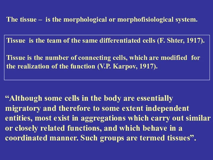
The tissue – is the morphological or morphofisiological system.
Tissue is the
The tissue – is the morphological or morphofisiological system.
Tissue is the
The tissues are systems of cells and noncellular structures characterized by
The tissues are systems of cells and noncellular structures characterized by
Differentiation – the morphofunctional exchange of the same organized cells.
The
Differentiation – the morphofunctional exchange of the same organized cells.
The

Layers
Stratum corneum
Stratum lucidum
Stratum granulosum
Stratum spinosum
Stratum basale
CONNECTIVE TISSUE
Time-differentiation – the stages of
Layers
Stratum corneum
Stratum lucidum
Stratum granulosum
Stratum spinosum
Stratum basale
CONNECTIVE TISSUE
Time-differentiation – the stages of
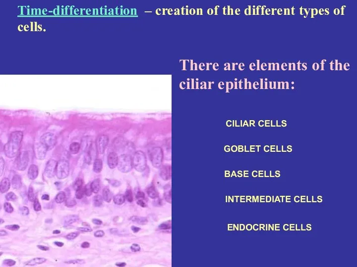
There are elements of the ciliar epithelium:
CILIAR CELLS
GOBLET CELLS
BASE CELLS
INTERMEDIATE CELLS
ENDOCRINE
There are elements of the ciliar epithelium:
CILIAR CELLS
GOBLET CELLS
BASE CELLS
INTERMEDIATE CELLS
ENDOCRINE
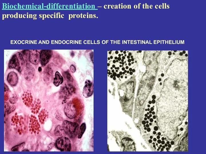
EXOCRINE AND ENDOCRINE CELLS OF THE INTESTINAL EPITHELIUM
Biochemical-differentiation – creation of
EXOCRINE AND ENDOCRINE CELLS OF THE INTESTINAL EPITHELIUM
Biochemical-differentiation – creation of
First of all start to differentiate the steam cells source the
First of all start to differentiate the steam cells source the
Regeneration – the capability of the tissue to recover itself after
Regeneration – the capability of the tissue to recover itself after
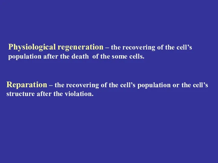
Physiological regeneration – the recovering of the cell’s population after the
Physiological regeneration – the recovering of the cell’s population after the

The History.
1665 год. Robert Hook was describe the “cell”.
1830 год. Jan
The History.
1665 год. Robert Hook was describe the “cell”.
1830 год. Jan
THE BASIC TYPES OF TISSUES
EPITHELIAL
CONNECTIVE (SUPPORT) AND BLOOD
MUSCLE
NERVOUS
THE BASIC TYPES OF TISSUES
EPITHELIAL
CONNECTIVE (SUPPORT) AND BLOOD
MUSCLE
NERVOUS
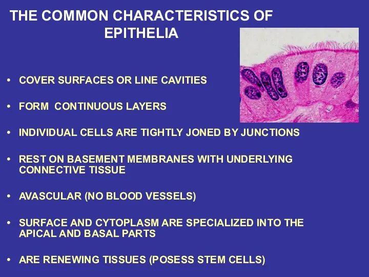
THE COMMON CHARACTERISTICS OF EPITHELIA
COVER SURFACES OR LINE CAVITIES
FORM CONTINUOUS LAYERS
THE COMMON CHARACTERISTICS OF EPITHELIA
COVER SURFACES OR LINE CAVITIES
FORM CONTINUOUS LAYERS
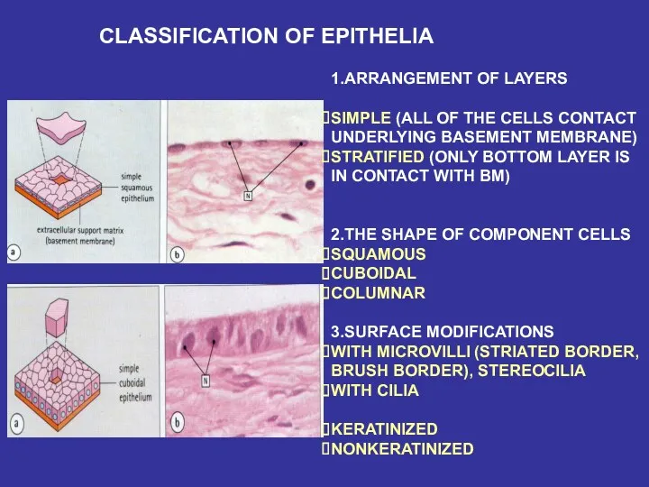
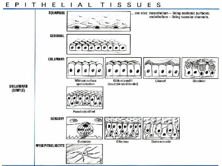
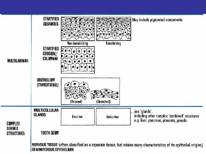
CLASSIFICATION OF EPITHELIA
1.ARRANGEMENT OF LAYERS
SIMPLE (ALL OF THE CELLS CONTACT
UNDERLYING
CLASSIFICATION OF EPITHELIA
1.ARRANGEMENT OF LAYERS
SIMPLE (ALL OF THE CELLS CONTACT
UNDERLYING
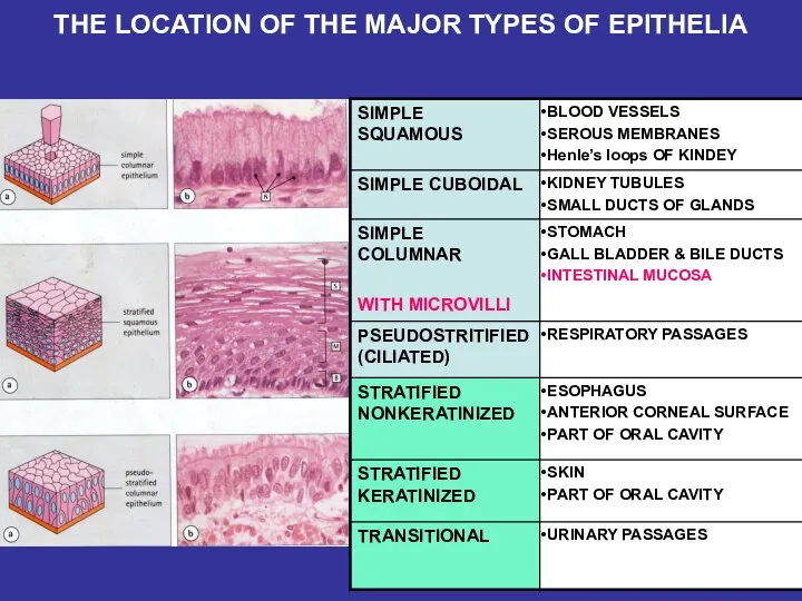
THE LOCATION OF THE MAJOR TYPES OF EPITHELIA
THE LOCATION OF THE MAJOR TYPES OF EPITHELIA
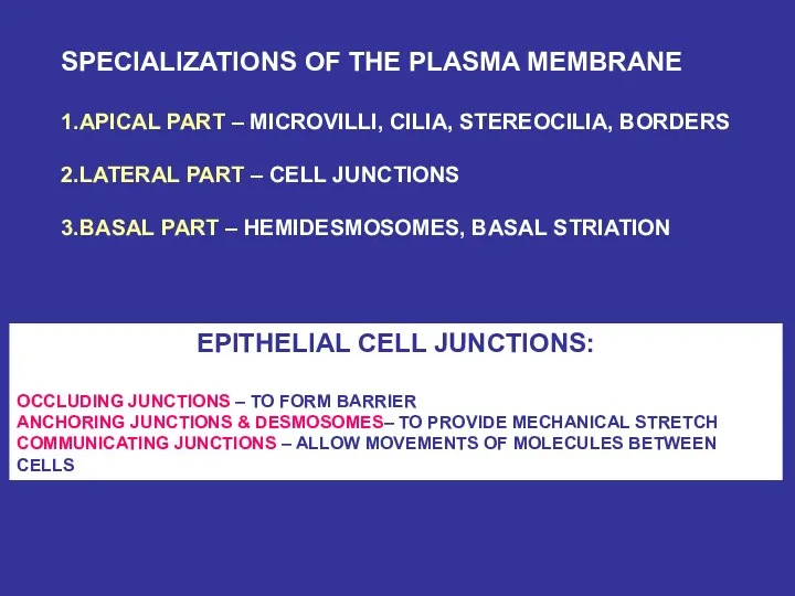
SPECIALIZATIONS OF THE PLASMA MEMBRANE
1.APICAL PART – MICROVILLI, CILIA, STEREOCILIA,
SPECIALIZATIONS OF THE PLASMA MEMBRANE
1.APICAL PART – MICROVILLI, CILIA, STEREOCILIA,
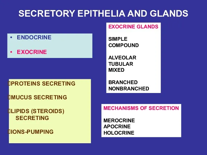
SECRETORY EPITHELIA AND GLANDS
ENDOCRINE
EXOCRINE
EXOCRINE GLANDS
SIMPLE
COMPOUND
ALVEOLAR
TUBULAR
MIXED
BRANCHED
NONBRANCHED
PROTEINS SECRETING
MUCUS SECRETING
LIPIDS (STEROIDS)
SECRETING
IONS-PUMPING
MECHANISMS OF
SECRETORY EPITHELIA AND GLANDS
ENDOCRINE
EXOCRINE
EXOCRINE GLANDS
SIMPLE
COMPOUND
ALVEOLAR
TUBULAR
MIXED
BRANCHED
NONBRANCHED
PROTEINS SECRETING
MUCUS SECRETING
LIPIDS (STEROIDS)
SECRETING
IONS-PUMPING
MECHANISMS OF
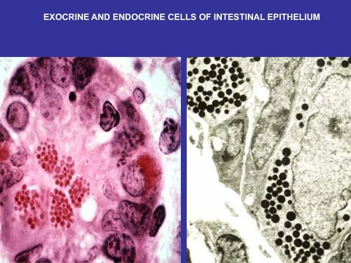
EXOCRINE AND ENDOCRINE CELLS OF INTESTINAL EPITHELIUM
EXOCRINE AND ENDOCRINE CELLS OF INTESTINAL EPITHELIUM
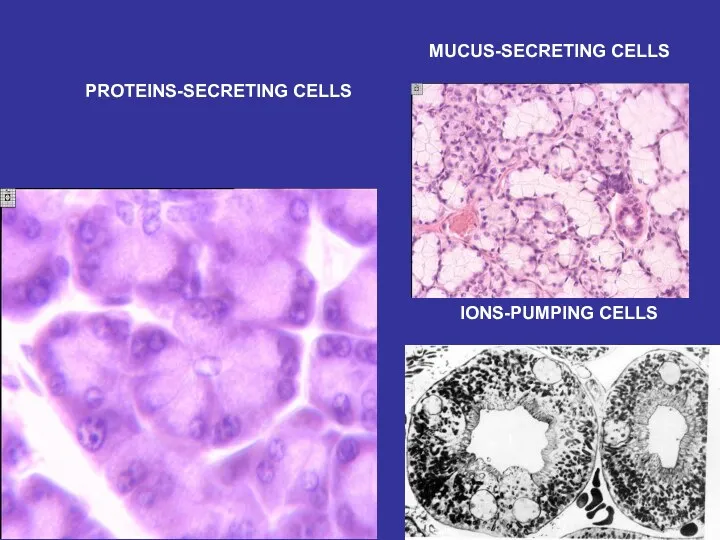
PROTEINS-SECRETING CELLS
MUCUS-SECRETING CELLS
IONS-PUMPING CELLS
PROTEINS-SECRETING CELLS
MUCUS-SECRETING CELLS
IONS-PUMPING CELLS
 Анатомия и физиология женских половых органов
 Вчення про тканини. Епітелій
 Антропология, как комплексное учение о человеке. Место человека в животном мире и этапы антропогенеза
 Строение стебля
 Анатомия внутреннего уха
 Физиология центральной нервной системы
 Генетически модифицированные организмы (ГМО)
 Презентация 1
 Лабораторная работа на тему Приспособленность организмов к их среде обитания
 Молодежный инновационный форум Воронежской области
 Нуклеиновые кислоты и их роль в жизнедеятельности клетки
 презентация к семинару
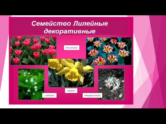 Презентация к уроку биологии 6 кл.
 Вредители и болезни газонных растений и меры борьбы с ними
 Химерные мыши
 Лишайники. Внешнее строение лишайников
 Общая характеристика подтипа Позвоночные. Часть1
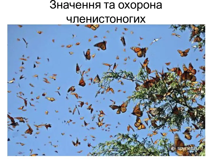 Значення та охорона членистоногих
 Животные Омского Прииртышья (насекомые). Занятие № 39
 Кровеносная система. Строение сердца. Строение сосудов
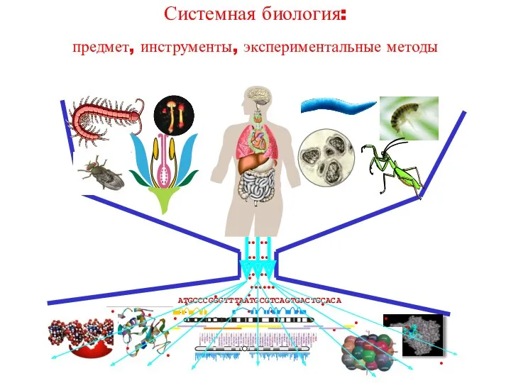 Системная биология: предмет, инструменты, экспериментальные методы
 Моторика (регуляция движений)
 История развития генетики. Основные понятия
 Режимы и способы хранения картофеля, плодов, и овощей. Лекция №3-4
 Тренировочные задания по биологии в формате ГИА
 Hedera helix
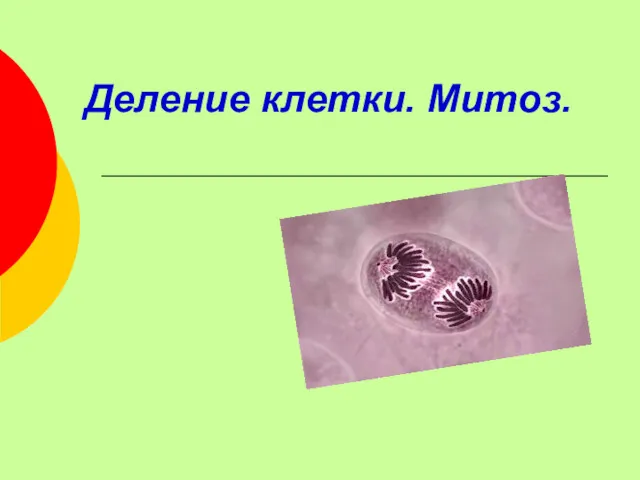 Деление клетки. Митоз. Жизненный цикл клетки
 Физиология памяти человека