- Transcription_and_translation

Содержание
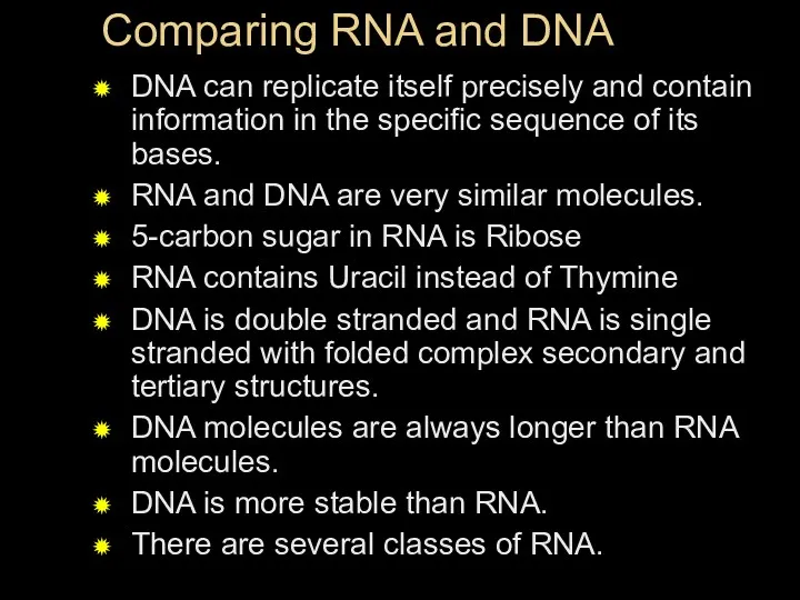
- 2. Comparing RNA and DNA DNA can replicate itself precisely and contain information in the specific sequence
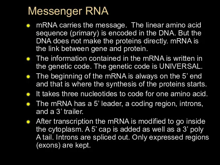
- 3. Messenger RNA mRNA carries the message. The linear amino acid sequence (primary) is encoded in the
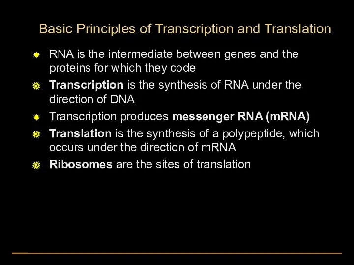
- 4. Basic Principles of Transcription and Translation RNA is the intermediate between genes and the proteins for
- 5. Fig. 17-2 RESULTS EXPERIMENT CONCLUSION Growth: Wild-type cells growing and dividing No growth: Mutant cells cannot
- 6. In prokaryotes, mRNA produced by transcription is immediately translated without more processing In a eukaryotic cell,
- 7. TRANSCRIPTION TRANSLATION DNA mRNA Ribosome Polypeptide (a) Bacterial cell Nuclear envelope TRANSCRIPTION RNA PROCESSING Pre-mRNA DNA
- 8. The Genetic Code The genetic code is the same for all organisms (universal) A codon is
- 9. (a) Tobacco plant expressing a firefly gene (b) Pig expressing a jellyfish gene
- 10. Key Words and definitions Transcription describes the synthesis of RNA on a DNA template Translation is
- 11. RNA Polymerase Binding and Initiation of Transcription Promoters signal the initiation of RNA synthesis Transcription factors
- 12. Promoters, terminators and start point
- 13. A eukaryotic promoter includes a TATA box 3 1 2 3 Promoter TATA box Start point
- 14. Only one strand of DNA is transcribed
- 15. Sense and antisense strands
- 17. Promoter Transcription unit Start point DNA RNA polymerase 5 5 3 3 Initiation 1 2 3
- 18. Protein-coding segment Polyadenylation signal 3 3 UTR 5 UTR 5 5 Cap Start codon Stop codon
- 20. Split Genes and RNA Splicing Most eukaryotic genes and their RNA transcripts have long noncoding stretches
- 21. Pre-mRNA mRNA Coding segment Introns cut out and exons spliced together 5 Cap Exon Intron 5
- 22. In some cases, RNA splicing is carried out by spliceosomes Spliceosomes consist of a variety of
- 23. RNA transcript (pre-mRNA) Exon 1 Exon 2 Intron Protein snRNA snRNPs Other proteins 5
- 24. RNA transcript (pre-mRNA) Exon 1 Exon 2 Intron Protein snRNA snRNPs Other proteins 5 5 Spliceosome
- 25. RNA transcript (pre-mRNA) Exon 1 Exon 2 Intron Protein snRNA snRNPs Other proteins 5 5 Spliceosome
- 26. Ribozymes Ribozymes are catalytic RNA molecules that function as enzymes and can splice RNA The discovery
- 27. Three properties of RNA enable it to function as an enzyme It can form a three-dimensional
- 28. The Functional and Evolutionary Importance of Introns Some genes can encode more than one kind of
- 29. Proteins often have a modular architecture consisting of discrete regions called domains In many cases, different
- 30. Fig. 17-12 Gene DNA Exon 1 Exon 2 Exon 3 Intron Intron Transcription RNA processing Translation
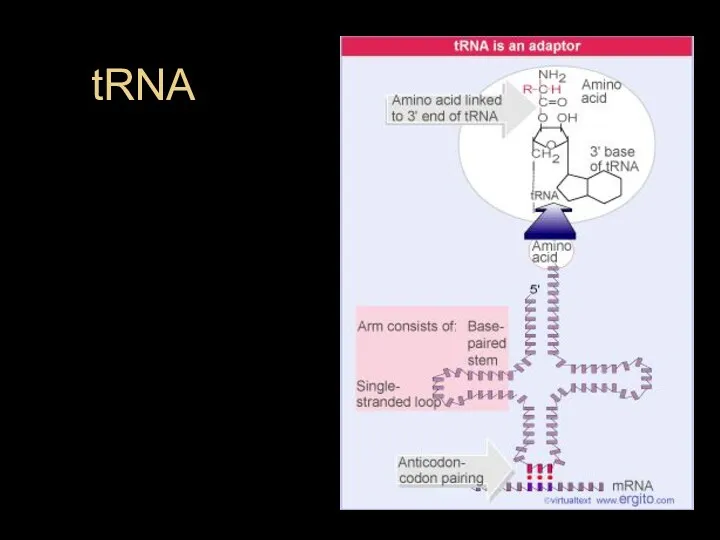
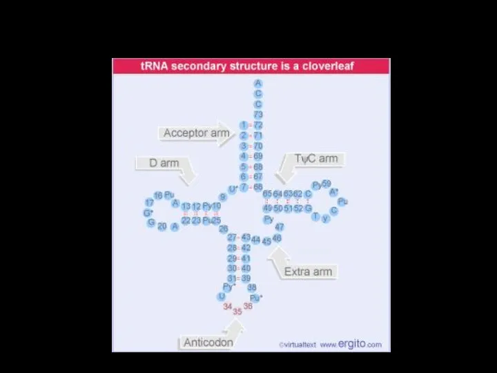
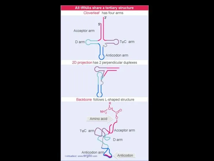
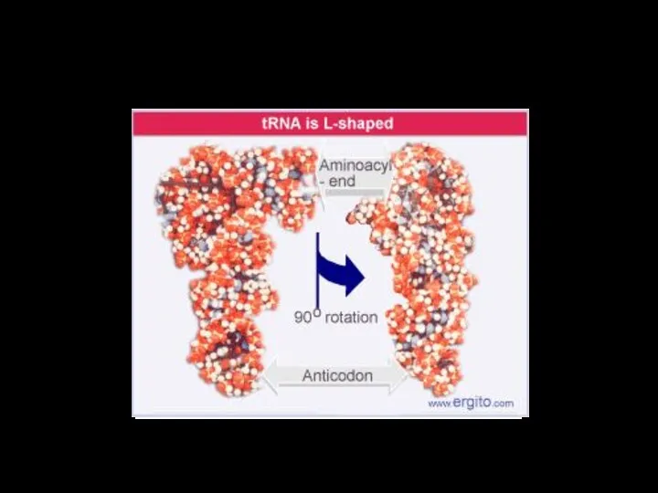
- 31. tRNA
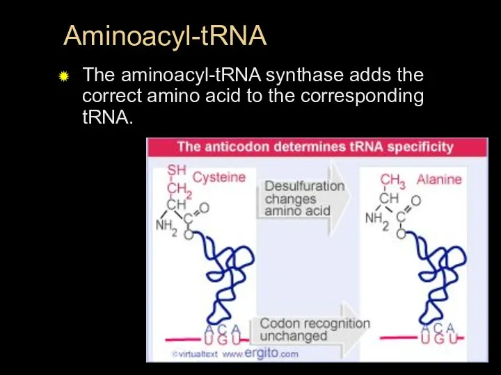
- 33. Aminoacyl-tRNA The aminoacyl-tRNA synthase adds the correct amino acid to the corresponding tRNA.
- 34. Fig. 17-15-4 Amino acid Aminoacyl-tRNA synthetase (enzyme) ATP Adenosine P P P Adenosine P P P
- 37. Ribosomal RNA Ribosomal RNA: contributes to the structure of Ribosomes. In eukaryotes rRNA is transcribed exclusively
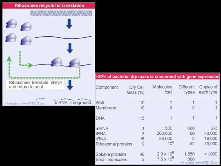
- 38. Ribosomes
- 40. In Prokaryotes
- 41. Fig. 17-16a Growing polypeptide Exit tunnel tRNA molecules Large subunit Small subunit (a) Computer model of
- 42. Fig. 17-16b P site (Peptidyl-tRNA binding site) A site (Aminoacyl- tRNA binding site) E site (Exit
- 43. A ribosome has three binding sites for tRNA: The P site holds the tRNA that carries
- 44. Ribosome Association and Initiation of Translation The initiation stage of translation brings together mRNA, a tRNA
- 45. Fig. 17-17 3′ 3′ 5′ 5′ U U A A C G Met GTP GDP Initiator
- 46. Elongation of the Polypeptide Chain During the elongation stage, amino acids are added one by one
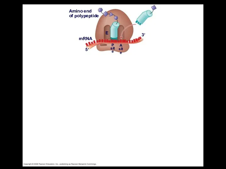
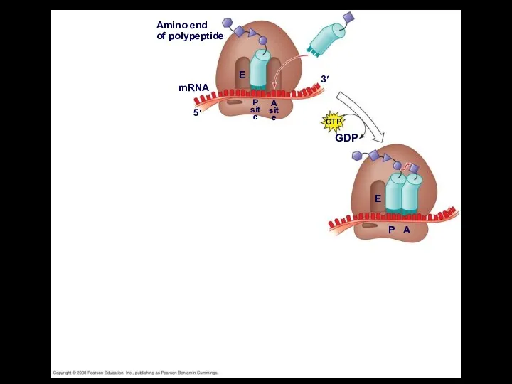
- 47. Amino end of polypeptide mRNA 5′ 3′ E P site A site
- 48. Amino end of polypeptide mRNA 5′ 3′ E P site A site GTP GDP E P
- 49. Fig. 17-18-3 Amino end of polypeptide mRNA 5′ 3′ E P site A site GTP GDP
- 50. Fig. 17-18-4 Amino end of polypeptide mRNA 5′ 3′ E P site A site GTP GDP
- 51. Termination of Translation Termination occurs when a stop codon in the mRNA reaches the A site
- 52. Fig. 17-19-1 Release factor 3′ 5′ Stop codon (UAG, UAA, or UGA)
- 53. Fig. 17-19-2 Release factor 3′ 5′ Stop codon (UAG, UAA, or UGA) 5′ 3′ 2 Free
- 54. Fig. 17-19-3 Release factor 3′ 5′ Stop codon (UAG, UAA, or UGA) 5′ 3′ 2 Free
- 55. Completing and Targeting the Functional Protein Often translation is not sufficient to make a functional protein
- 56. Protein Folding and Post-Translational Modifications During and after synthesis, a polypeptide chain spontaneously coils and folds
- 57. Targeting Polypeptides to Specific Locations Two populations of ribosomes are evident in cells: free ribsomes (in
- 58. Polypeptide synthesis always begins in the cytosol Synthesis finishes in the cytosol unless the polypeptide signals
- 59. Fig. 17-21 Ribosome mRNA Signal peptide Signal- recognition particle (SRP) CYTOSOL Translocation complex SRP receptor protein
- 61. Скачать презентацию
Comparing RNA and DNA
DNA can replicate itself precisely and contain information
Comparing RNA and DNA
DNA can replicate itself precisely and contain information
Messenger RNA
mRNA carries the message. The linear amino acid sequence (primary)
Messenger RNA
mRNA carries the message. The linear amino acid sequence (primary)
Basic Principles of Transcription and Translation
RNA is the intermediate between genes
Basic Principles of Transcription and Translation
RNA is the intermediate between genes
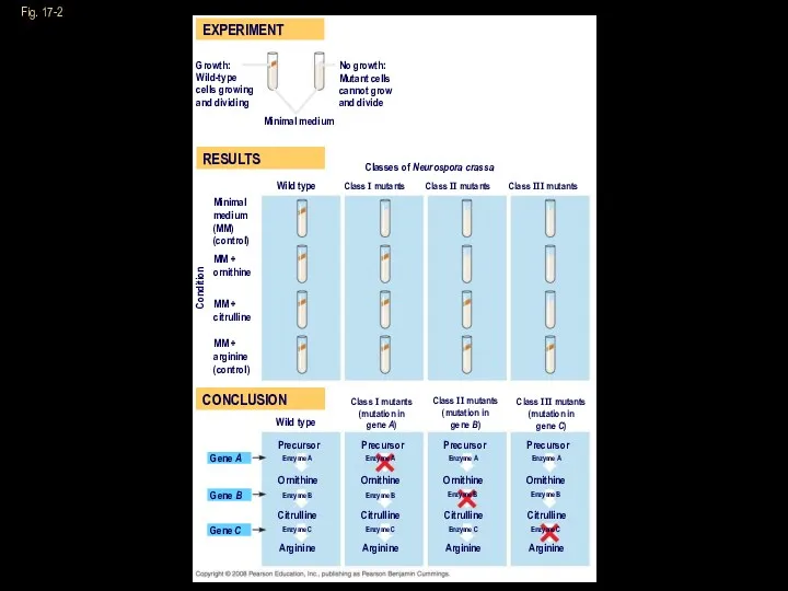
Fig. 17-2
RESULTS
EXPERIMENT
CONCLUSION
Growth:
Wild-type
cells growing
and dividing
No growth:
Mutant cells
cannot grow
and divide
Minimal medium
Classes
Fig. 17-2
RESULTS
EXPERIMENT
CONCLUSION
Growth:
Wild-type
cells growing
and dividing
No growth:
Mutant cells
cannot grow
and divide
Minimal medium
Classes
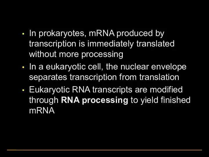
In prokaryotes, mRNA produced by transcription is immediately translated without more
In prokaryotes, mRNA produced by transcription is immediately translated without more
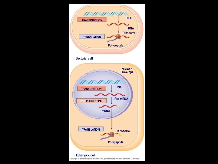
TRANSCRIPTION
TRANSLATION
DNA
mRNA
Ribosome
Polypeptide
(a) Bacterial cell
Nuclear
envelope
TRANSCRIPTION
RNA PROCESSING
Pre-mRNA
DNA
mRNA
TRANSLATION
Ribosome
Polypeptide
(b) Eukaryotic cell
TRANSCRIPTION
TRANSLATION
DNA
mRNA
Ribosome
Polypeptide
(a) Bacterial cell
Nuclear
envelope
TRANSCRIPTION
RNA PROCESSING
Pre-mRNA
DNA
mRNA
TRANSLATION
Ribosome
Polypeptide
(b) Eukaryotic cell
The Genetic Code
The genetic code is the same for all organisms
The Genetic Code
The genetic code is the same for all organisms
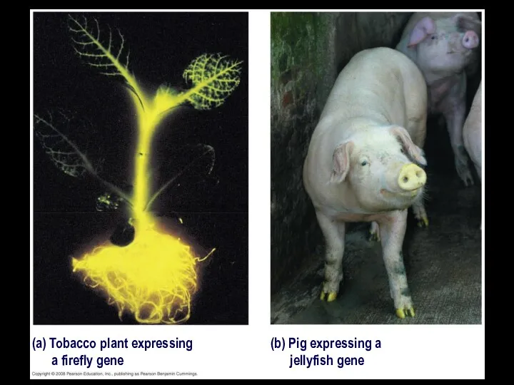
(a) Tobacco plant expressing
a firefly gene
(b) Pig expressing a
jellyfish
(a) Tobacco plant expressing
a firefly gene
(b) Pig expressing a
jellyfish
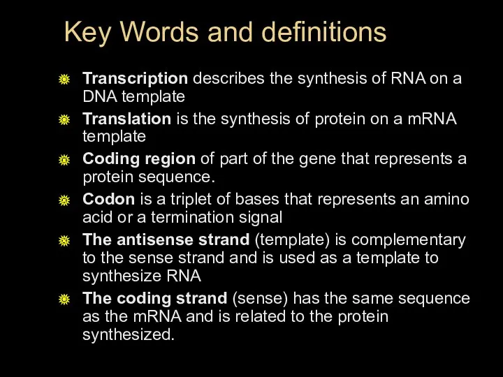
Key Words and definitions
Transcription describes the synthesis of RNA on a
Key Words and definitions
Transcription describes the synthesis of RNA on a
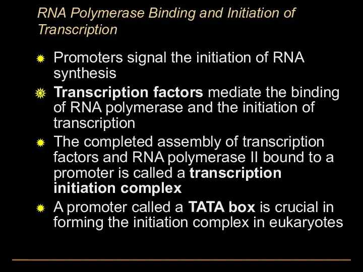
RNA Polymerase Binding and Initiation of Transcription
Promoters signal the initiation of
RNA Polymerase Binding and Initiation of Transcription
Promoters signal the initiation of
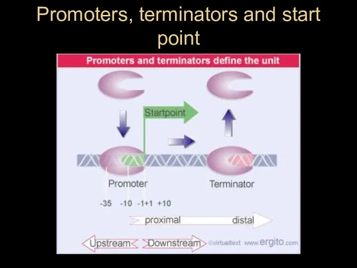
Promoters, terminators and start point
Promoters, terminators and start point
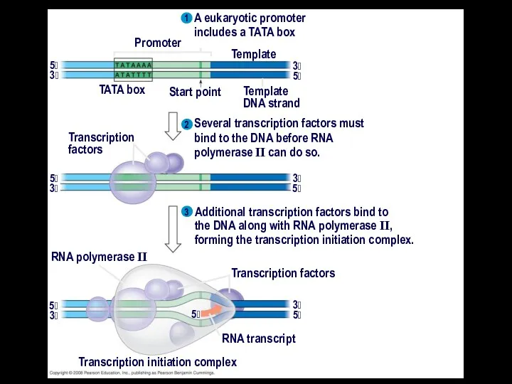
A eukaryotic promoter
includes a TATA box
3
1
2
3
Promoter
TATA box
Start point
Template
Template
DNA strand
5
3
5
Transcription
factors
Several transcription factors
A eukaryotic promoter
includes a TATA box
3
1
2
3
Promoter
TATA box
Start point
Template
Template
DNA strand
5
3
5
Transcription
factors
Several transcription factors
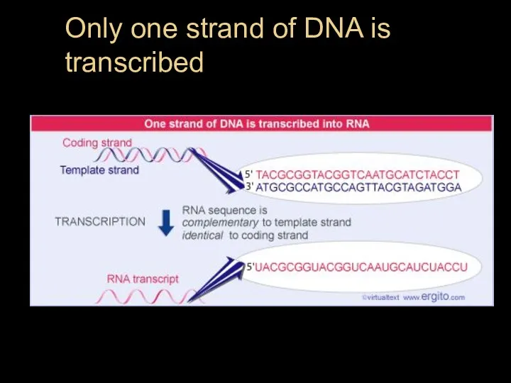
Only one strand of DNA is transcribed
Only one strand of DNA is transcribed
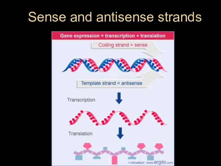
Sense and antisense strands
Sense and antisense strands
Promoter
Transcription unit
Start point
DNA
RNA polymerase
5
5
3
3
Initiation
1
2
3
5
5
3
3
Unwound
DNA
RNA
transcript
Template strand
of DNA
Elongation
Rewound
DNA
5
5
5
5
5
3
3
3
3
RNA
transcript
Termination
5
5
3
3
3
5
Completed RNA transcript
Newly made
RNA
Template
strand of DNA
Direction
Promoter
Transcription unit
Start point
DNA
RNA polymerase
5
5
3
3
Initiation
1
2
3
5
5
3
3
Unwound
DNA
RNA
transcript
Template strand
of DNA
Elongation
Rewound
DNA
5
5
5
5
5
3
3
3
3
RNA
transcript
Termination
5
5
3
3
3
5
Completed RNA transcript
Newly made
RNA
Template
strand of DNA
Direction

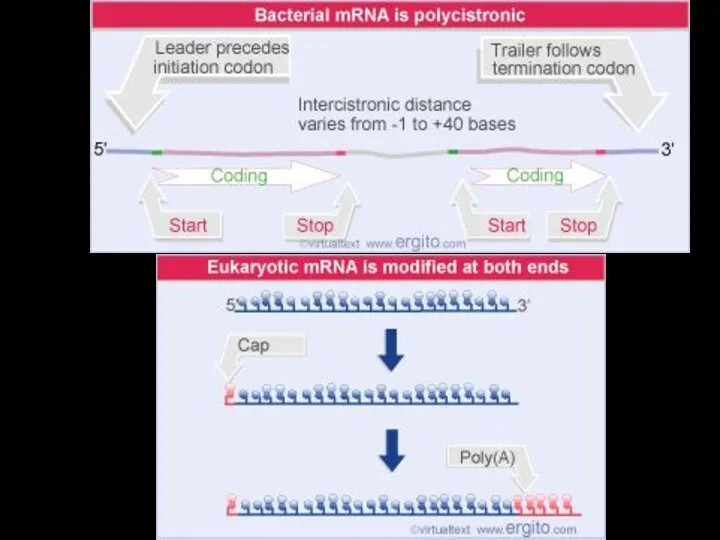
Protein-coding segment
Polyadenylation signal
3
3 UTR
5 UTR
5
5 Cap
Start codon
Stop codon
Poly-A tail
G
P
P
P
AAUAAA
AAA
AAA
…
Protein-coding segment
Polyadenylation signal
3
3 UTR
5 UTR
5
5 Cap
Start codon
Stop codon
Poly-A tail
G
P
P
P
AAUAAA
AAA
AAA
…
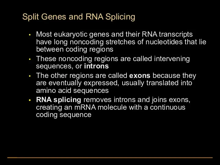
Split Genes and RNA Splicing
Most eukaryotic genes and their RNA transcripts
Split Genes and RNA Splicing
Most eukaryotic genes and their RNA transcripts
Pre-mRNA
mRNA
Coding
segment
Introns cut out and
exons spliced together
5 Cap
Exon
Intron
5
1
30
31
104
Exon
Intron
105
Exon
146
3
Poly-A tail
Poly-A tail
5 Cap
5 UTR
3
Pre-mRNA
mRNA
Coding
segment
Introns cut out and
exons spliced together
5 Cap
Exon
Intron
5
1
30
31
104
Exon
Intron
105
Exon
146
3
Poly-A tail
Poly-A tail
5 Cap
5 UTR
3
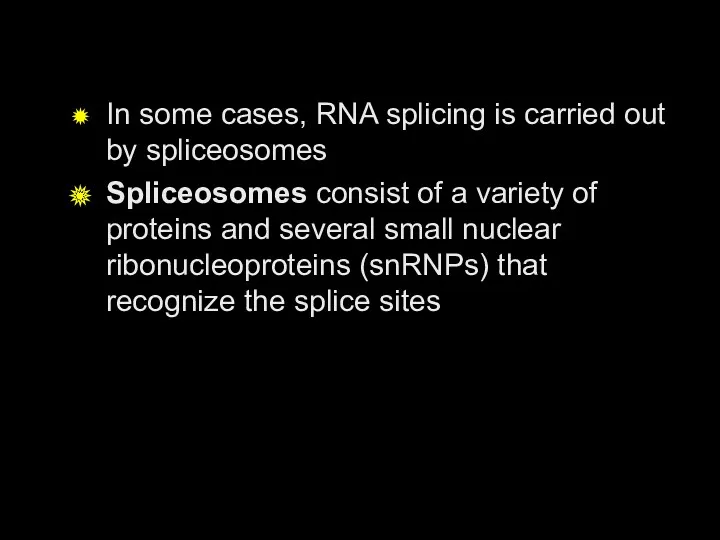
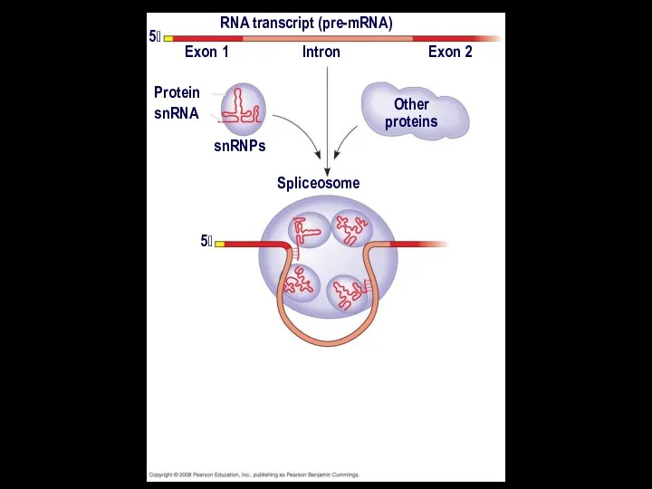
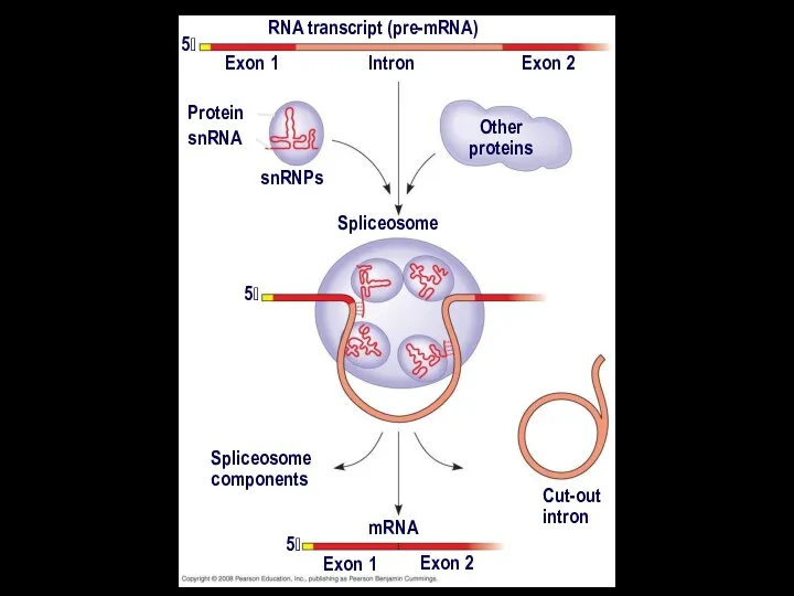
In some cases, RNA splicing is carried out by spliceosomes
Spliceosomes consist
In some cases, RNA splicing is carried out by spliceosomes
Spliceosomes consist
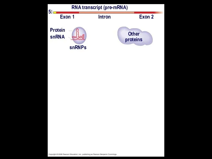
RNA transcript (pre-mRNA)
Exon 1
Exon 2
Intron
Protein
snRNA
snRNPs
Other
proteins
5
RNA transcript (pre-mRNA)
Exon 1
Exon 2
Intron
Protein
snRNA
snRNPs
Other
proteins
5
RNA transcript (pre-mRNA)
Exon 1
Exon 2
Intron
Protein
snRNA
snRNPs
Other
proteins
5
5
Spliceosome
RNA transcript (pre-mRNA)
Exon 1
Exon 2
Intron
Protein
snRNA
snRNPs
Other
proteins
5
5
Spliceosome
RNA transcript (pre-mRNA)
Exon 1
Exon 2
Intron
Protein
snRNA
snRNPs
Other
proteins
5
5
Spliceosome
Spliceosome
components
Cut-out
intron
mRNA
Exon 1
Exon 2
5
RNA transcript (pre-mRNA)
Exon 1
Exon 2
Intron
Protein
snRNA
snRNPs
Other
proteins
5
5
Spliceosome
Spliceosome
components
Cut-out
intron
mRNA
Exon 1
Exon 2
5
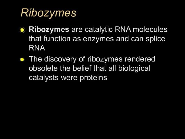
Ribozymes
Ribozymes are catalytic RNA molecules that function as enzymes and can
Ribozymes
Ribozymes are catalytic RNA molecules that function as enzymes and can
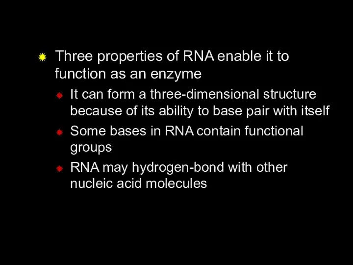
Three properties of RNA enable it to function as an enzyme
It
Three properties of RNA enable it to function as an enzyme
It
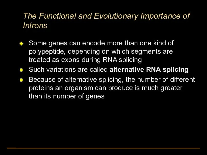
The Functional and Evolutionary Importance of Introns
Some genes can encode more
The Functional and Evolutionary Importance of Introns
Some genes can encode more
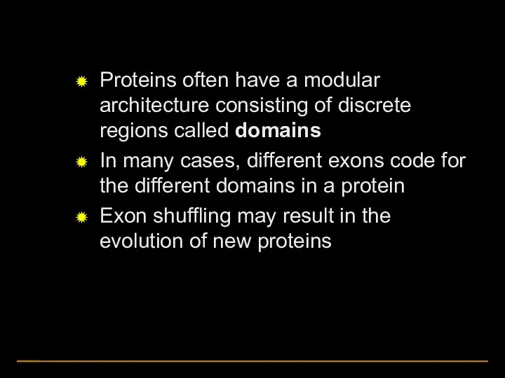
Proteins often have a modular architecture consisting of discrete regions called
Proteins often have a modular architecture consisting of discrete regions called
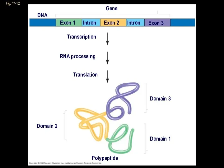
Fig. 17-12
Gene
DNA
Exon 1
Exon 2
Exon 3
Intron
Intron
Transcription
RNA processing
Translation
Domain 2
Domain 3
Domain 1
Polypeptide
Fig. 17-12
Gene
DNA
Exon 1
Exon 2
Exon 3
Intron
Intron
Transcription
RNA processing
Translation
Domain 2
Domain 3
Domain 1
Polypeptide
tRNA
tRNA
Aminoacyl-tRNA
The aminoacyl-tRNA synthase adds the correct amino acid to the corresponding
Aminoacyl-tRNA
The aminoacyl-tRNA synthase adds the correct amino acid to the corresponding
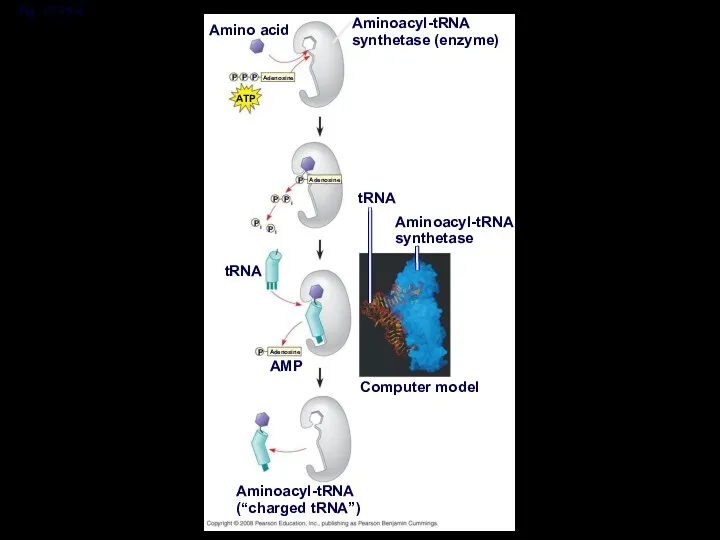
Fig. 17-15-4
Amino acid
Aminoacyl-tRNA
synthetase (enzyme)
ATP
Adenosine
P
P
P
Adenosine
P
P
P
i
P
P
i
i
tRNA
tRNA
Aminoacyl-tRNA
synthetase
Computer model
AMP
Adenosine
P
Aminoacyl-tRNA
(“charged tRNA”)
Fig. 17-15-4
Amino acid
Aminoacyl-tRNA
synthetase (enzyme)
ATP
Adenosine
P
P
P
Adenosine
P
P
P
i
P
P
i
i
tRNA
tRNA
Aminoacyl-tRNA
synthetase
Computer model
AMP
Adenosine
P
Aminoacyl-tRNA
(“charged tRNA”)
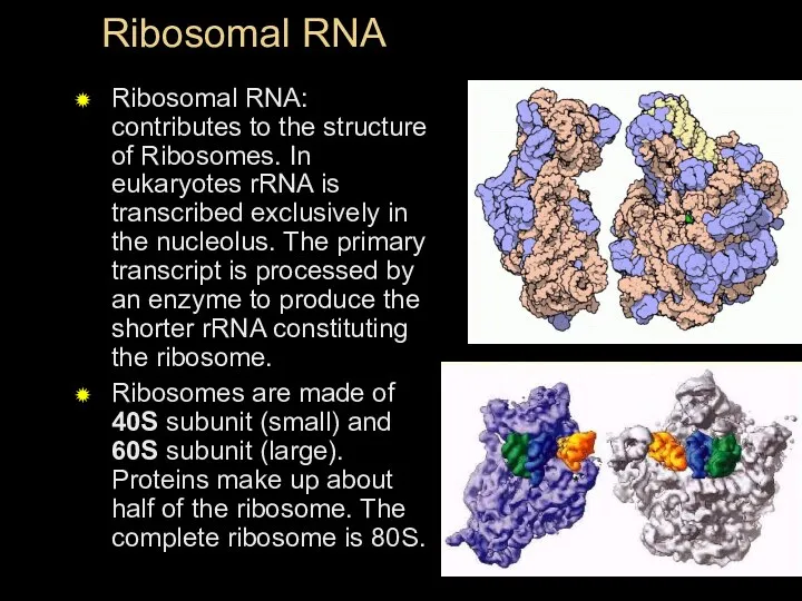
Ribosomal RNA
Ribosomal RNA: contributes to the structure of Ribosomes. In eukaryotes
Ribosomal RNA
Ribosomal RNA: contributes to the structure of Ribosomes. In eukaryotes
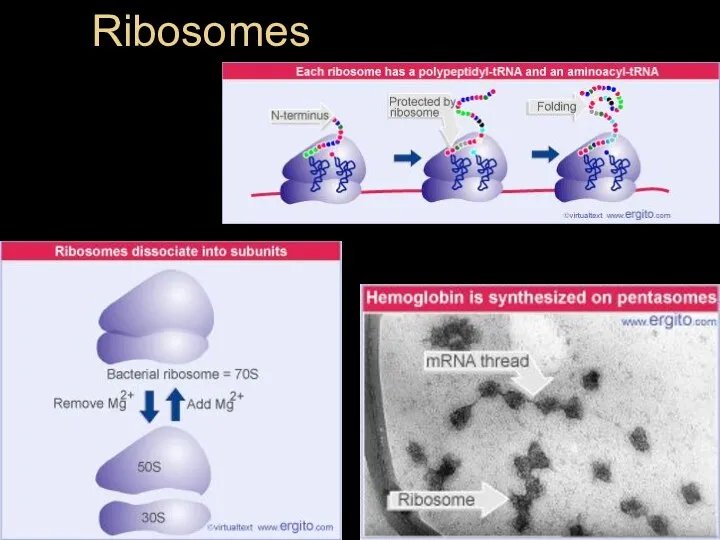
Ribosomes
Ribosomes
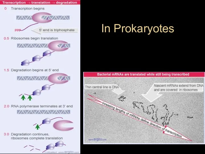
In Prokaryotes
In Prokaryotes
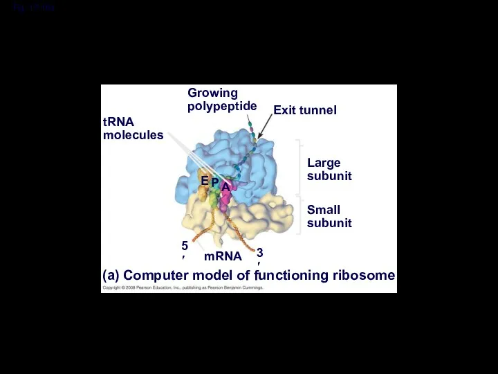
Fig. 17-16a
Growing
polypeptide
Exit tunnel
tRNA
molecules
Large
subunit
Small
subunit
(a) Computer model of functioning ribosome
mRNA
E
P
A
5′
3′
Fig. 17-16a
Growing
polypeptide
Exit tunnel
tRNA
molecules
Large
subunit
Small
subunit
(a) Computer model of functioning ribosome
mRNA
E
P
A
5′
3′
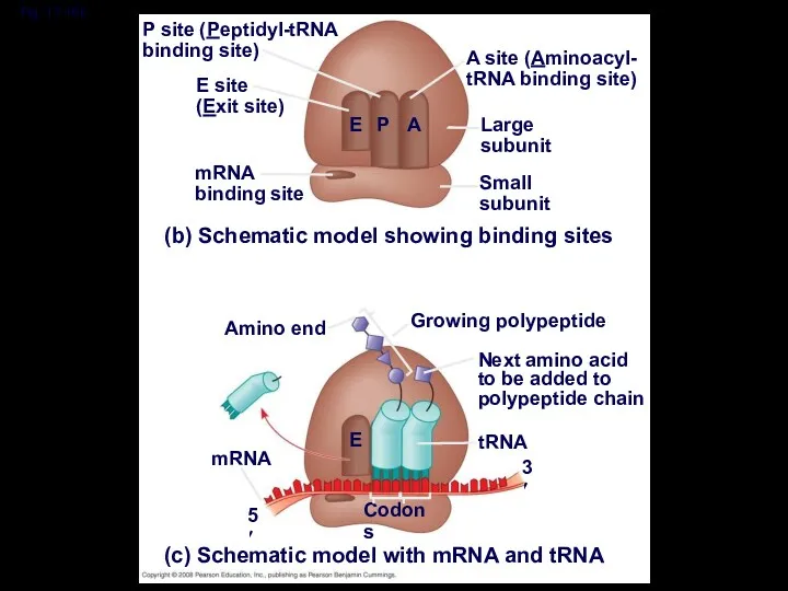
Fig. 17-16b
P site (Peptidyl-tRNA
binding site)
A site (Aminoacyl-
tRNA binding site)
E site
(Exit site)
mRNA
binding
Fig. 17-16b
P site (Peptidyl-tRNA
binding site)
A site (Aminoacyl-
tRNA binding site)
E site
(Exit site)
mRNA
binding
A ribosome has three binding sites for tRNA:
The P site holds
A ribosome has three binding sites for tRNA:
The P site holds
Ribosome Association and Initiation of Translation
The initiation stage of translation brings
Ribosome Association and Initiation of Translation
The initiation stage of translation brings
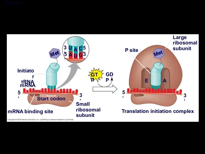
Fig. 17-17
3′
3′
5′
5′
U
U
A
A
C
G
Met
GTP
GDP
Initiator
tRNA
mRNA
5′
3′
Start codon
mRNA binding site
Small
ribosomal
subunit
5′
P site
Translation initiation complex
3′
E
A
Met
Large
ribosomal
subunit
Fig. 17-17
3′
3′
5′
5′
U
U
A
A
C
G
Met
GTP
GDP
Initiator
tRNA
mRNA
5′
3′
Start codon
mRNA binding site
Small
ribosomal
subunit
5′
P site
Translation initiation complex
3′
E
A
Met
Large
ribosomal
subunit
Elongation of the Polypeptide Chain
During the elongation stage, amino acids are
Elongation of the Polypeptide Chain
During the elongation stage, amino acids are
Amino end
of polypeptide
mRNA
5′
3′
E
P
site
A
site
Amino end
of polypeptide
mRNA
5′
3′
E
P
site
A
site
Amino end
of polypeptide
mRNA
5′
3′
E
P
site
A
site
GTP
GDP
E
P
A
Amino end
of polypeptide
mRNA
5′
3′
E
P
site
A
site
GTP
GDP
E
P
A
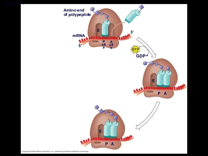
Fig. 17-18-3
Amino end
of polypeptide
mRNA
5′
3′
E
P
site
A
site
GTP
GDP
E
P
A
E
P
A
Fig. 17-18-3
Amino end
of polypeptide
mRNA
5′
3′
E
P
site
A
site
GTP
GDP
E
P
A
E
P
A
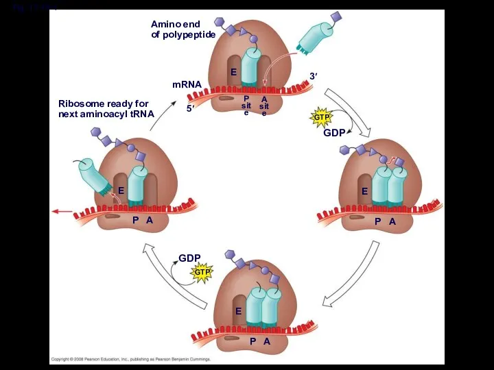
Fig. 17-18-4
Amino end
of polypeptide
mRNA
5′
3′
E
P
site
A
site
GTP
GDP
E
P
A
E
P
A
GDP
GTP
Ribosome ready for
next aminoacyl tRNA
E
P
A
Fig. 17-18-4
Amino end
of polypeptide
mRNA
5′
3′
E
P
site
A
site
GTP
GDP
E
P
A
E
P
A
GDP
GTP
Ribosome ready for
next aminoacyl tRNA
E
P
A
Termination of Translation
Termination occurs when a stop codon in the mRNA
Termination of Translation
Termination occurs when a stop codon in the mRNA
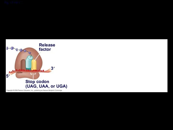
Fig. 17-19-1
Release
factor
3′
5′
Stop codon
(UAG, UAA, or UGA)
Fig. 17-19-1
Release
factor
3′
5′
Stop codon
(UAG, UAA, or UGA)
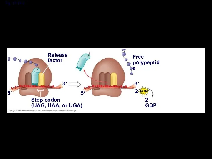
Fig. 17-19-2
Release
factor
3′
5′
Stop codon
(UAG, UAA, or UGA)
5′
3′
2
Free
polypeptide
2 GDP
GTP
Fig. 17-19-2
Release
factor
3′
5′
Stop codon
(UAG, UAA, or UGA)
5′
3′
2
Free
polypeptide
2 GDP
GTP
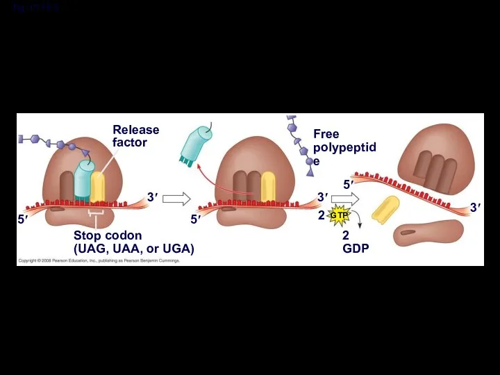
Fig. 17-19-3
Release
factor
3′
5′
Stop codon
(UAG, UAA, or UGA)
5′
3′
2
Free
polypeptide
2 GDP
GTP
5′
3′
Fig. 17-19-3
Release
factor
3′
5′
Stop codon
(UAG, UAA, or UGA)
5′
3′
2
Free
polypeptide
2 GDP
GTP
5′
3′
Completing and Targeting the Functional Protein
Often translation is not sufficient to
Completing and Targeting the Functional Protein
Often translation is not sufficient to
Protein Folding and Post-Translational Modifications
During and after synthesis, a polypeptide chain
Protein Folding and Post-Translational Modifications
During and after synthesis, a polypeptide chain
Targeting Polypeptides to Specific Locations
Two populations of ribosomes are evident in
Targeting Polypeptides to Specific Locations
Two populations of ribosomes are evident in
Polypeptide synthesis always begins in the cytosol
Synthesis finishes in the cytosol
Polypeptide synthesis always begins in the cytosol
Synthesis finishes in the cytosol
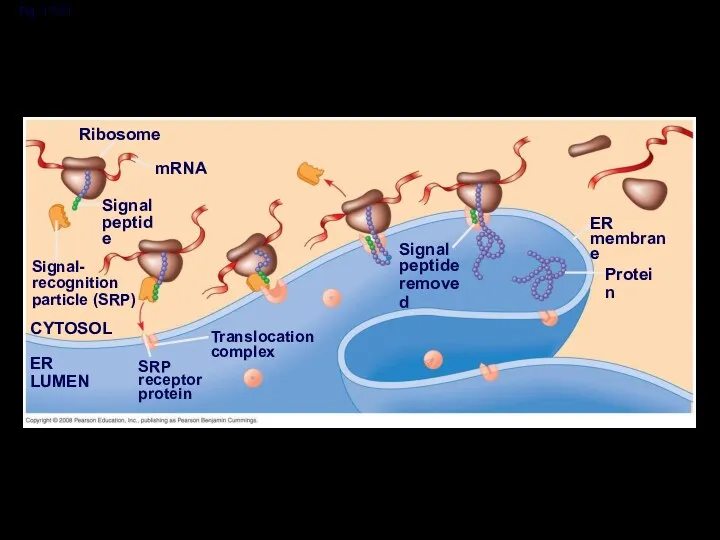
Fig. 17-21
Ribosome
mRNA
Signal
peptide
Signal-
recognition
particle (SRP)
CYTOSOL
Translocation
complex
SRP
receptor
protein
ER LUMEN
Signal
peptide
removed
ER
membrane
Protein
Fig. 17-21
Ribosome
mRNA
Signal
peptide
Signal-
recognition
particle (SRP)
CYTOSOL
Translocation
complex
SRP
receptor
protein
ER LUMEN
Signal
peptide
removed
ER
membrane
Protein
 Чудесное превращение. Появление комара
 Цели, задачи и методы науки селекции
 Север - богатый край.
 Акселерация және ретардация реактивтілік және организмнің резистенттілігі
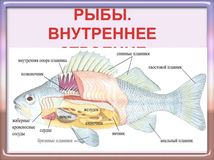 Рыбы. Внутреннее строение
 Загальна характеристика біосфери. Вчення Вернадського про біосферу. Роль живих організмів у біосфері. Біомаса
 Презентация Дигибридное скрещивание. Третий закон Менделя.
 Кожа и её производные
 Отдел Моховидные. Общая характеристика, значение
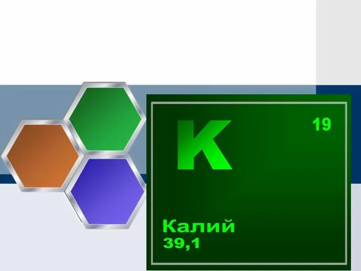 Презентация Значение калия в организме
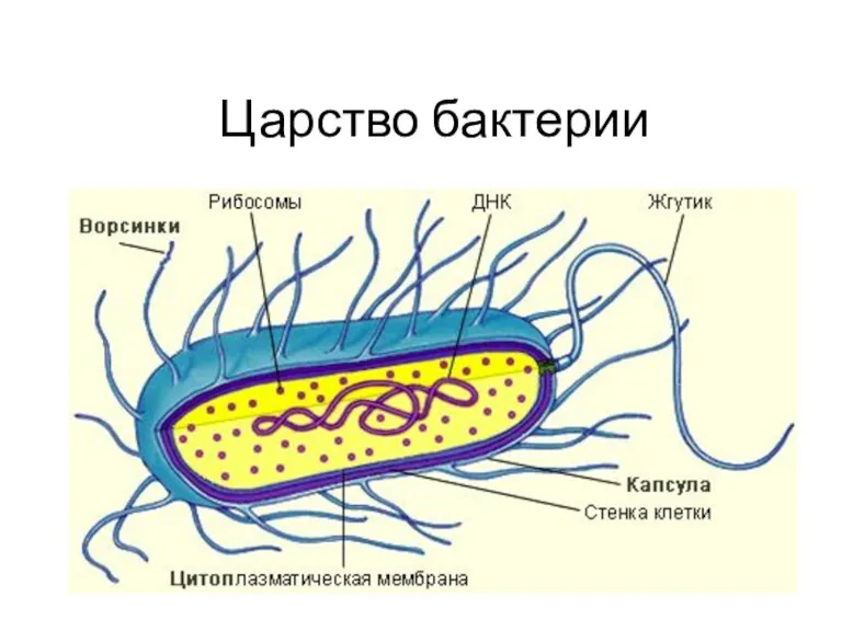 Царство Бактерии. 6 класс
 презентация Цветок
 Пищевая микробиология. Превращения азотсодержащих веществ
 Экология и природопользование. Экосистемы
 Кожа. Наружный покров
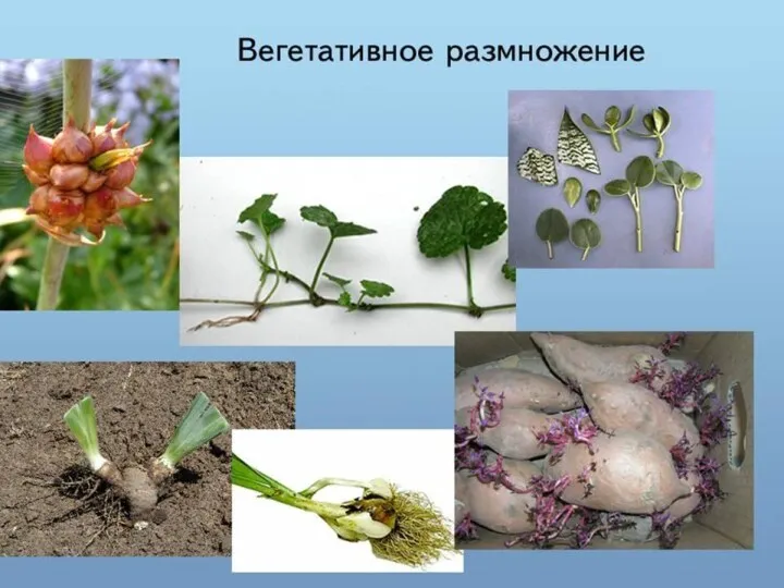 ВИДЫ ВЕГЕТАТИВНОГО РАЗМНОЖЕНИЯ РАСТЕНИИ
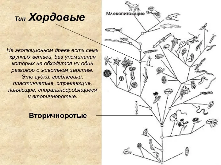 Тип Хордовые
 Какие бывают животные? Окружающий мир. 2 класс
 Вегетативное размножение растений
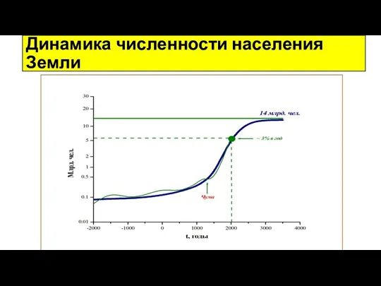 Динамика численности населения Земли
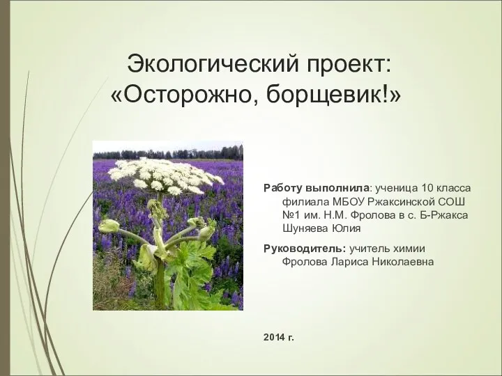 Осторожно, борщевик!
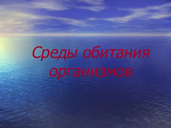 Среды обитания организмов
 Малина звичайна
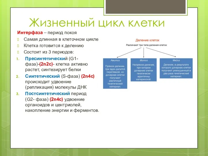 Жизненный цикл клетки
 Болотные птицы
 Общая характеристика грибов. Многообразие
 Растения болот
 Международная классификация микроорганизмов по Берги