- Fibrous proteins and their functions. Membrane proteins and their functions

Содержание
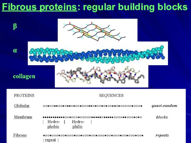
- 2. Globular proteins Fibrous proteins H-bonds (NH:::OC) & hydrophobic forces Membrane proteins
- 3. Fibrous proteins: regular building blocks ____________________________________ Here, we will not consider fibrous proteins made of globules
- 4. Fibrous proteins: regular building blocks β α collagen
- 5. Silk fibroin ×~50 β 4.8A
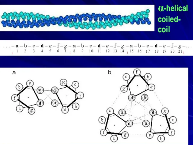
- 6. α-helical coiled- coil
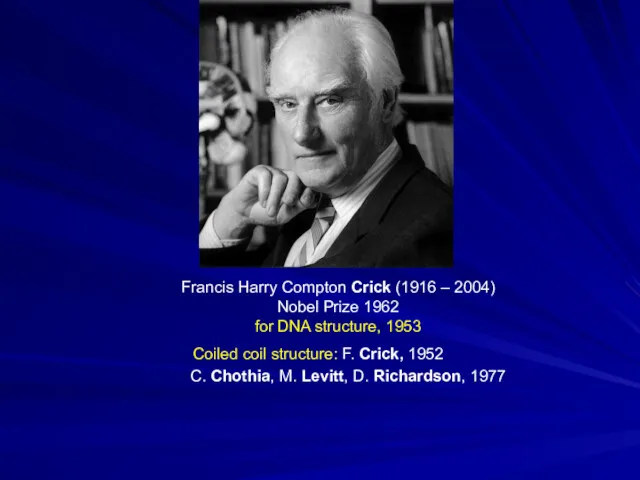
- 7. Francis Harry Compton Crick (1916 – 2004) Nobel Prize 1962 for DNA structure, 1953 Coiled coil
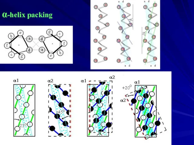
- 8. α-helix packing
- 9. collagen triple helix: 3 chains ≈ [Gly-X-Pro]≈500
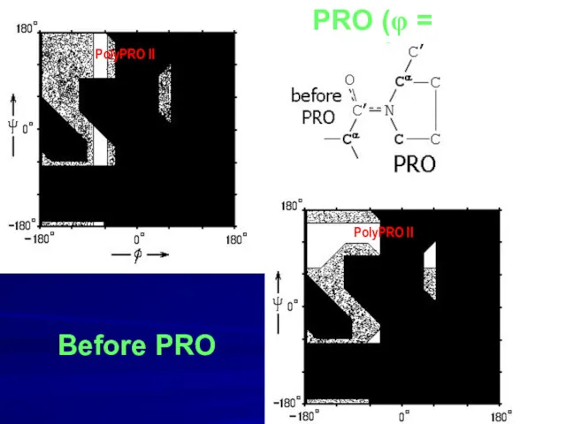
- 10. PRO (φ = -70o) Before PRO PolyPRO II PolyPRO II
- 11. Collagen: assisted folding
- 12. Kuru: a mysterious disease, later demonstrated to be infectious prion disease. Daniel Carleton Gajdusek (1923 –2008)
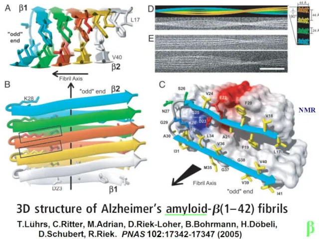
- 13. β ______ NMR
- 14. Lu J.X., Qiang W., Yau W.M., Schwieters C.D., Meredith S.C., Tycko R. Molecular structure of β-amyloid
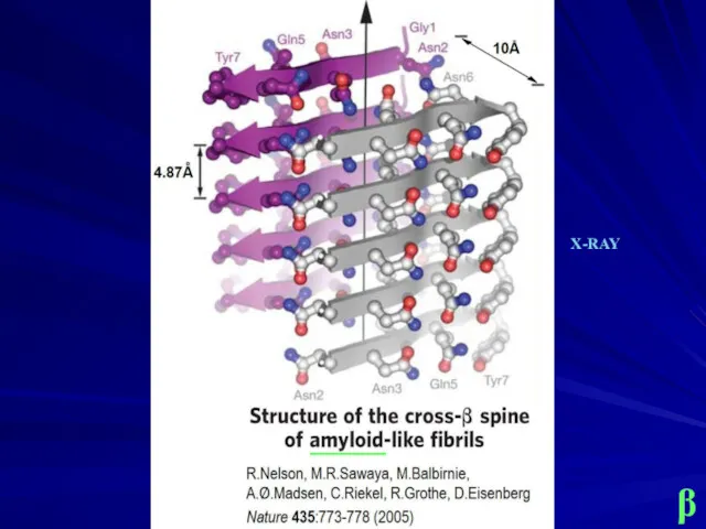
- 15. β _____ X-RAY
- 17. Growth of amyloids Dovidchenko N.V., Finkelstein A.V., Galzitskaya O.V. 2014. How to determine the size of
- 18. Oligomers Protofibrils Mature amyloid fibrils Relini A., Marano N., Gliozzi A. 2014. Misfolding of amyloidogenic proteins
- 19. Elastin: Matrix protein. Short repeats. Poor secondary structure. Chains are linked by chemically modified Lys residues.
- 20. H-bonds & hydrophobics Membrane proteins: transmitters ____ heads (polar) tails tails heads (polar)
- 21. H+ strong binding H+ inside H+ H+ H+ H+ H+ H+ H+ H+ H+ H+ H+
- 22. Porin Transport of polar molecules β
- 23. Membrane protein in vivo: Folding is assisted by “directing factors” - chaperones
- 24. MANY OF SIMPLE MEMBRANE PROTEINS REFOLD IN VITRO IN THE PRESENCE OF PHOSPHOLIPID VESICLES OR SURFACTANT
- 25. Pore in membrane: SELECTIVITY Free energy of a charge in the non-charged non-polar pore: ~ q2
- 26. Photo- synthetic center Robert Huber, 1937. Nobel prize 1988
- 27. Pigments in photo- synthetic center: Electron transfer chlorophyll ? ? Light ?
- 29. Скачать презентацию
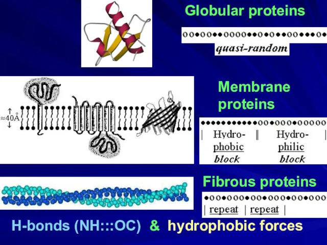
Globular proteins
Fibrous proteins
H-bonds (NH:::OC) & hydrophobic forces
Membrane
proteins
Globular proteins
Fibrous proteins
H-bonds (NH:::OC) & hydrophobic forces
Membrane
proteins
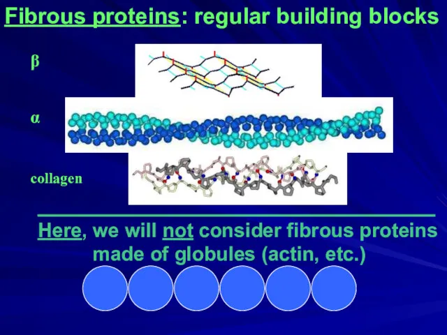
Fibrous proteins: regular building blocks
____________________________________
Here, we will not consider fibrous proteins
Fibrous proteins: regular building blocks
____________________________________
Here, we will not consider fibrous proteins
Fibrous proteins: regular building blocks
β
α
collagen
Fibrous proteins: regular building blocks
β
α
collagen
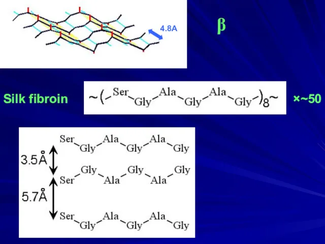
Silk fibroin
×~50
β
4.8A
Silk fibroin
×~50
β
4.8A
α-helical
coiled-
coil
α-helical
coiled-
coil
Francis Harry Compton Crick (1916 – 2004)
Nobel Prize 1962
for DNA structure, 1953
Coiled
Francis Harry Compton Crick (1916 – 2004)
Nobel Prize 1962
for DNA structure, 1953
Coiled
α-helix packing
α-helix packing
![collagen triple helix: 3 chains ≈ [Gly-X-Pro]≈500](/_ipx/f_webp&q_80&fit_contain&s_1440x1080/imagesDir/jpg/141138/slide-8.jpg)
collagen triple helix: 3 chains ≈ [Gly-X-Pro]≈500
collagen triple helix: 3 chains ≈ [Gly-X-Pro]≈500
PRO (φ = -70o)
Before PRO
PolyPRO II
PolyPRO II
PRO (φ = -70o)
Before PRO
PolyPRO II
PolyPRO II
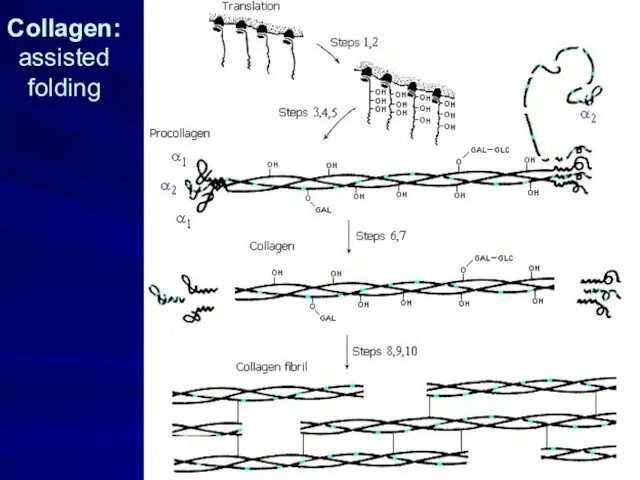
Collagen: assisted
folding
Collagen: assisted
folding
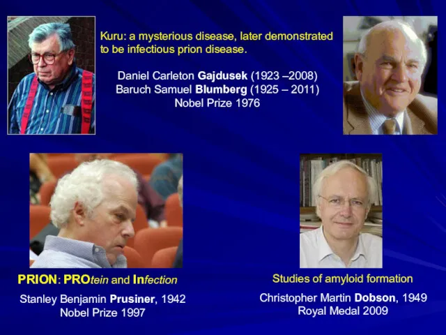
Kuru: a mysterious disease, later demonstrated to be infectious prion disease.
Daniel
Kuru: a mysterious disease, later demonstrated to be infectious prion disease.
Daniel
β
______
NMR
β
______
NMR
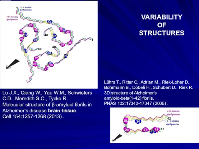
Lu J.X., Qiang W., Yau W.M., Schwieters C.D., Meredith S.C., Tycko
Lu J.X., Qiang W., Yau W.M., Schwieters C.D., Meredith S.C., Tycko
β
_____
X-RAY
β
_____
X-RAY
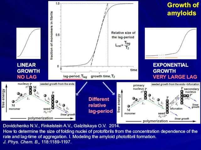
Growth of
amyloids
Dovidchenko N.V., Finkelstein A.V., Galzitskaya O.V. 2014.
How to determine
Growth of
amyloids
Dovidchenko N.V., Finkelstein A.V., Galzitskaya O.V. 2014.
How to determine
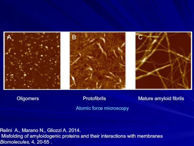
Oligomers Protofibrils Mature amyloid fibrils
Relini A., Marano N., Gliozzi A.
Oligomers Protofibrils Mature amyloid fibrils
Relini A., Marano N., Gliozzi A.
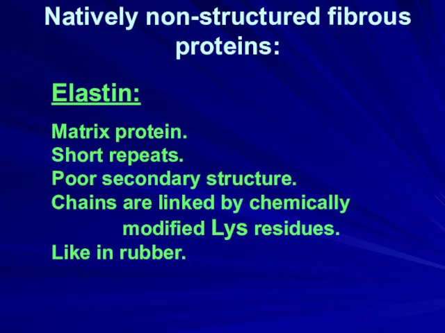
Elastin:
Matrix protein.
Short repeats.
Poor secondary structure.
Chains are linked by
Elastin:
Matrix protein.
Short repeats.
Poor secondary structure.
Chains are linked by
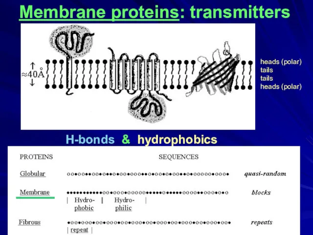
H-bonds & hydrophobics
Membrane proteins: transmitters
____
heads (polar)
tails
tails
heads (polar)
H-bonds & hydrophobics
Membrane proteins: transmitters
____
heads (polar)
tails
tails
heads (polar)
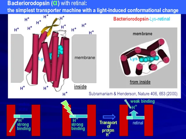
H+
strong
binding
H+
inside
H+
H+
H+
H+
H+
H+
H+
H+
H+
H+
H+
H+
H+
weak binding
H+
strong
binding
stable
state
Transport
H+
strong
binding
H+
inside
H+
H+
H+
H+
H+
H+
H+
H+
H+
H+
H+
H+
H+
weak binding
H+
strong
binding
stable
state
Transport

Porin
Transport of polar molecules
β
Porin
Transport of polar molecules
β
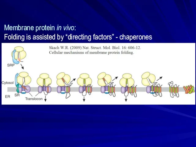
Membrane protein in vivo:
Folding is assisted by “directing factors” -
Membrane protein in vivo:
Folding is assisted by “directing factors” -
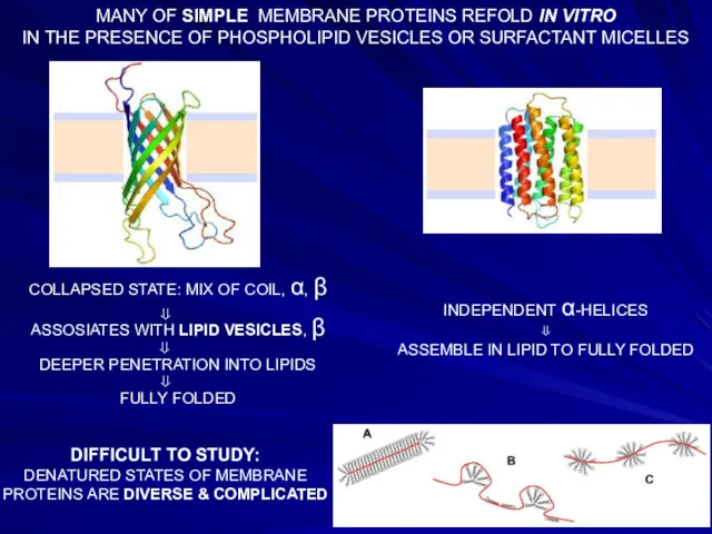
MANY OF SIMPLE MEMBRANE PROTEINS REFOLD IN VITRO
IN THE PRESENCE OF
MANY OF SIMPLE MEMBRANE PROTEINS REFOLD IN VITRO
IN THE PRESENCE OF
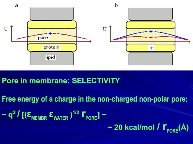
Pore in membrane: SELECTIVITY
Free energy of a charge in the non-charged
Pore in membrane: SELECTIVITY
Free energy of a charge in the non-charged
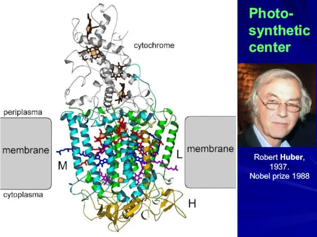
Photo-
synthetic
center
Robert Huber, 1937.
Nobel prize 1988
Photo-
synthetic
center
Robert Huber, 1937.
Nobel prize 1988
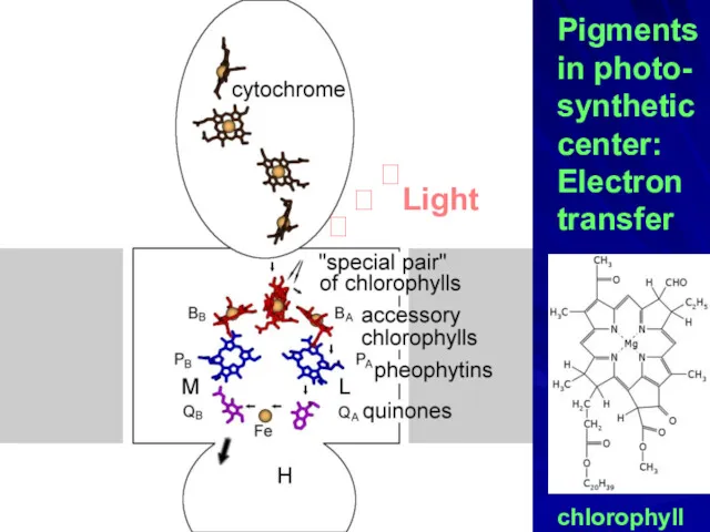
Pigments
in photo-
synthetic
center:
Electron
transfer
chlorophyll
?
? Light
?
Pigments
in photo-
synthetic
center:
Electron
transfer
chlorophyll
?
? Light
?
 Үйкеліс күші
 Дозиметрия ионизирующих излучений. Лекция 1
 Mechanical System
 Методы наблюдения и регистрации элементарных частиц
 Технология ультразвукового контроля узлов и агрегатов электропоезда
 X6000平台电子电气系统介绍. Внедрение электронной и электрической системы платформы X6000
 Коробка передач (4AT,JATCO). Составные части
 Рентгеновские лучи. Свойства, дифракция, устройство, применение рентгеновских лучей
 Лекции 3-4 курса Ф -3 2020 — копия
 Термодинамические циклы холодильных машин
 Состав молекулы. Современные представления о строении атома
 Электромагнитные переходные процессы в электроэнергетических системах
 презентация урока Выталкивающая сила
 Отчет о лабораторной работе №14 Изучение явления электромагнитной индукции
 Магнитное поле
 Решение задач. Магнитное поле и проводник с током
 Построение изображений в тонких линзах
 Взаимное притяжение и отталкивание молекул
 Электрические явления
 Демонстрационные задания. Физика. 8 класс
 Прямолинейное равноускоренное движение. Ускорение
 Буксовый узел грузового вагона. Техническая ревизия буксового узла колесный пары РУ1-Ш
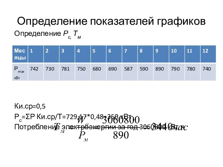 Расчет электрических нагрузок жилых и общественных зданий. (Лекция 4)
 ТО системи пуску двигунів автомобілів. ТО акумуляторних батарей. ТО генераторів та реле-регуляторів
 Окружающий мир как иерархическая система
 Тепловое излучение
 Газовые законы
 Призначення, технічна характеристика, загальна будова системи живлення двигуна УТД-20С1 паливом