- Protein structure: prediction engineering design

Содержание
- 2. Homology - - - - - -
- 3. SEQUENCE ALIGNMENT: BIOINFORMATICS PREDICTION FROM HOMOLOGY SIMILAR SEQUENCES ?? SIMILAR FOLDS ______ __________________ _______
- 4. N0 ?TWILIGHT? ======= GOOD PREDICTION =======
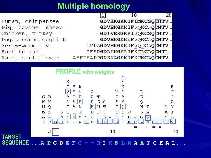
- 5. Multiple homology PROFILE with weights TARGET SEQUENCE ...A P G D E F G - -
- 6. Multiple homology PROFILE with weights TARGET SEQUENCE ...A P G D E F G - -
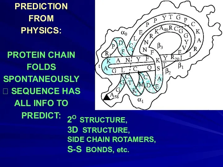
- 7. PREDICTION FROM PHYSICS: PROTEIN CHAIN FOLDS SPONTANEOUSLY ? SEQUENCE HAS ALL INFO TO PREDICT: 2O STRUCTURE,
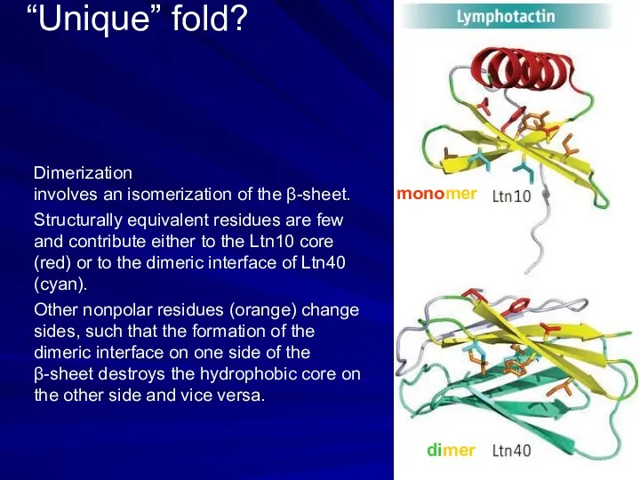
- 8. “Unique” fold? monomer dimer Dimerization involves an isomerization of the β-sheet. Structurally equivalent residues are few
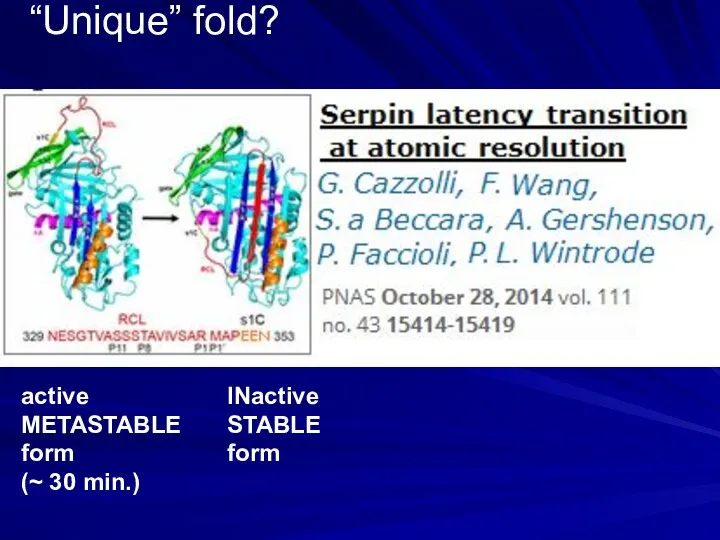
- 9. “Unique” fold? active METASTABLE form (~ 30 min.) INactive STABLE form
- 10. no Cβ: coil Сβ, ≤1 γ: α, β, coil Сβ, 2 γ: β imino: coil, turn
- 11. no Сβ: coil Сβ, ≤1 γ: α, β, coil imino: coil, turn, αN Сβ, 2 γ:
- 12. non-polar: core polar: surface
- 13. non_polar: in the core polar: at the surface
- 14. charged −: coil, α_N charged +: coil, α_C Half-charged: active sites
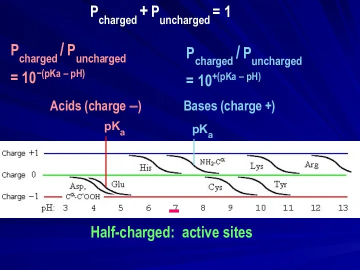
- 15. − Half-charged: active sites pKa | | pKa | | | | Pcharged / Puncharged =
- 16. charged −: coil, α_N ==== charged +: coil, α_C ==== - + - + NOT (-
- 17. PREDICTION FROM PHYSICS (OR PROTEIN STATISTICS) 2O STRUCTURES USUALLY, THIS WORKS WELL, BUT…
- 18. A B C D .---different--- Prediction, 1985 X-ray str.,1990 β γ
- 19. THREADING helps, when sequence identity is low ( BIOINFORMATICS Finkelstein, Reva, 1990-91 (Nature); Bowie, Lüthy, Eisenberg,
- 20. choice of one structure out of zillions: REQUIRES very precise estimate of interactions choice of one
- 21. HOT POINTS IN PROTEIN PHYSICS The Nobel Prize in Chemistry 2013 Martin Karplus, Michael Levitt, Arieh
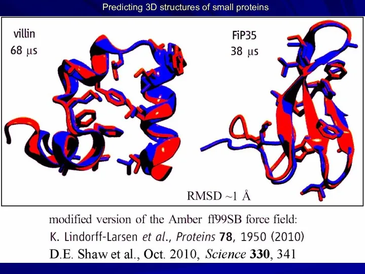
- 22. Predicting 3D structures of small proteins
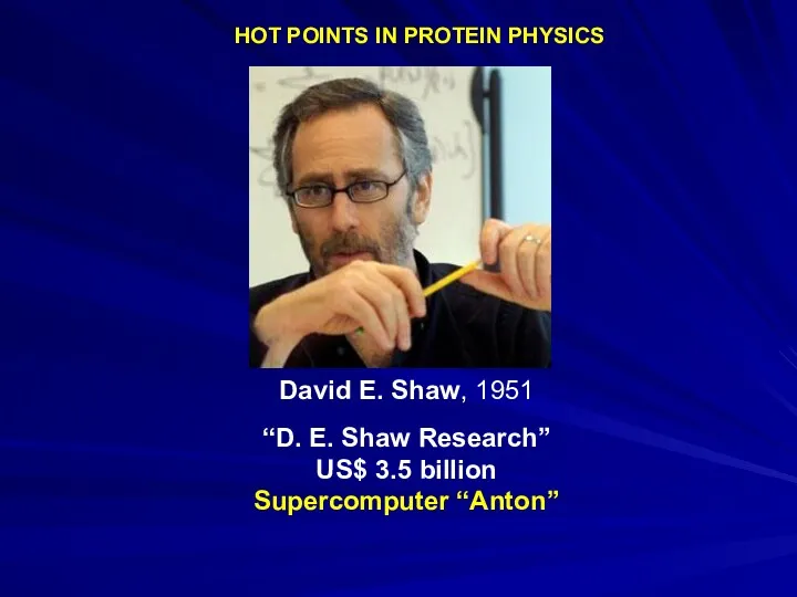
- 23. HOT POINTS IN PROTEIN PHYSICS David E. Shaw, 1951 “D. E. Shaw Research” US$ 3.5 billion
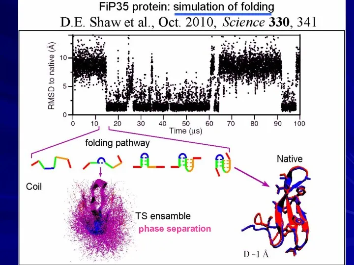
- 24. phase separation
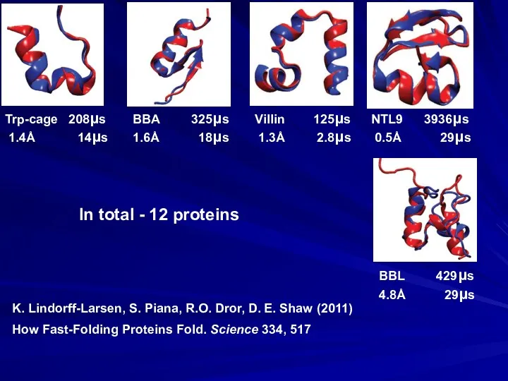
- 25. How Fast-Folding Proteins Fold. Science 334, 517 K. Lindorff-Larsen, S. Piana, R.O. Dror, D. E. Shaw
- 26. BUT: comparison of experimental and simulation-derived unfolding enthalpies shows very large differences… S. Piana, J.L. Klepeis,
- 27. Protein engineering Wanted: new protein with additional salt bridge (e.g., His+:::Asp-)
- 28. 2008 David Baker
- 29. DOES NOT MELT ! MOLTEN GLOBULE… + ION BINDING ? SOLID DeGrado, 1989 DESIGN
- 30. DESIGN Designed without ion: Mayo, 1997 Natural protein (with Zn ion) Stephen L. Mayo Later, in
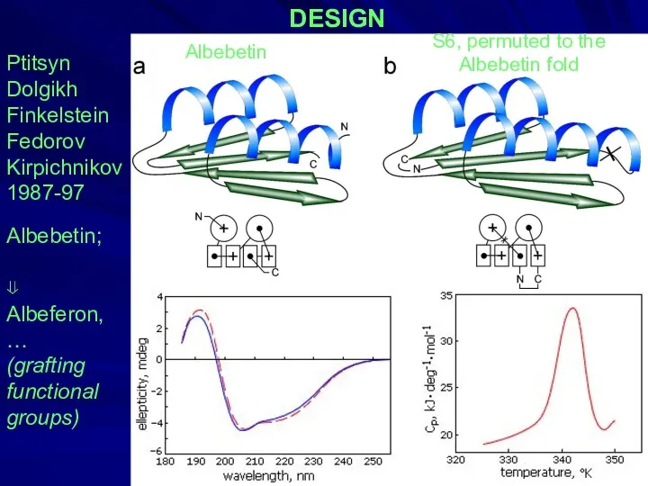
- 31. DESIGN Ptitsyn Dolgikh Finkelstein Fedorov Kirpichnikov 1987-97 Albebetin; ⇓ Albeferon, … (grafting functional groups) Albebetin S6,
- 32. DESIGN OF A “HAMELION” PROTEIN: Direct single-molecule observation of a protein living in two opposed native
- 33. Y.He, Y.Chen, P.Alexander, P.N.Bryan, J.Orban PNAS, 2008, 105, 14412-7 NMR structures of two designed proteins with
- 35. Скачать презентацию
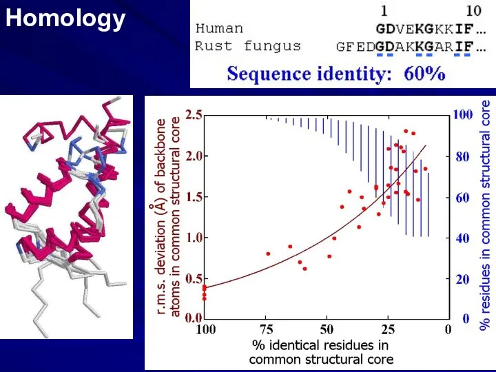
Homology
- - - - - -
Homology
- - - - - -
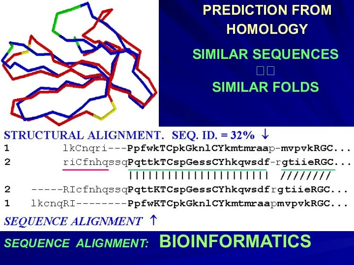
SEQUENCE ALIGNMENT: BIOINFORMATICS
PREDICTION FROM
HOMOLOGY
SIMILAR SEQUENCES ??
SIMILAR FOLDS
______ __________________ _______
SEQUENCE ALIGNMENT: BIOINFORMATICS
PREDICTION FROM
HOMOLOGY
SIMILAR SEQUENCES ??
SIMILAR FOLDS
______ __________________ _______
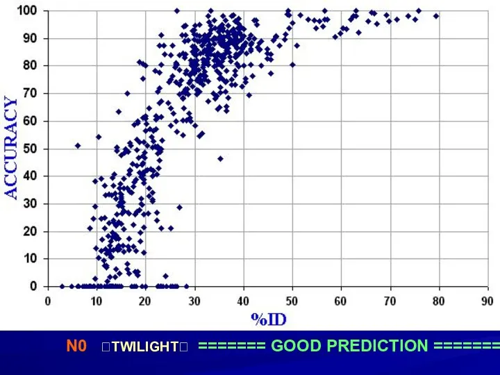
N0 ?TWILIGHT? ======= GOOD PREDICTION =======
N0 ?TWILIGHT? ======= GOOD PREDICTION =======
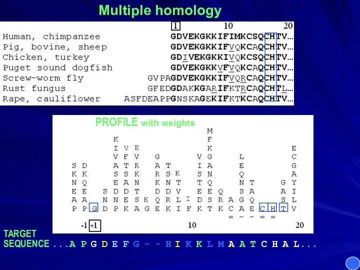
Multiple homology
PROFILE with weights
TARGET
SEQUENCE ...A P G D E F
Multiple homology
PROFILE with weights
TARGET
SEQUENCE ...A P G D E F
Multiple homology
PROFILE with weights
TARGET
SEQUENCE ...A P G D E F
Multiple homology
PROFILE with weights
TARGET
SEQUENCE ...A P G D E F
PREDICTION
FROM
PHYSICS:
PROTEIN CHAIN
FOLDS
SPONTANEOUSLY
? SEQUENCE HAS
ALL INFO TO
PREDICT:
2O STRUCTURE,
3D STRUCTURE,
SIDE CHAIN
PREDICTION
FROM
PHYSICS:
PROTEIN CHAIN
FOLDS
SPONTANEOUSLY
? SEQUENCE HAS
ALL INFO TO
PREDICT:
2O STRUCTURE,
3D STRUCTURE,
SIDE CHAIN
“Unique” fold?
monomer
dimer
Dimerization
involves an isomerization of the β-sheet.
Structurally equivalent residues
“Unique” fold?
monomer
dimer
Dimerization
involves an isomerization of the β-sheet.
Structurally equivalent residues
“Unique” fold?
active
METASTABLE
form
(~ 30 min.)
INactive
STABLE
form
“Unique” fold?
active
METASTABLE
form
(~ 30 min.)
INactive
STABLE
form
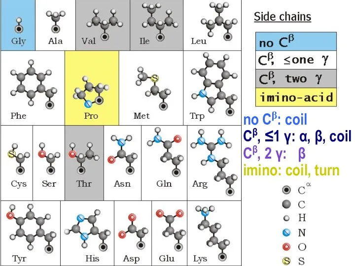
no Cβ: coil
Сβ, ≤1 γ: α, β, coil
Сβ, 2 γ: β
imino:
no Cβ: coil
Сβ, ≤1 γ: α, β, coil
Сβ, 2 γ: β
imino:
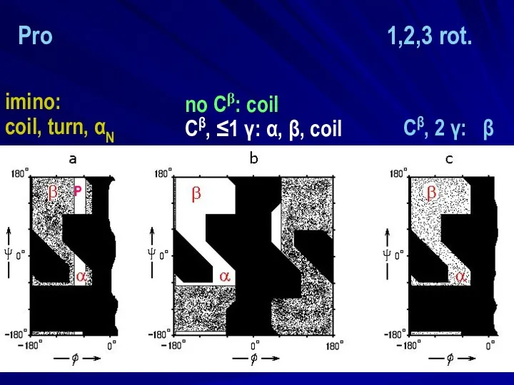
no Сβ: coil
Сβ, ≤1 γ: α, β, coil
imino:
coil, turn, αN
no Сβ: coil
Сβ, ≤1 γ: α, β, coil
imino:
coil, turn, αN
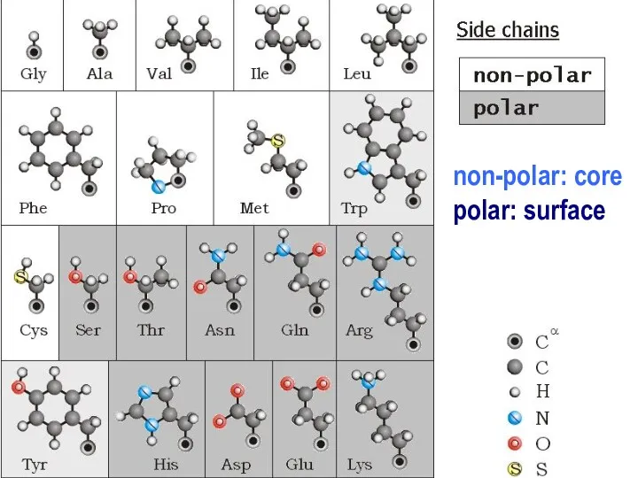
non-polar: core
polar: surface
non-polar: core
polar: surface
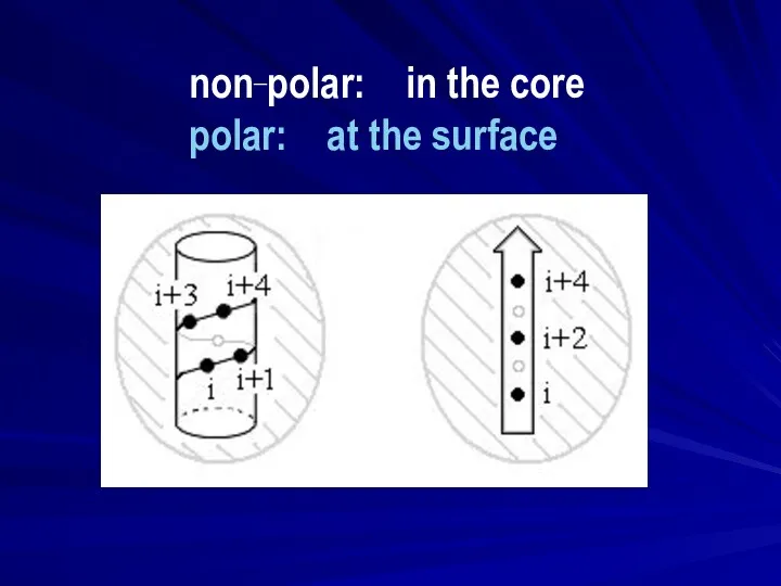
non_polar: in the core
polar: at the surface
non_polar: in the core
polar: at the surface
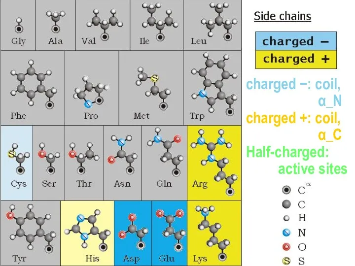
charged −: coil,
α_N
charged +: coil,
α_C
Half-charged:
active
charged −: coil,
α_N
charged +: coil,
α_C
Half-charged:
active
−
Half-charged: active sites
pKa
|
|
pKa
|
|
|
|
Pcharged / Puncharged
= 10−(pKa – pH)
Acids (charge −)
−
Half-charged: active sites
pKa
|
|
pKa
|
|
|
|
Pcharged / Puncharged
= 10−(pKa – pH)
Acids (charge −)
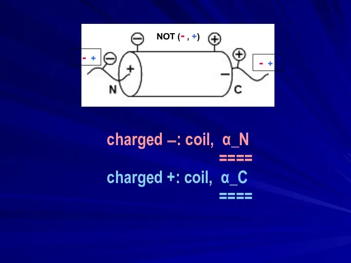
charged −: coil, α_N
====
charged +: coil, α_C
====
- +
- +
NOT
charged −: coil, α_N
====
charged +: coil, α_C
====
- +
- +
NOT
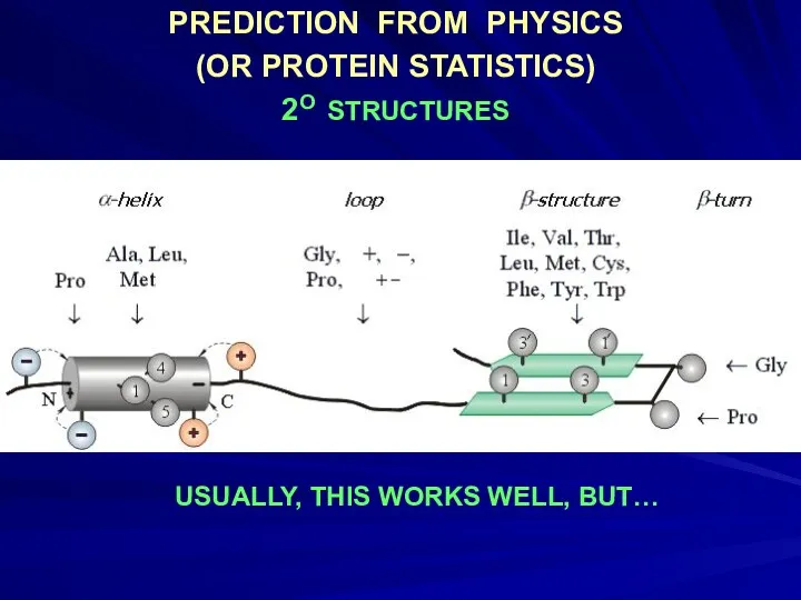
PREDICTION FROM PHYSICS
(OR PROTEIN STATISTICS)
2O STRUCTURES
USUALLY, THIS WORKS WELL, BUT…
PREDICTION FROM PHYSICS
(OR PROTEIN STATISTICS)
2O STRUCTURES
USUALLY, THIS WORKS WELL, BUT…
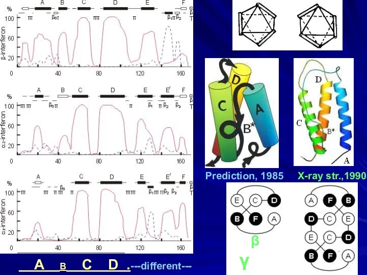
A B C D .---different---
Prediction, 1985 X-ray str.,1990
β γ
A B C D .---different---
Prediction, 1985 X-ray str.,1990
β γ
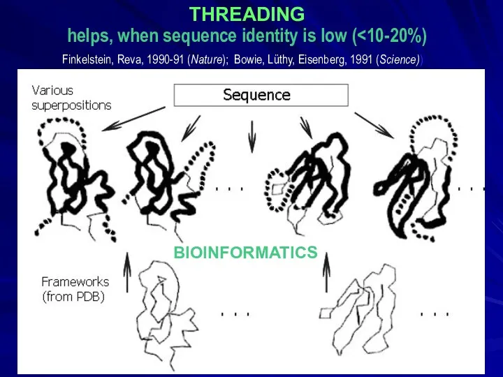
THREADING
helps, when sequence identity is low (<10-20%)
BIOINFORMATICS
Finkelstein, Reva, 1990-91 (Nature);
THREADING
helps, when sequence identity is low (<10-20%)
BIOINFORMATICS
Finkelstein, Reva, 1990-91 (Nature);
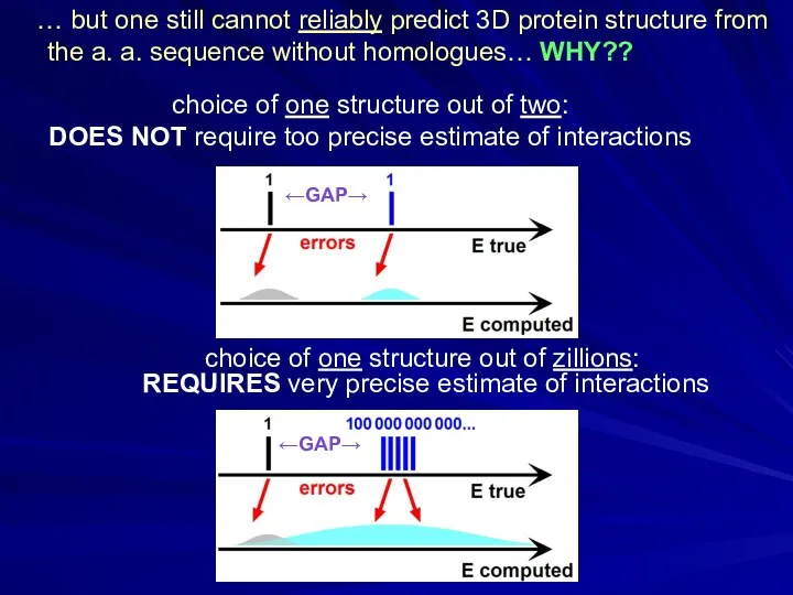
choice of one structure out of zillions:
REQUIRES very precise estimate
choice of one structure out of zillions: REQUIRES very precise estimate
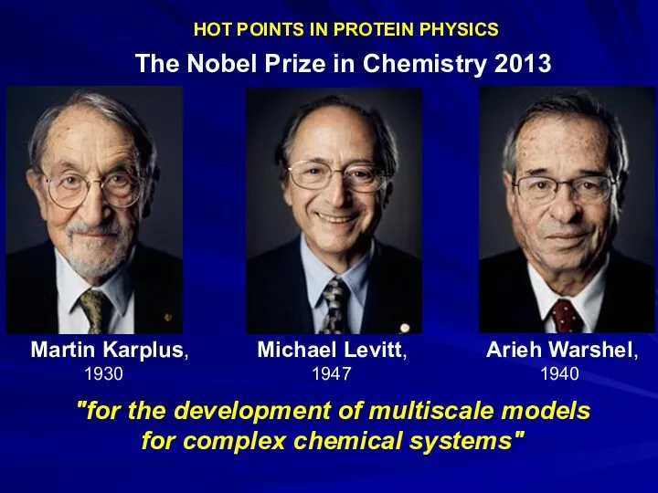
HOT POINTS IN PROTEIN PHYSICS
The Nobel Prize in Chemistry 2013
HOT POINTS IN PROTEIN PHYSICS
The Nobel Prize in Chemistry 2013
Predicting 3D structures of small proteins
Predicting 3D structures of small proteins
HOT POINTS IN PROTEIN PHYSICS
David E. Shaw, 1951
“D. E. Shaw Research”
US$ 3.5
HOT POINTS IN PROTEIN PHYSICS
David E. Shaw, 1951
“D. E. Shaw Research”
US$ 3.5
phase separation
phase separation
How Fast-Folding Proteins Fold. Science 334, 517
K. Lindorff-Larsen, S. Piana, R.O.
How Fast-Folding Proteins Fold. Science 334, 517
K. Lindorff-Larsen, S. Piana, R.O.
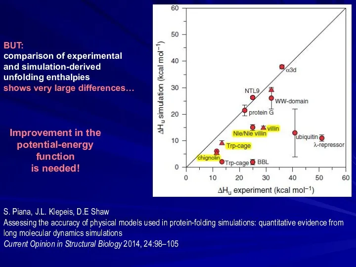
BUT:
comparison of experimental
and simulation-derived
unfolding enthalpies
shows very large differences…
S. Piana,
BUT:
comparison of experimental
and simulation-derived
unfolding enthalpies
shows very large differences…
S. Piana,
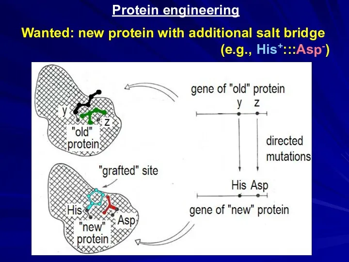
Protein engineering
Wanted: new protein with additional salt bridge
(e.g., His+:::Asp-)
Protein engineering
Wanted: new protein with additional salt bridge
(e.g., His+:::Asp-)
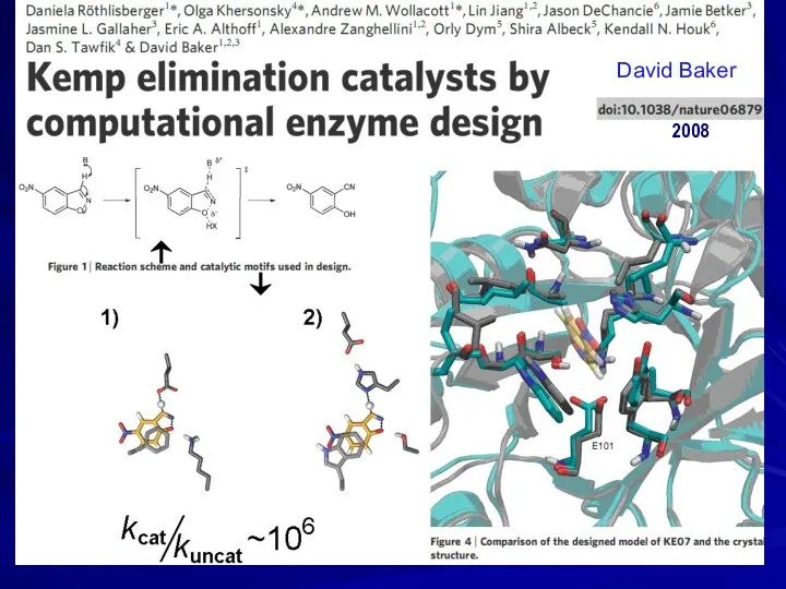
2008
David Baker
2008
David Baker
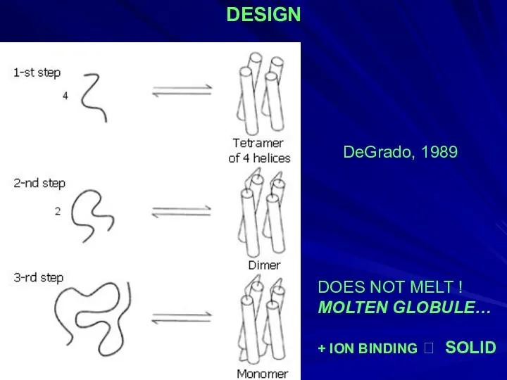
DOES NOT MELT !
MOLTEN GLOBULE…
+ ION BINDING ? SOLID
DeGrado, 1989
DESIGN
DOES NOT MELT !
MOLTEN GLOBULE…
+ ION BINDING ? SOLID
DeGrado, 1989
DESIGN
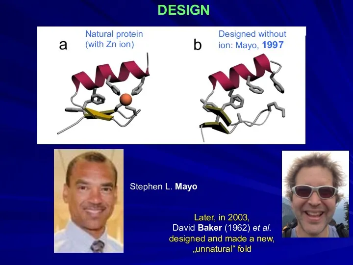
DESIGN
Designed without
ion: Mayo, 1997
Natural protein
(with Zn ion)
Stephen L. Mayo
Later, in 2003,
David Baker (1962)
DESIGN
Designed without
ion: Mayo, 1997
Natural protein
(with Zn ion)
Stephen L. Mayo
Later, in 2003,
David Baker (1962)
DESIGN
Ptitsyn
Dolgikh
Finkelstein
Fedorov
Kirpichnikov
1987-97
Albebetin;
⇓
Albeferon,
…
(grafting
functional
groups)
Albebetin
S6, permuted to the
Albebetin fold
DESIGN
Ptitsyn
Dolgikh
Finkelstein
Fedorov
Kirpichnikov
1987-97
Albebetin;
⇓
Albeferon,
…
(grafting
functional
groups)
Albebetin
S6, permuted to the
Albebetin fold
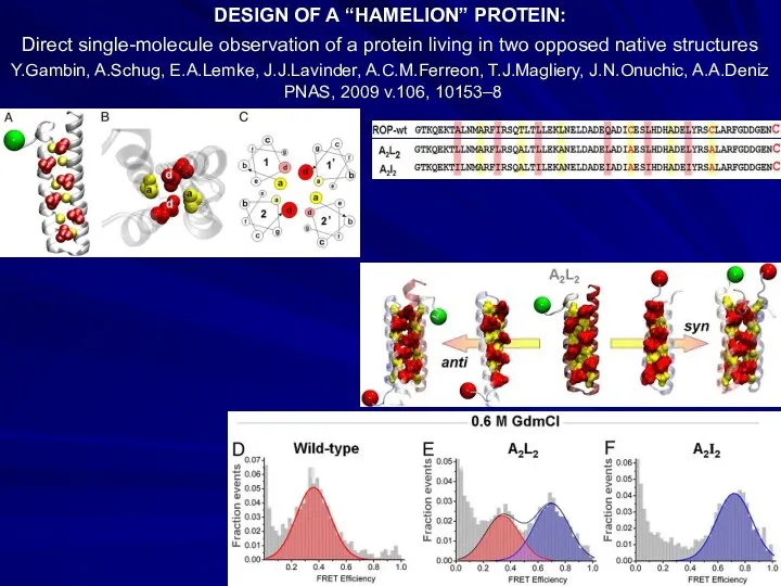
DESIGN OF A “HAMELION” PROTEIN:
Direct single-molecule observation of a protein living
DESIGN OF A “HAMELION” PROTEIN:
Direct single-molecule observation of a protein living
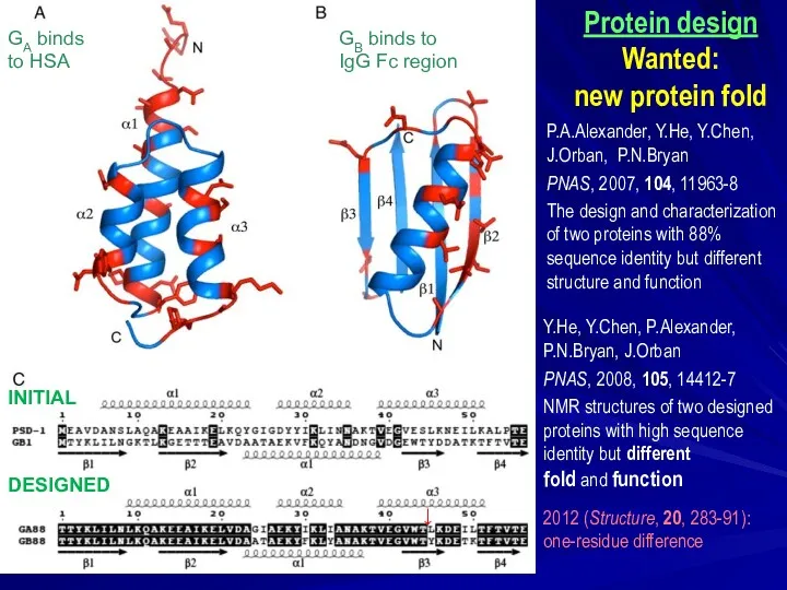
Y.He, Y.Chen, P.Alexander,
P.N.Bryan, J.Orban
PNAS, 2008, 105, 14412-7
NMR structures
Y.He, Y.Chen, P.Alexander,
P.N.Bryan, J.Orban
PNAS, 2008, 105, 14412-7
NMR structures
 Электромагниттік сәуле шығару
 Особенности заданий ЕГЭ. Колебания и волны
 Линзы. Построение изображения в собирающей линзе
 Остов, кривошипно-шатунний та газорозподільний механізми ДВЗ. (Лекція 3.1)
 Структура и принципы интеграции МС
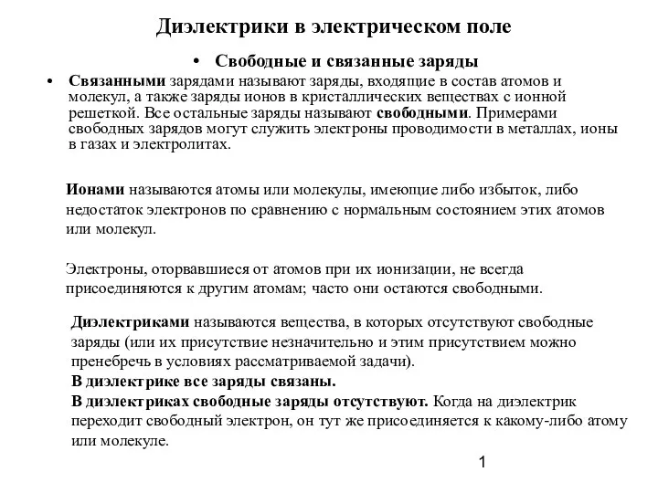 Диэлектрики в электрическом поле
 Перемещение при прямолинейном равномерном движении
 Основные положения молекулярно-кинетической теории и их опытные подтверждения
 Электронная проводимость металлов. Электрический ток и его характеристики
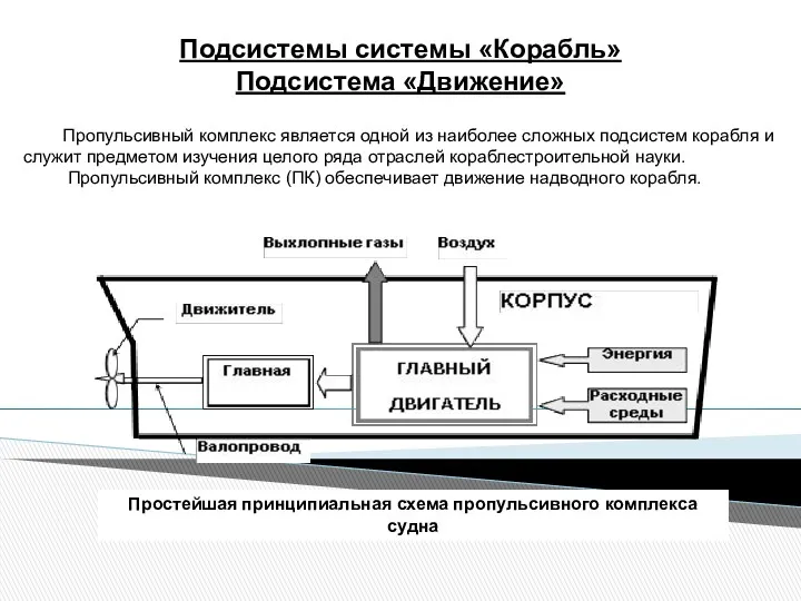 Подсистемы системы Корабль. Подсистема Движение
 Известные физики
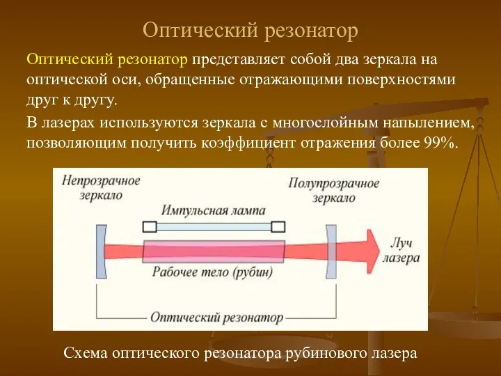 Оптический резонатор
 Презентация 10 класса Газовые законы
 Специальная теория относительности
 Презентация Интерференция света
 Презентация доклада системнодеятельностного подхода на уроках физики
 Уравнения Максвелла
 Движение по окружности
 Занятие № 1 Методы повышения эффективности усвоения понятийпри изучении темы:Кинематика
 Детекторы нейтронов
 Презентация 8 класс. Количество теплоты. Едельная теплоемкость
 Водородная бомба
 Решение физических задач с применением производной функции
 Презентация Броуновское движение. Диффузия. Взаимодействие молекул
 Сила трения. Особенности сил трения
 Способы герметизации клепаных швов и изделий
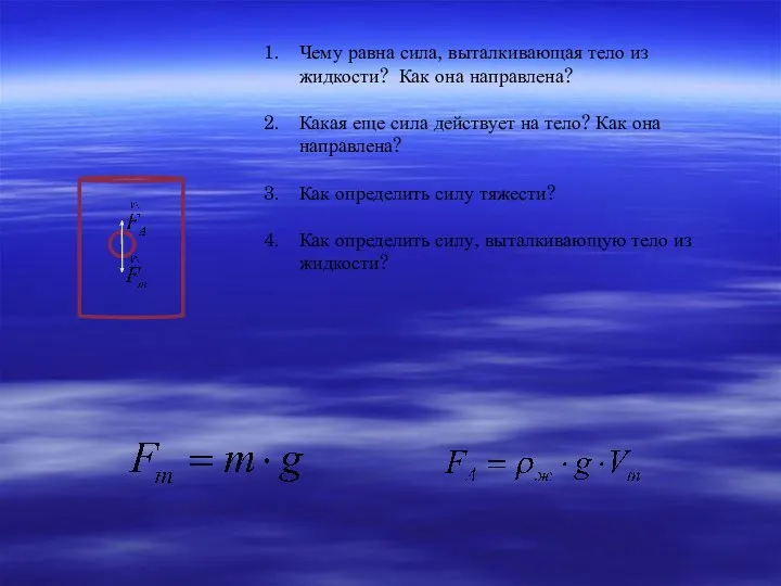 Урок и презентация по теме Плавание тел 7 класс
 Биологическое действие радиации. Закон радиоактивного распада