- Radiation

Содержание
- 2. Definition of Radiation “Radiation is an energy in the form of electro-magnetic waves or particulate matter,
- 3. Forces: There are many interactions among nuclei. It turns out that there are forces other than
- 4. Radioactivity: Elements & Atoms Atoms are composed of smaller particles referred to as: Protons Neutrons Electrons
- 5. Basic Model of a Neutral Atom. Electrons (-) orbiting nucleus of protons (+) and neutrons. Same
- 7. Radioactivity If a nucleus is unstable for any reason, it will emit and absorb particles. There
- 8. Ionization Ionizing radiation is produced by unstable atoms. Unstable atoms differ from stable atoms because they
- 13. Types or Products of Ionizing Radiation β α γ or X-ray neutron
- 14. Radioactive Atom X-ray gamma ray
- 15. The electro-magnetic waves vary in their length and frequency along a very wide spectrum.
- 19. Types of Radiation Radiation is classified into: Ionizing radiation Non-ionizing radiation
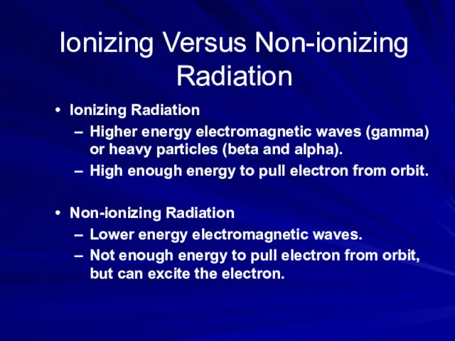
- 20. Ionizing Versus Non-ionizing Radiation Ionizing Radiation Higher energy electromagnetic waves (gamma) or heavy particles (beta and
- 21. Ionizing Radiation Definition: “ It is a type of radiation that is able to disrupt atoms
- 22. Another Definition Ionizing radiation A radiation is said to be ionizing when it has enough energy
- 23. Primary Types of Ionizing Radiation Alpha particles Beta particles Gamma rays (or photons) X-Rays (or photons)
- 24. Alpha Particles: 2 neutrons and 2 protons They travel short distances, have large mass Only a
- 25. Alpha Particles (or Alpha Radiation): Helium nucleus (2 neutrons and 2 protons); +2 charge; heavy (4
- 26. Beta Particles Beta Particles: Electrons or positrons having small mass and variable energy. Electrons form when
- 27. Beta Particles: High speed electron ejected from nucleus; -1 charge, light 0.00055 AMU; Typical Energy =
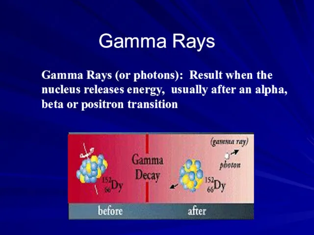
- 29. Gamma Rays Gamma Rays (or photons): Result when the nucleus releases energy, usually after an alpha,
- 30. X-Rays X-Rays: Occur whenever an inner shell orbital electron is removed and rearrangement of the atomic
- 31. X- and Gamma Rays: X-rays are photons (Electromagnetic radiations) emitted from electron orbits. Gamma rays are
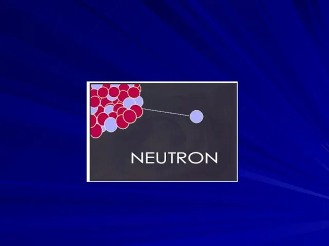
- 32. Neutrons Neutrons: Have the same mass as protons but are uncharged
- 35. QUANTIFICATION OF RADIATION A. Quantifying Radioactive Decay B. Quantifying Exposure and Dose
- 36. A. Quantifying Radioactive Decay Measurement of Activity in disintegrations per second (dps); 1 Becquerel (Bq) =
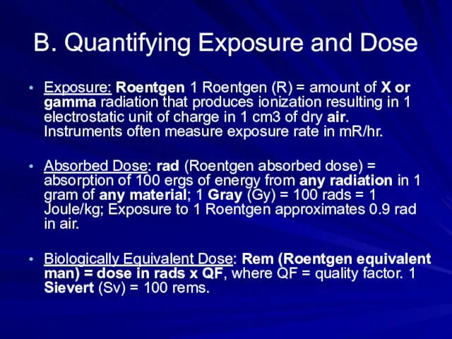
- 37. B. Quantifying Exposure and Dose Exposure: Roentgen 1 Roentgen (R) = amount of X or gamma
- 38. Half Life Calculation
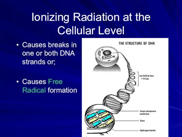
- 39. Ionizing Radiation at the Cellular Level Causes breaks in one or both DNA strands or; Causes
- 40. Exposure Limits OSHA Limits: Whole body limit = 1.25 rem/qtr or 5 rem (50 mSv) per
- 41. External/Internal Exposure Limits for Occupationally Exposed Individuals Annual Dose Limits *Effective dose equivalent
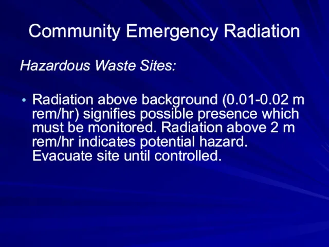
- 43. Community Emergency Radiation Hazardous Waste Sites: Radiation above background (0.01-0.02 m rem/hr) signifies possible presence which
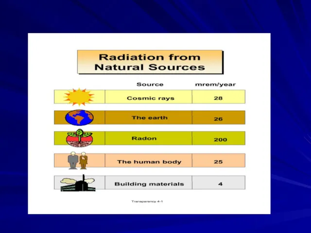
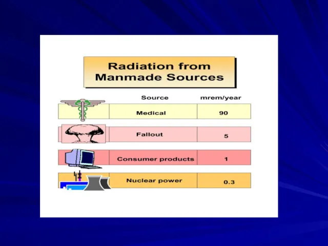
- 44. Your Annual Exposure
- 45. HEALTH EFFECTS Generalizations: Biological effects are due to the ionization process that destroys the capacity for
- 46. ACUTE DOSE(RAD) EFFECT
- 47. Delayed Somatic Effects: Delayed effects to exposed person include: Cancer, leukemia, cataracts, life shortening from organ
- 48. Critical Organs: Organs generally most susceptible to radiation damage include: Lymphocytes, bone marrow, gastro-intestinal, gonads, and
- 49. Non-ionizing Radiation Definition: “ They are electromagnetic waves incapable of producing ions while passing through matter,
- 50. All earth surface system components emit radiation---the sun and the earth are the components we are
- 52. Path of incoming solar radiation
- 53. Albedo: a measure of how well a surface reflects insolation
- 54. Examples on Non-ionizing Radiation Sources Visible light Microwaves Radios Video Display Terminals Power lines Radiofrequency Diathermy
- 55. Other Manmade Sources of Non-Ionizing Radiation
- 58. Effects Radiofrequency Ranges (10 kHz to 300 GHz) Effects only possible at ten times the permissible
- 59. RADIATION CONTROLS A. Basic Control Methods for External Radiation Decrease Time Increase Distance Increase Shielding
- 60. Time: Minimize time of exposure to minimize total dose. Rotate employees to restrict individual dose. Distance:
- 62. B. Monitoring Personal Dosimeters: Normally they do not prevent exposures (no alarm), just record it. They
- 66. Direct Reading Survey Meters and Counters: Useful in identifying source of exposures recorded by personal dosimeters,
- 68. Continuous Monitors: Continuous direct reading ionization detectors (same detectors as above) can provide read-out and/or alarm
- 69. Elements of Radiation Protection Program Monitoring of exposures: Personal, area, and screening measurements; Medical/biologic monitoring. Task-Specific
- 71. Скачать презентацию

Definition of Radiation
“Radiation is an energy in the form of electro-magnetic
Definition of Radiation
“Radiation is an energy in the form of electro-magnetic
Forces: There are many interactions among nuclei. It turns out that
Forces: There are many interactions among nuclei. It turns out that
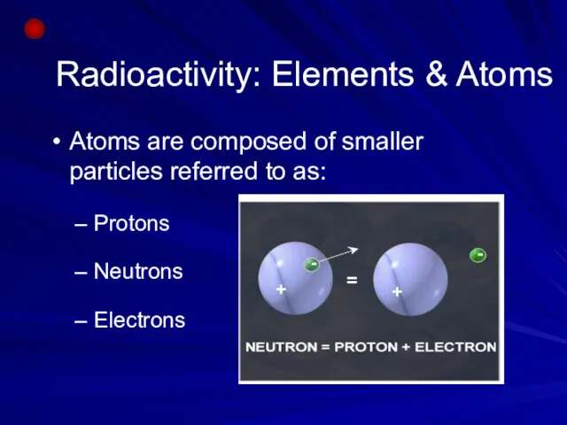
Radioactivity: Elements & Atoms
Atoms are composed of smaller particles referred to
Radioactivity: Elements & Atoms
Atoms are composed of smaller particles referred to
Basic Model of a Neutral Atom.
Electrons (-) orbiting nucleus of protons
Basic Model of a Neutral Atom.
Electrons (-) orbiting nucleus of protons

Radioactivity
If a nucleus is unstable for any reason, it will emit
Radioactivity
If a nucleus is unstable for any reason, it will emit

Ionization
Ionizing radiation is produced by unstable atoms. Unstable atoms differ
Ionization
Ionizing radiation is produced by unstable atoms. Unstable atoms differ


Types or Products of Ionizing Radiation
β
α
γ or X-ray
neutron
Types or Products of Ionizing Radiation
β
α
γ or X-ray
neutron

Radioactive Atom
X-ray
gamma ray
Radioactive Atom
X-ray
gamma ray

The electro-magnetic waves vary in their length and frequency along a
The electro-magnetic waves vary in their length and frequency along a


Types of Radiation
Radiation is classified into:
Ionizing radiation
Non-ionizing radiation
Types of Radiation
Radiation is classified into:
Ionizing radiation
Non-ionizing radiation
Ionizing Versus Non-ionizing Radiation
Ionizing Radiation
Higher energy electromagnetic waves (gamma) or heavy
Ionizing Versus Non-ionizing Radiation
Ionizing Radiation
Higher energy electromagnetic waves (gamma) or heavy
Ionizing Radiation
Definition:
“ It is a type of radiation that
Ionizing Radiation
Definition:
“ It is a type of radiation that

Another Definition
Ionizing radiation
A radiation is said to be ionizing when
Another Definition
Ionizing radiation
A radiation is said to be ionizing when

Primary Types of Ionizing Radiation
Alpha particles
Beta particles
Gamma rays (or photons)
X-Rays (or
Primary Types of Ionizing Radiation
Alpha particles
Beta particles
Gamma rays (or photons)
X-Rays (or

Alpha Particles: 2 neutrons and 2 protons
They travel short distances, have
Alpha Particles: 2 neutrons and 2 protons
They travel short distances, have

Alpha Particles (or Alpha Radiation): Helium nucleus (2 neutrons and 2
Alpha Particles (or Alpha Radiation): Helium nucleus (2 neutrons and 2

Beta Particles
Beta Particles: Electrons or positrons having small mass and variable
Beta Particles
Beta Particles: Electrons or positrons having small mass and variable

Beta Particles: High speed electron ejected from nucleus; -1 charge, light
Beta Particles: High speed electron ejected from nucleus; -1 charge, light

Gamma Rays
Gamma Rays (or photons): Result when the nucleus releases energy,
Gamma Rays
Gamma Rays (or photons): Result when the nucleus releases energy,
X-Rays
X-Rays: Occur whenever an inner shell orbital electron is removed and
X-Rays
X-Rays: Occur whenever an inner shell orbital electron is removed and

X- and Gamma Rays: X-rays are photons (Electromagnetic radiations) emitted from
X- and Gamma Rays: X-rays are photons (Electromagnetic radiations) emitted from
Neutrons
Neutrons: Have the same mass as protons but are uncharged
Neutrons
Neutrons: Have the same mass as protons but are uncharged

QUANTIFICATION OF RADIATION
A. Quantifying Radioactive Decay
B. Quantifying Exposure and Dose
QUANTIFICATION OF RADIATION
A. Quantifying Radioactive Decay
B. Quantifying Exposure and Dose

A. Quantifying Radioactive Decay
Measurement of Activity in disintegrations per second (dps);
A. Quantifying Radioactive Decay
Measurement of Activity in disintegrations per second (dps);
B. Quantifying Exposure and Dose
Exposure: Roentgen 1 Roentgen (R) = amount
B. Quantifying Exposure and Dose
Exposure: Roentgen 1 Roentgen (R) = amount
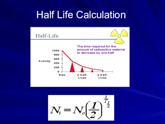
Half Life Calculation
Half Life Calculation
Ionizing Radiation at the Cellular Level
Causes breaks in one or both
Ionizing Radiation at the Cellular Level
Causes breaks in one or both

Exposure Limits
OSHA Limits: Whole body limit = 1.25 rem/qtr or 5
Exposure Limits
OSHA Limits: Whole body limit = 1.25 rem/qtr or 5

External/Internal Exposure Limits for Occupationally Exposed Individuals
Annual Dose Limits
*Effective dose equivalent
External/Internal Exposure Limits for Occupationally Exposed Individuals
Annual Dose Limits
*Effective dose equivalent

Community Emergency Radiation
Hazardous Waste Sites:
Radiation above background (0.01-0.02 m rem/hr)
Community Emergency Radiation
Hazardous Waste Sites:
Radiation above background (0.01-0.02 m rem/hr)
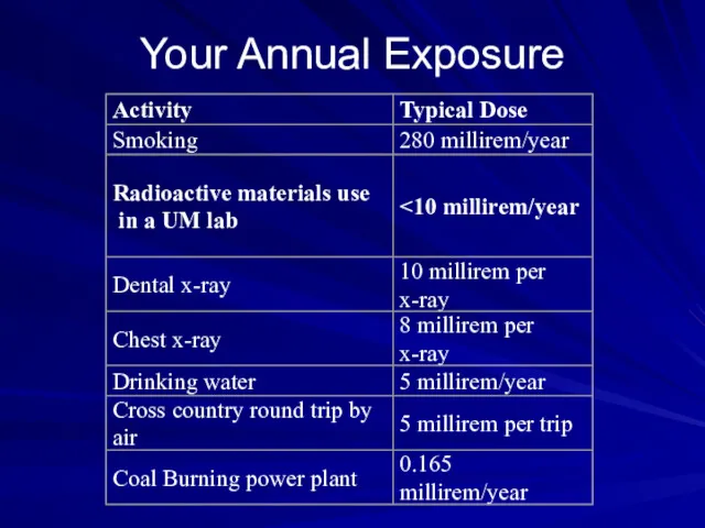
Your Annual Exposure
Your Annual Exposure
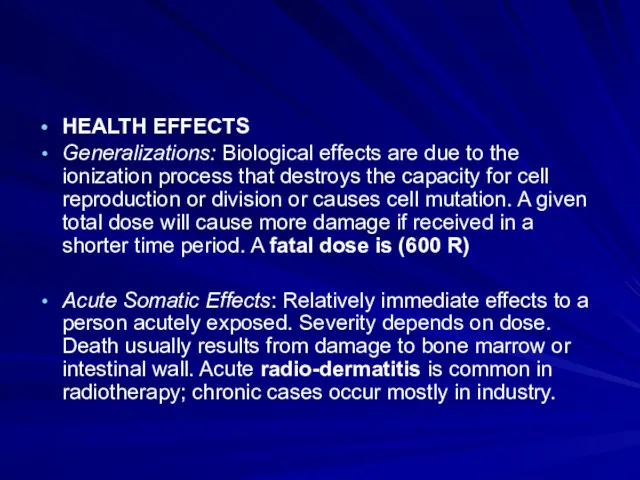
HEALTH EFFECTS
Generalizations: Biological effects are due to the ionization process
HEALTH EFFECTS
Generalizations: Biological effects are due to the ionization process

ACUTE DOSE(RAD) EFFECT
ACUTE DOSE(RAD) EFFECT

Delayed Somatic Effects: Delayed effects to exposed person include: Cancer, leukemia,
Delayed Somatic Effects: Delayed effects to exposed person include: Cancer, leukemia,
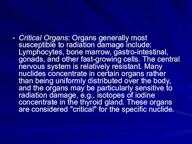
Critical Organs: Organs generally most susceptible to radiation damage include: Lymphocytes,
Critical Organs: Organs generally most susceptible to radiation damage include: Lymphocytes,
Non-ionizing Radiation
Definition:
“ They are electromagnetic waves incapable of producing ions while
Non-ionizing Radiation
Definition:
“ They are electromagnetic waves incapable of producing ions while

All earth surface system components emit radiation---the sun and the earth
All earth surface system components emit radiation---the sun and the earth


Path of incoming solar radiation
Path of incoming solar radiation
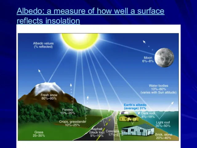
Albedo: a measure of how well a surface reflects insolation
Albedo: a measure of how well a surface reflects insolation

Examples on Non-ionizing Radiation Sources
Visible light
Microwaves
Radios
Video Display Terminals
Power lines
Radiofrequency Diathermy (Physical
Examples on Non-ionizing Radiation Sources
Visible light
Microwaves
Radios
Video Display Terminals
Power lines
Radiofrequency Diathermy (Physical
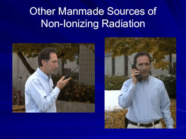
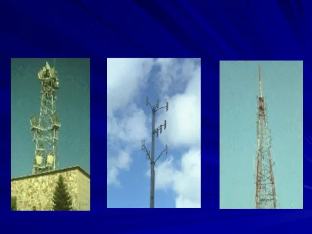
Other Manmade Sources of Non-Ionizing Radiation
Other Manmade Sources of Non-Ionizing Radiation

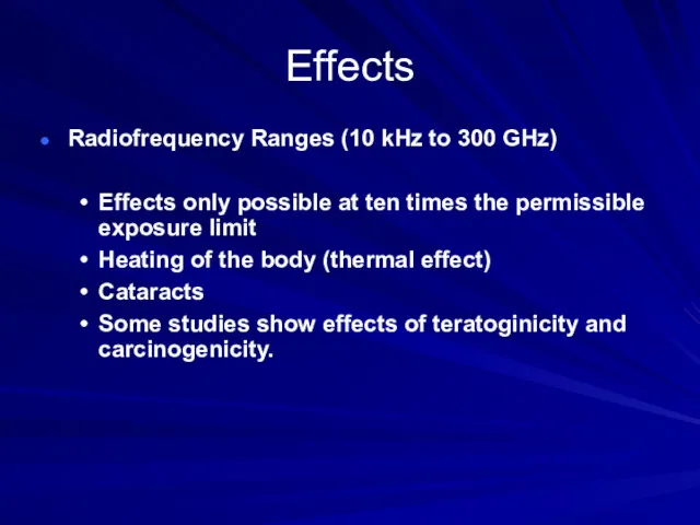
Effects
Radiofrequency Ranges (10 kHz to 300 GHz)
Effects only possible at
Effects
Radiofrequency Ranges (10 kHz to 300 GHz)
Effects only possible at
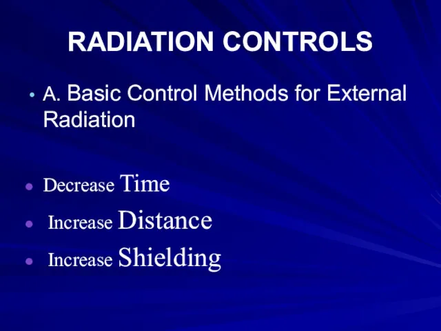
RADIATION CONTROLS
A. Basic Control Methods for External Radiation
Decrease Time
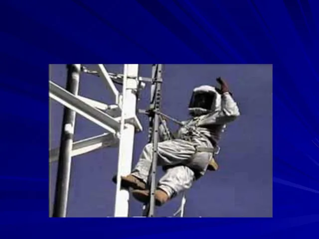
Increase
RADIATION CONTROLS
A. Basic Control Methods for External Radiation
Decrease Time
Increase

Time: Minimize time of exposure to minimize total dose. Rotate employees
Time: Minimize time of exposure to minimize total dose. Rotate employees

B. Monitoring
Personal Dosimeters: Normally they do not prevent exposures (no
B. Monitoring
Personal Dosimeters: Normally they do not prevent exposures (no


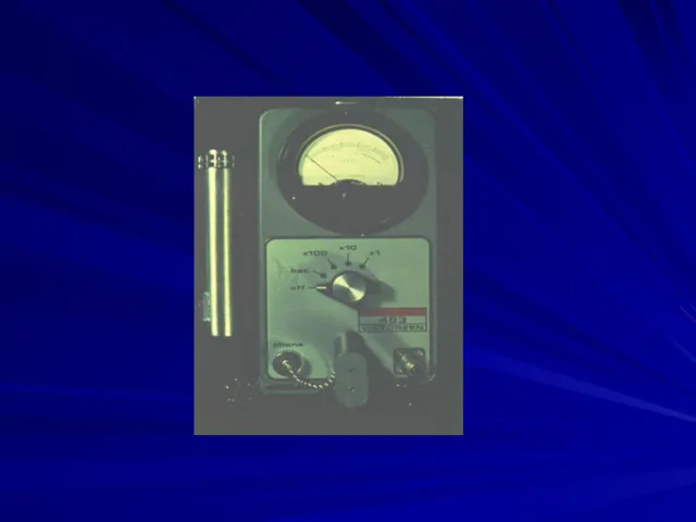
Direct Reading Survey Meters and Counters: Useful in identifying source of
Direct Reading Survey Meters and Counters: Useful in identifying source of

Continuous Monitors: Continuous direct reading ionization detectors (same detectors as above)
Continuous Monitors: Continuous direct reading ionization detectors (same detectors as above)

Elements of Radiation Protection Program
Monitoring of exposures: Personal, area, and
Elements of Radiation Protection Program
Monitoring of exposures: Personal, area, and
 Измерение давления в покоящейся жидкости
 Physics basics (Unit 1)
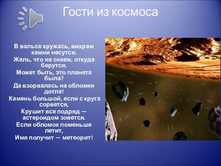 Презентация. Гости из космоса
 Властивості радіохвиль. Розподіл спектру радіохвиль. Особливості розповсюдження радіохвиль (Лекція 2.1)
 Машины и оборудование для свайных работ. Лекция 6
 Электромагнитные переходные процессы в электроэнергетических системах
 Открытое мероприятие Добро пожаловать в космос
 Применение сообщающихся сосудов
 Презентация по теме Решение задач по кинематике с помощью квадратных уравнений.
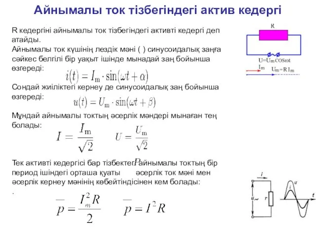 Айнымалы ток тізбегіндегі актив кедергі. (Лекция 14)
 Своя игра. Строения атома. (11 класс)
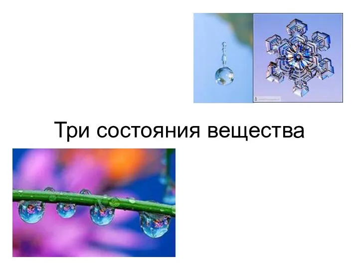 Три состояния вещества
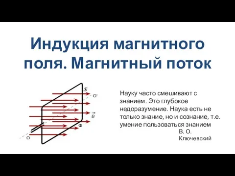 Индукция магнитного поля. Магнитный поток
 Газовые законы
 Автокөліктерді жөндеу технологиясы
 Зеленый дом
 Энтропия в техносфере
 Раздел 3. Оценка технического состояния, ТО и Р автомобилей. Урок №72. Тема ТО рулевого управления
 Ядерные реакции. Радиоактивность
 Пучковые технологии. Вакуум: физические свойства, получение, измерение
 Тепловой расчет сферического носка ЛА. Вариант 34
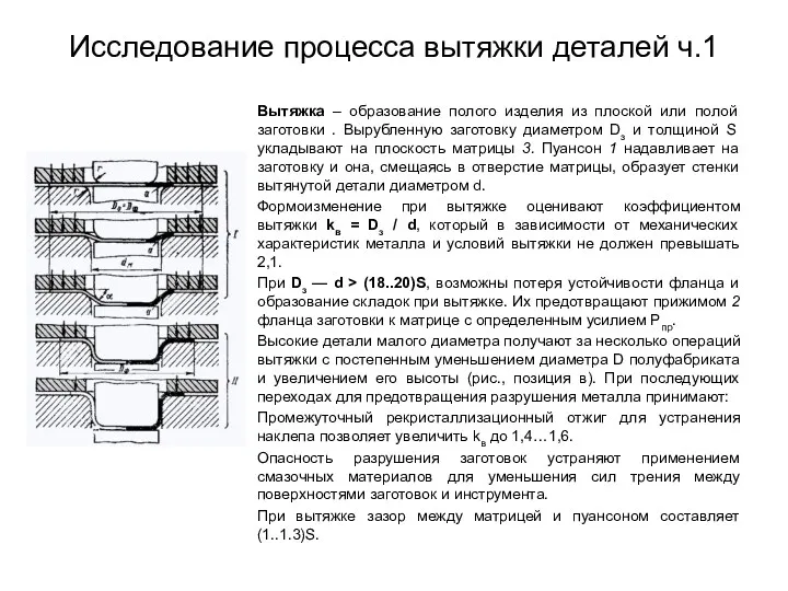 Исследование процесса вытяжки деталей ч.1
 Магнитное поле
 Работа и мощность электрического тока
 Своя игра по физике
 Техническое обслуживание и ремонт сцепления автомобиля ВАЗ 2107
 Робототехника. Робот-дворник
 Коррозионные процессы