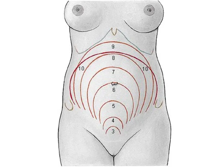
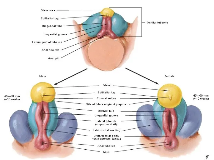
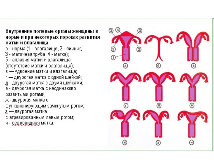
- Classification of female genitals

Содержание
- 6. Uterus, Uterus; uterine (fallopian) tube, Tuba uterina; ap-radiograph after injection of a contrast medium through the
- 12. 3 month 5 month 7 month
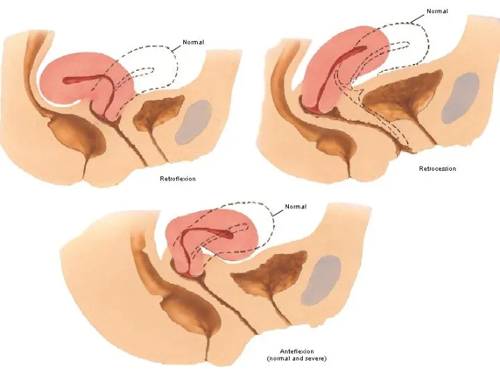
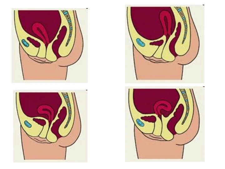
- 13. Uterus, Uterus, Vagina, Vagina, normal angles between the vagina, the cervix and the corpus of the
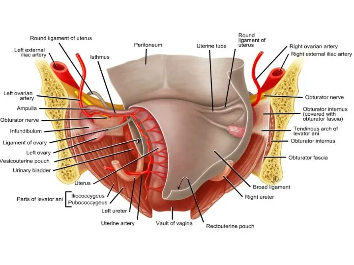
- 20. Cardinal ligament, Parametrium* Cervix uteri Lig. pubovesicale Pararectal space, Paraproctium* Paravesical space, Paracystium* Petropubical space (=
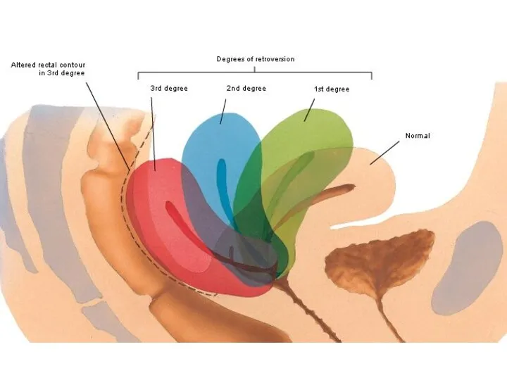
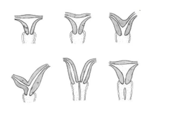
- 31. Bipartite uterus. Note the absence of the fundus of the uterus and in it’s place a
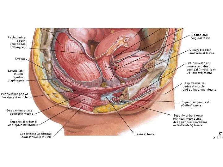
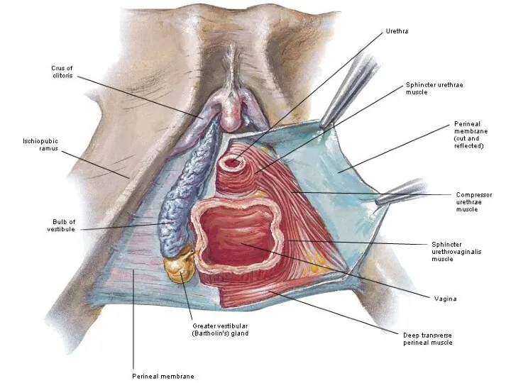
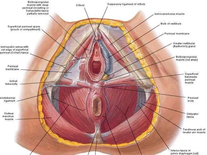
- 35. Perineum Diafragma urogenitale Diafragma pelvis Trigonum urogenitale Trigonum anale
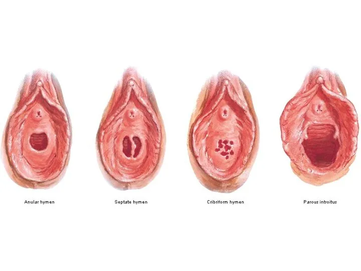
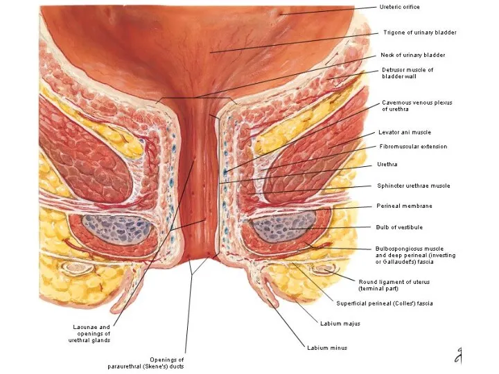
- 37. External urethral sphincter, urethrovaginal sphincter, and compressor urethrae. Left, male; right female.
- 47. Скачать презентацию
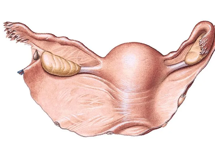
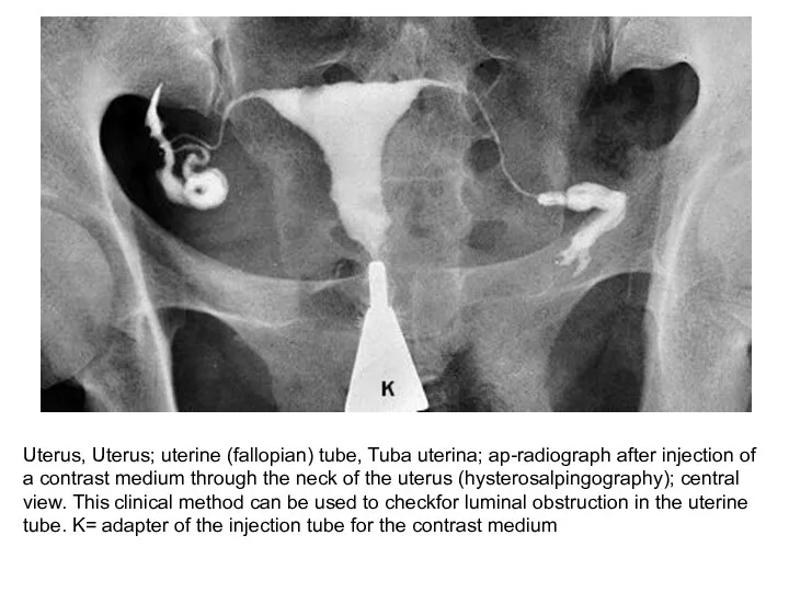
Uterus, Uterus; uterine (fallopian) tube, Tuba uterina; ap-radiograph after injection of
Uterus, Uterus; uterine (fallopian) tube, Tuba uterina; ap-radiograph after injection of

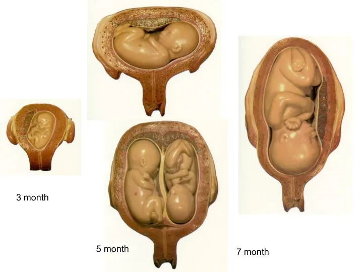
3 month
5 month
7 month
3 month
5 month
7 month
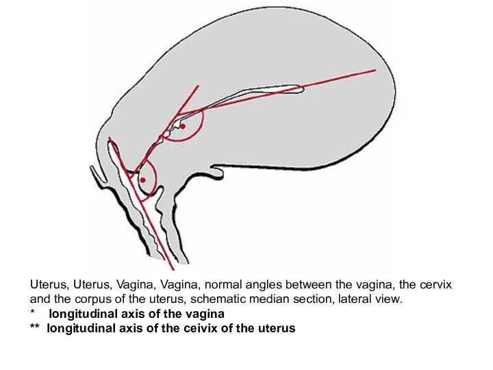
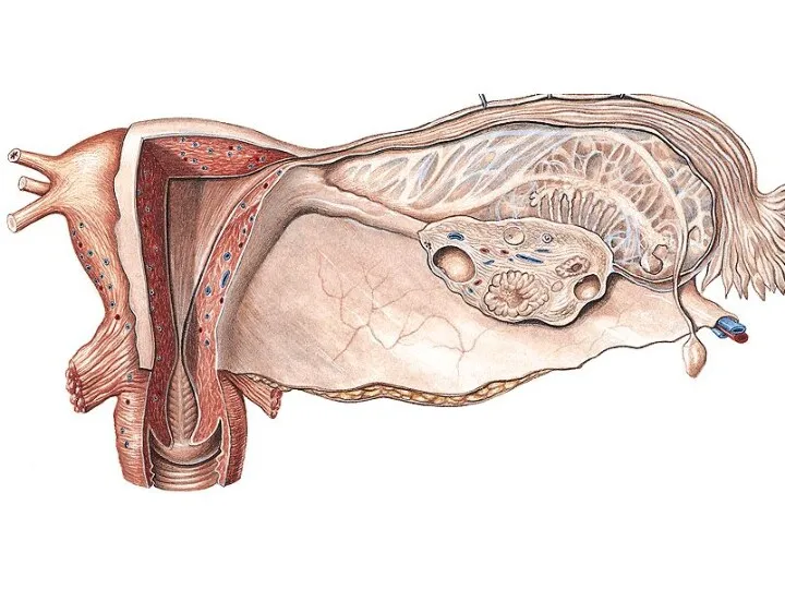
Uterus, Uterus, Vagina, Vagina, normal angles between the vagina, the cervix
Uterus, Uterus, Vagina, Vagina, normal angles between the vagina, the cervix
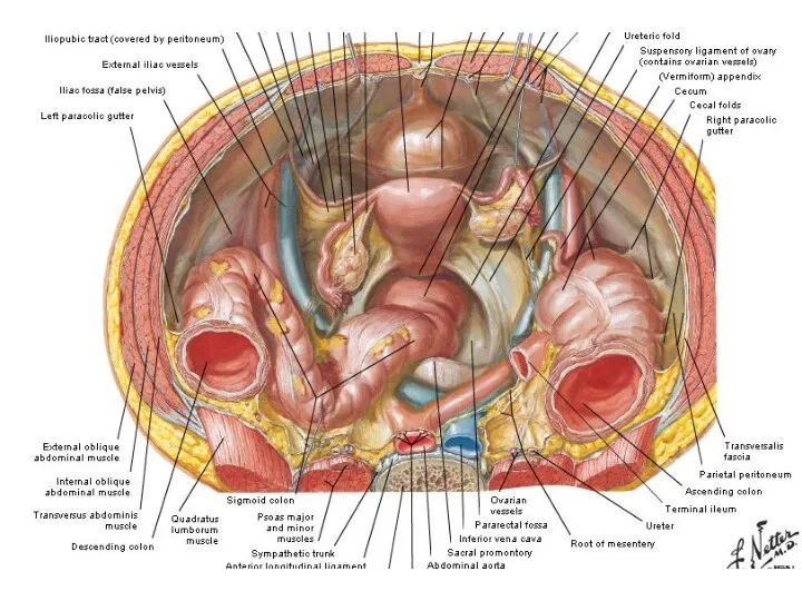

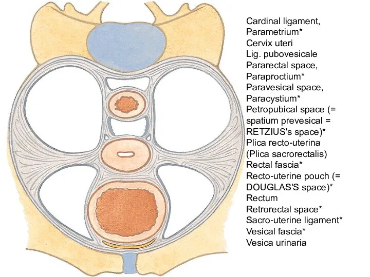
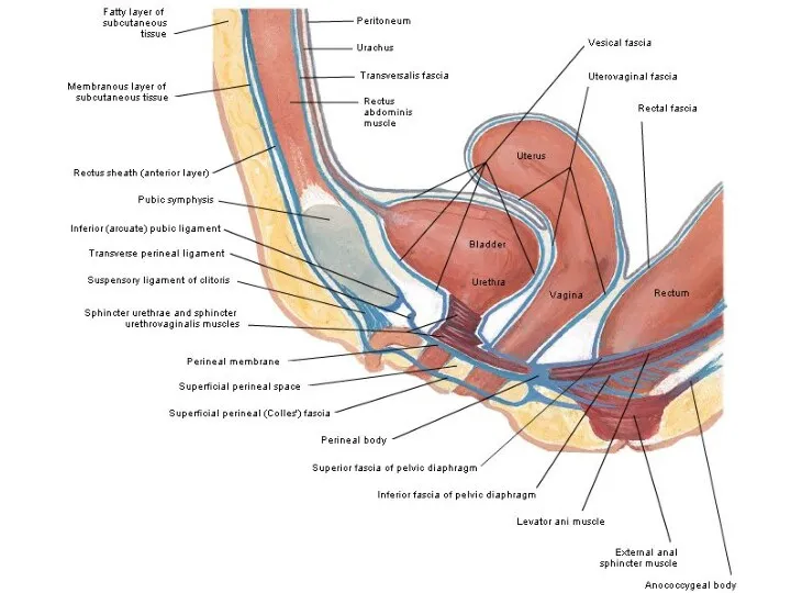
Cardinal ligament, Parametrium*
Cervix uteri
Lig. pubovesicale
Pararectal space, Paraproctium*
Paravesical space, Paracystium*
Petropubical space (=
Cardinal ligament, Parametrium*
Cervix uteri
Lig. pubovesicale
Pararectal space, Paraproctium*
Paravesical space, Paracystium*
Petropubical space (=


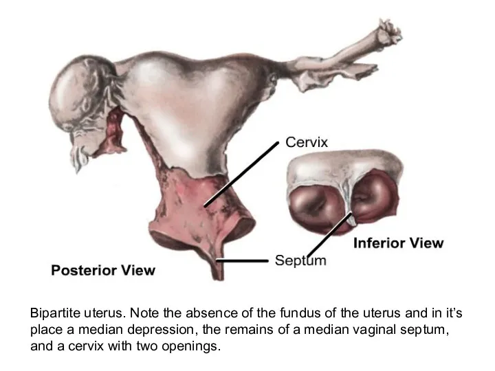
Bipartite uterus. Note the absence of the fundus of the uterus
Bipartite uterus. Note the absence of the fundus of the uterus
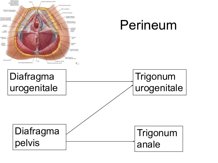
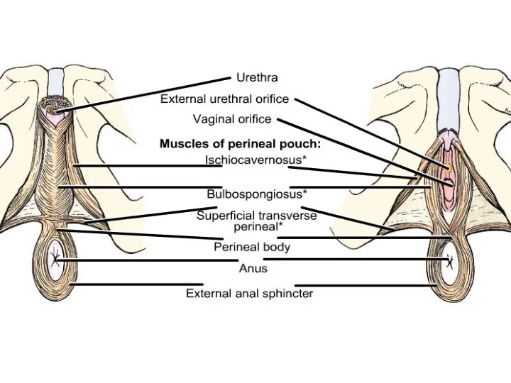
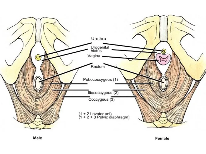
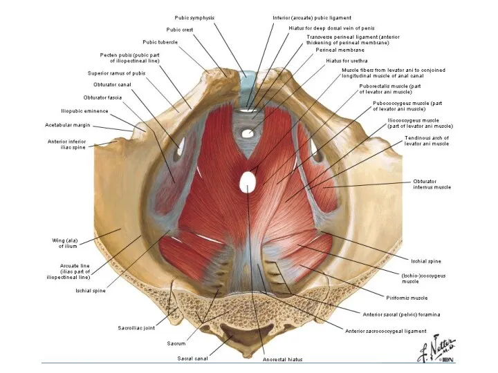
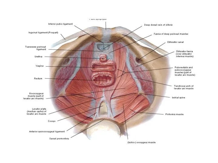
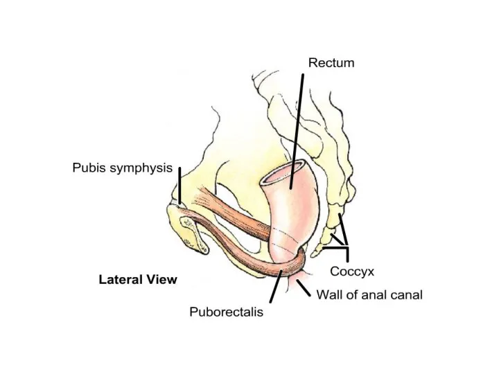
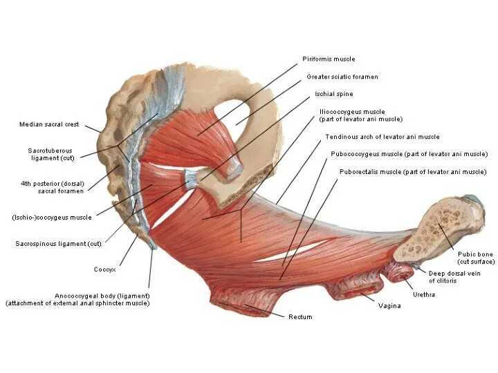
Perineum
Diafragma urogenitale
Diafragma pelvis
Trigonum urogenitale
Trigonum anale
Perineum
Diafragma urogenitale
Diafragma pelvis
Trigonum urogenitale
Trigonum anale
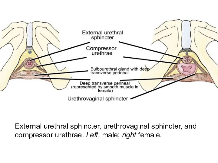
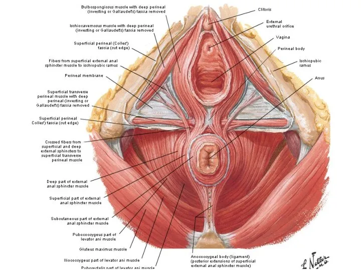
External urethral sphincter, urethrovaginal sphincter, and compressor urethrae. Left, male; right
External urethral sphincter, urethrovaginal sphincter, and compressor urethrae. Left, male; right
 Лекарственные средства, используемые при патологии ССС
 Дифференциальная диагностика кашля у детей
 Патологическая стираемость твердых тканей зубов
 История изучения высшей нервной деятельности
 Артроскопия. Тізе буынының артроскопиясы
 Профілактика анемії у дітей
 Менингит. Классификация. Этиология. Патогенез. Клиника
 Злокачественные опухоли женских половых органов
 Анализ заболеваемости бронхиальной астмой в г. Новочеркасске. Роль медицинской сестры в уходе за пациентом с бронхиальной астмой
 Диафрагмальные грыжи
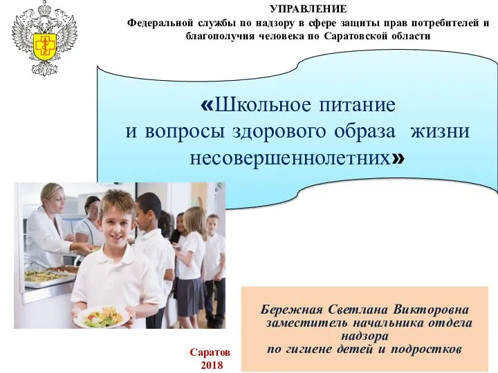 Школьное питание и вопросы здорового образа жизни несовершеннолетних
 Filling’s material: permanent & temporary
 Классификация антибиотиков и механизмы их действия. БЛРС
 Возбудители вирусных гепатитов
 Пищевые отравления
 Modal verbs
 Туберкулинодиагностика
 Болезнь Виллебранда. Клинический случай
 Гигиена питания
 Дети с особенностями развития
 Принципы лечения злокачественных новообразований
 Виды биопсии шейки матки
 Жедел коронарлы синдромның асқынуы. Ауруханаға дейінгі этапта емдеу алгоритмы
 Жедел ревматикалық қызба
 Қояншық мінезді тыныс тұншықпасында тұншығу синдромы
 Алкалоидтар. Алкалоидтарға жалпы сипаттама, жіктелуінің негізгі принциптері, өсімдіктерде атқаратын қызметтері
 Мази №2
 Сүйек және буын туберкулезі