- Diseases of immune system

Содержание
- 2. Reactions of hypersensitiveness Autoimmune diseases Immunodeficiency syndromes Amyloidosis Tumors of the lymphatic system Pathology of the
- 3. Autoimmunity it is an immune reaction against "self AG“. Auto-AB can be formed in response to
- 4. Organospecific - single organ (or single cell) type disorders - specific immune reactions directed against one
- 5. Multisystem diseases, characterized by lesions in many organs, associated usually with a multiplicity of auto-AB or
- 6. Immune tolerance is defined as a state in which the individual is incapable of developing an
- 7. Protection from self "protectors." Deletion of auto-reactive clones appears to be the major mechanism of self-tolerance
- 8. Breakdown of one or more of the mechanisms of self-tolerance can unleash an immunologic attack on
- 9. Autoimmunity results from multiple factors, including susceptibility genes that may interfere with self-tolerance and environmental triggers
- 10. 1. Loss of Self-Tolerance Bypass of T-Helper Tolerance. AB responses against most self-AG require collaboration between
- 11. 2. Genetic Factors in Autoimmunity proved by: 1.Familial clustering of human autoimmune diseases such as systemic
- 12. 3. Microbial Agents in Autoimmunity Bacteria, mycoplasmas, and viruses, have been implicated in triggering autoimmunity. Microbes
- 13. The immunodeficiency's can be subdivided into: primary diseases of genetic origin secondary to some underlying disorder
- 14. It is inadequacy of immune answer because of an innate defect in the immune system (defect
- 15. It is a failure of pre-B cells to differentiate into mature B cells. Clinical recognition -
- 16. Characteristics of the classic form of this disease: B-cells are absent or remarkably decreased, and the
- 17. It is a lack of thymic influence on the immune system. The thymus is usually rudimentary
- 18. It represents a constellation of syndromes all having in common variable defects in both humoral and
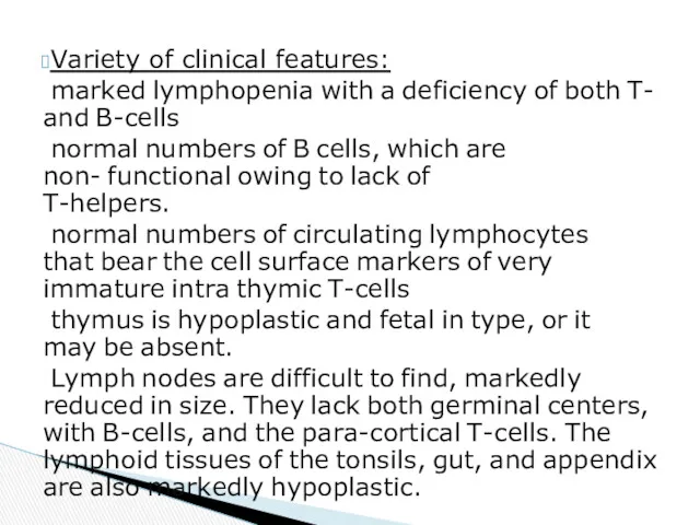
- 19. Variety of clinical features: marked lymphopenia with a deficiency of both T- and B-cells normal numbers
- 20. It is the commonest of all the primary immunodeficiency diseases (1/700). Both serum and secretory IgA
- 21. most persons are asymptomatic, some present with a variety of symptoms: respiratory infections, chronic diarrhea, and
- 22. It is acquired inadequacy of immune answer because of fatigue or damage of the normally formed
- 23. Main reasons of development: Infecting HIV-virus with development of AIDS. Ionizing irradiation or incorporation of radionuclide.
- 24. Infectious diseases with the defeat of lymphocytes and macrophages (cytomegalovirus and herpetic infections, hepatitis B, other).
- 25. Amyloid is an abnormal proteinaceous substance that is deposited between cells in many tissues and organs
- 26. Two major chemical classes of amyloid have been identified: composed of immunoglobulin light chains called AL
- 27. Immunocyte dyscrasias with amyloidosis (primary amyloidosis): Multiple myeloma and other monoclonal B-cell proliferations Reactive systemic amyloidosis
- 28. II. Anatomic distribution: 1. Systemic (Generalized) 2. Localized III. Chemical composition of amyloid : 1. AL
- 29. Small amounts are not recognized until the surface of the cut organ is painted with iodine
- 30. Histologically, the deposition begins between cells, often closely adjacent to basement membranes. In time the depositions
- 31. is the most common and the most serious involvement in the disease. Grossly, the kidney may
- 32. often causes moderate or even marked enlargement (200 to 800 gm). The deposits may be virtually
- 33. Histologically, the deposits appear first in the space of Disse and then progressively enlarge to the
- 34. Macroscopic characteristic: gray-pink, dewdrop- like subendocardial elevations, particularly in the atrial chambers. Amyloidosis of the heart
- 36. Скачать презентацию
Reactions of hypersensitiveness
Autoimmune diseases
Immunodeficiency syndromes
Amyloidosis
Tumors of the lymphatic system
Pathology of the
Reactions of hypersensitiveness
Autoimmune diseases
Immunodeficiency syndromes
Amyloidosis
Tumors of the lymphatic system
Pathology of the
Autoimmunity it is an immune reaction against "self AG“.
Auto-AB can be
Autoimmunity it is an immune reaction against "self AG“.
Auto-AB can be
Organospecific - single organ (or single cell) type disorders - specific
Organospecific - single organ (or single cell) type disorders - specific
Multisystem diseases, characterized by lesions in many organs, associated usually with
Multisystem diseases, characterized by lesions in many organs, associated usually with
Immune tolerance is defined as a state in which the individual
Immune tolerance is defined as a state in which the individual
Protection from self "protectors." Deletion of auto-reactive clones appears to be
Protection from self "protectors." Deletion of auto-reactive clones appears to be
Breakdown of one or more of the mechanisms of self-tolerance can
Breakdown of one or more of the mechanisms of self-tolerance can
Autoimmunity results from multiple factors, including susceptibility genes that may interfere
Autoimmunity results from multiple factors, including susceptibility genes that may interfere
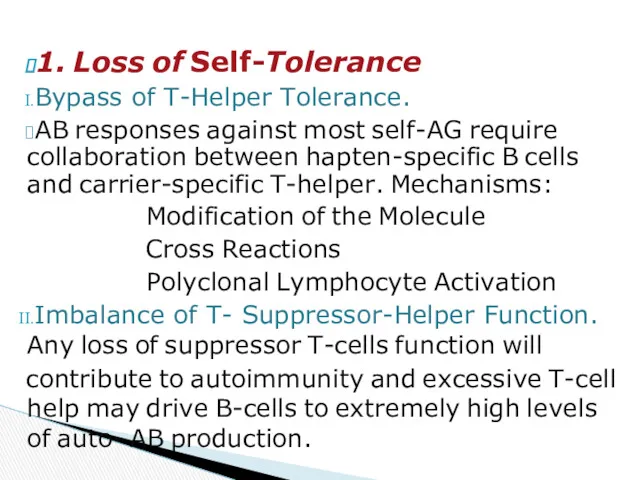
1. Loss of Self-Tolerance
Bypass of T-Helper Tolerance.
AB responses against most self-AG
1. Loss of Self-Tolerance
Bypass of T-Helper Tolerance.
AB responses against most self-AG
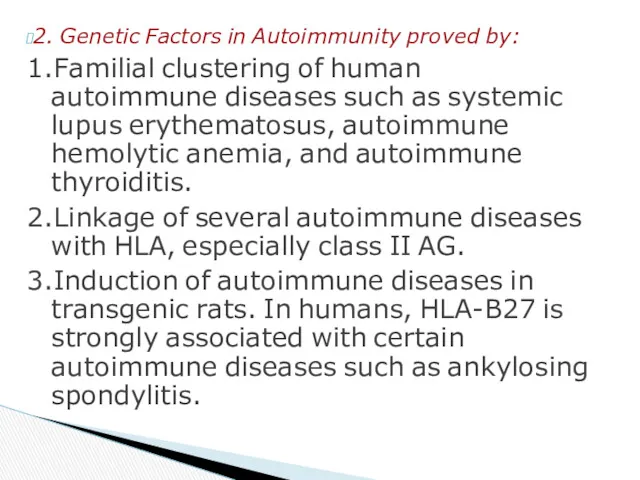
2. Genetic Factors in Autoimmunity proved by:
1.Familial clustering of human autoimmune
2. Genetic Factors in Autoimmunity proved by:
1.Familial clustering of human autoimmune
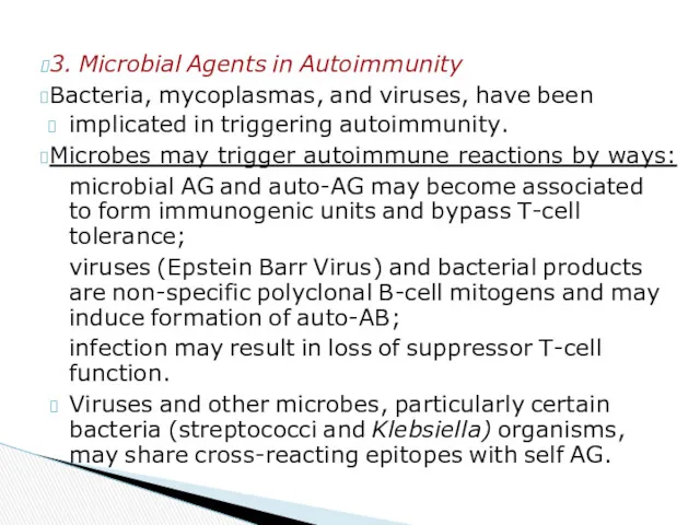
3. Microbial Agents in Autoimmunity
Bacteria, mycoplasmas, and viruses, have been
implicated in
3. Microbial Agents in Autoimmunity
Bacteria, mycoplasmas, and viruses, have been
implicated in
The immunodeficiency's can be subdivided into:
primary diseases of genetic origin
secondary to
The immunodeficiency's can be subdivided into:
primary diseases of genetic origin
secondary to

It is inadequacy of immune answer because of an innate defect
It is inadequacy of immune answer because of an innate defect
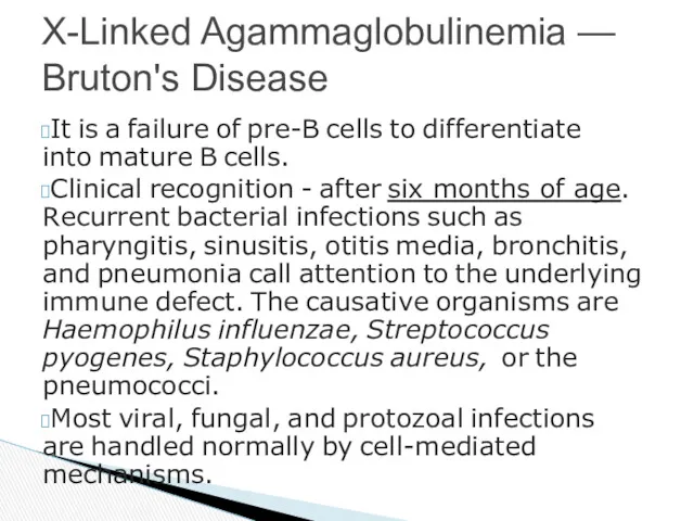
It is a failure of pre-B cells to differentiate into mature
It is a failure of pre-B cells to differentiate into mature
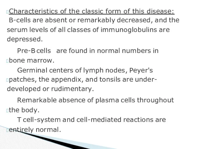
Characteristics of the classic form of this disease:
B-cells are absent or
Characteristics of the classic form of this disease:
B-cells are absent or
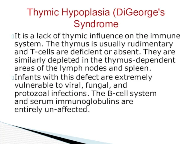
It is a lack of thymic influence on the immune system.
It is a lack of thymic influence on the immune system.
It represents a constellation of syndromes all having in common variable
It represents a constellation of syndromes all having in common variable
Variety of clinical features:
marked lymphopenia with a deficiency of both T-
Variety of clinical features:
marked lymphopenia with a deficiency of both T-
It is the commonest of all the primary immunodeficiency diseases (1/700).
Both
It is the commonest of all the primary immunodeficiency diseases (1/700).
Both
most persons are asymptomatic,
some present with a variety of symptoms: respiratory
most persons are asymptomatic,
some present with a variety of symptoms: respiratory
It is acquired inadequacy of immune answer because of fatigue or
It is acquired inadequacy of immune answer because of fatigue or
Main reasons of development:
Infecting HIV-virus with development of AIDS.
Ionizing irradiation or
Main reasons of development:
Infecting HIV-virus with development of AIDS.
Ionizing irradiation or
Infectious diseases with the defeat of lymphocytes and macrophages (cytomegalovirus and
Infectious diseases with the defeat of lymphocytes and macrophages (cytomegalovirus and
Amyloid is an abnormal proteinaceous substance that is deposited between cells
Amyloid is an abnormal proteinaceous substance that is deposited between cells
Two major chemical classes of amyloid have
been identified:
composed of immunoglobulin light
Two major chemical classes of amyloid have
been identified:
composed of immunoglobulin light
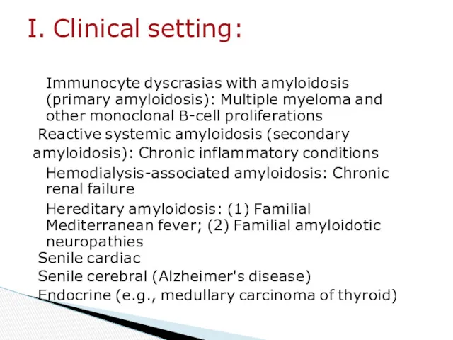
Immunocyte dyscrasias with amyloidosis (primary amyloidosis): Multiple myeloma and other monoclonal
Immunocyte dyscrasias with amyloidosis (primary amyloidosis): Multiple myeloma and other monoclonal
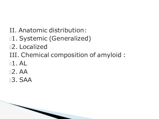
II. Anatomic distribution:
1. Systemic (Generalized)
2. Localized
III. Chemical composition of amyloid :
1.
II. Anatomic distribution:
1. Systemic (Generalized)
2. Localized
III. Chemical composition of amyloid :
1.
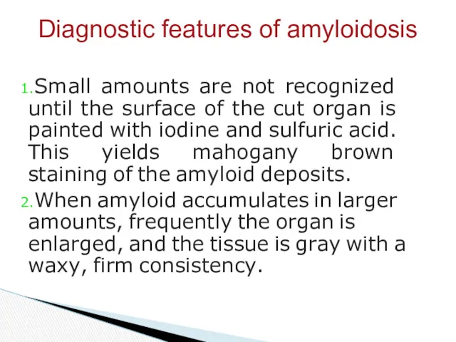
Small amounts are not recognized until the surface of the cut
Small amounts are not recognized until the surface of the cut
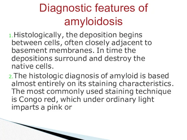
Histologically, the deposition begins between cells, often closely adjacent to basement
Histologically, the deposition begins between cells, often closely adjacent to basement
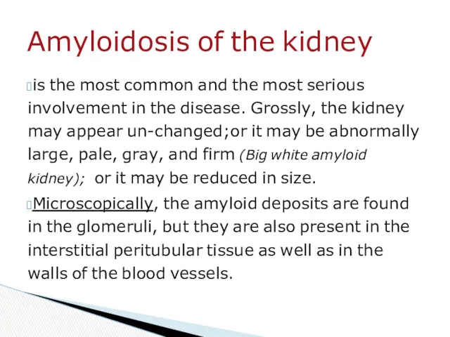
is the most common and the most serious involvement in the
is the most common and the most serious involvement in the
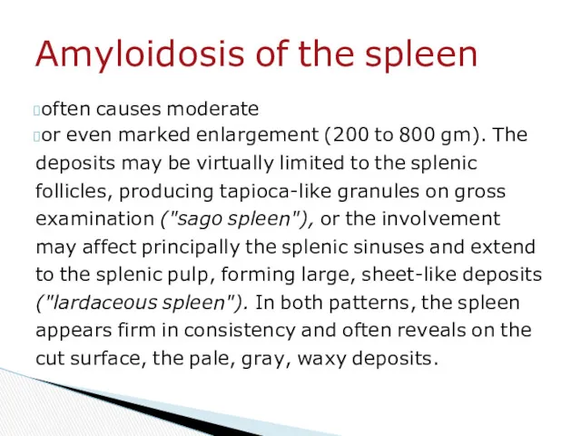
often causes moderate
or even marked enlargement (200 to 800 gm). The
often causes moderate
or even marked enlargement (200 to 800 gm). The
Histologically, the deposits appear first in the space of Disse and
Histologically, the deposits appear first in the space of Disse and
Macroscopic characteristic: gray-pink, dewdrop- like subendocardial elevations, particularly in the atrial
Macroscopic characteristic: gray-pink, dewdrop- like subendocardial elevations, particularly in the atrial
 Этиология, патогенез, классификация и клиника эпилепсии
 Инфекции, связанные с оказанием медицинской помощи
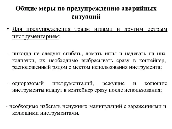 Общие меры по предупреждению аварийных ситуаций
 Повернення до нормальної вентиляції після абдомінальних операцій у хворих з ожирінням
 Пневмония. Классификация
 Дәрігердің науқаспен вербальды және вербальды емес қатынас
 Острый живот в гинекологии
 Инновационные лекарственные средства
 Витамины. Значение витаминов. Водорастворимые и жирорастворимые витамины. Несовместимость витаминов
 How to properly take care of the body
 Иммуномодуляторы. Иммунокорректоры
 Санитарное законодательство РФ
 Исследование расстройств пищевого поведения детей и подростков
 Старость. Старение организма. (Лекция 37)
 История развития сестринского дела в России
 ФР после артроскопической аутопластики связок КС
 Рестриктивные кардиомиопатии. Формы РКМП
 Сердечная недостаточность с сохраненной фракцией выброса левого желудочка
 Актуальные вопросы медицины и права
 Общие сведения о геморрагических высококонтагиозных лихорадках
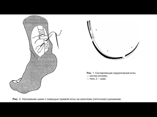 Иглы и шовные материалы
 Физиотерапия при бронхолёгочной патологии
 Смерть мозга
 Предоперационная подготовка гинекологических больных
 Медикаментозная и немедикаментозная коррекция дислипидемий
 Первая медицинская помощь при кровотечениях и травмах. Использование подручных средств
 Гемотрансфузионный шок
 Глюкокортикостероиды