- Electrical Processes of the Heart

Содержание
- 2. Terminology 1 – Cardiac Mechanism https://quizlet.com/173937887/chapter-7-cardiac-cycle-conduction-system-of-the-heart-flash-cards/ Flash Cards
- 3. 2 chambers 3 chambers 2 chambers Fish 1-atrium 1-ventricle 4 chambers 3 chambers Amphibian/reptile 2-atrium 1-ventricle
- 4. Blood travels through the heart twice before returning to the body Double Circulatory System
- 5. Heart Double Pump Pulmonary – lungs Systemic - body
- 6. -Short videofragmet beating isolated rat heart: http://www.youtube.com/watch?v=CzIMSr-8Ko0
- 7. Voluntary Striated Multinucleated Non-branched Involuntary Non-striated Mononucleated Tapered Involuntary Striated Intercalculated disks Mononucleated Branched Three Types
- 8. The action potential travels though all cells connected together forming a functional syncytium in which cells
- 10. Define the following terms Functional syncytium –the heart consists of individual cells, the entire mass normally
- 11. The heart is myogenic – it contracts on its own without stimulus from the nervous system.
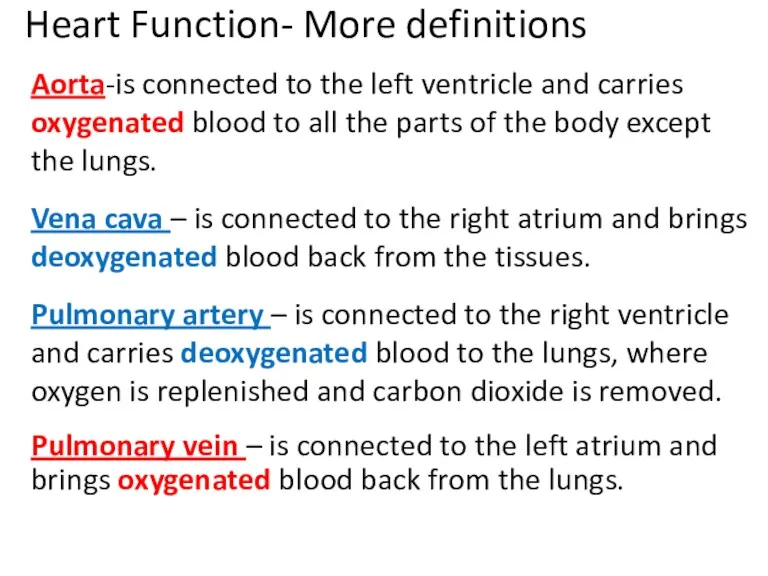
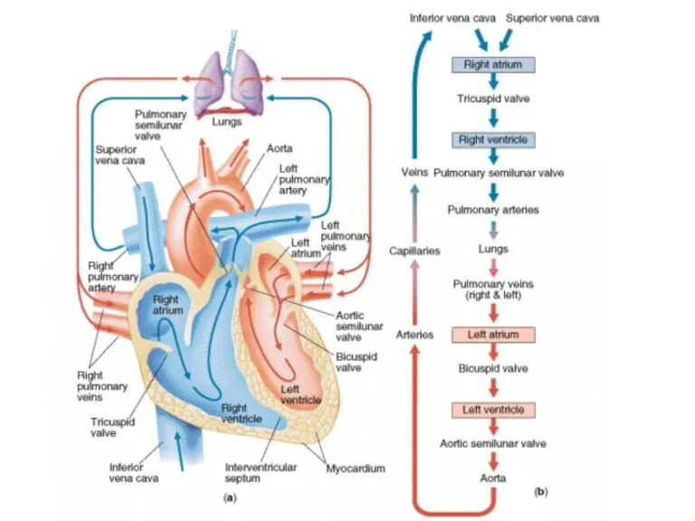
- 12. Heart Function- More definitions Aorta-is connected to the left ventricle and carries oxygenated blood to all
- 13. Label Heart - 1 min
- 14. External Features-Label Superior Inferior Left Right
- 15. Transverse section of the heart apex -Left ventricle is thicker because it needs to pump blood
- 17. Deoxygenated blood from body to RA through vena cava Blood from RA to RV through tri-cuspid
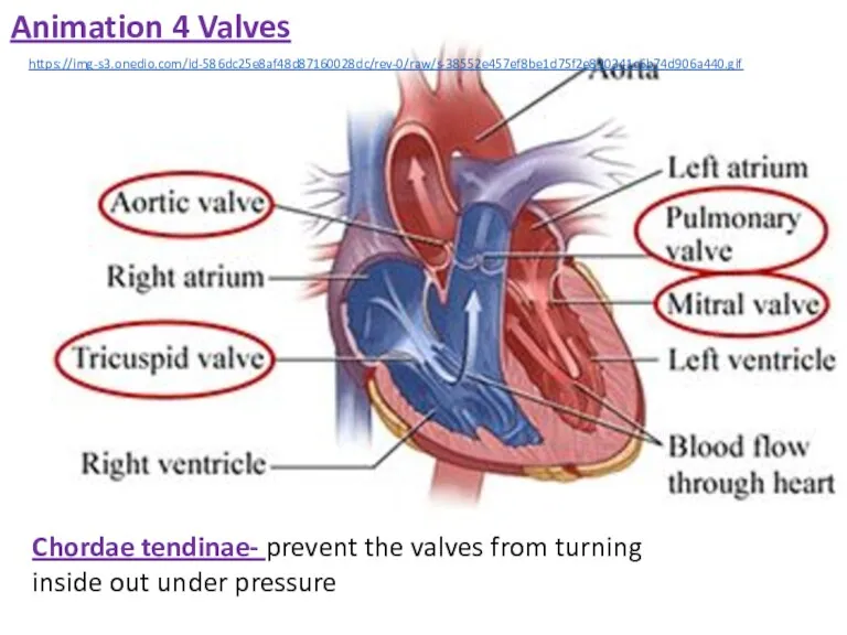
- 18. Chordae tendinae- prevent the valves from turning inside out under pressure. 4 Valves of the heart,
- 19. Chordae tendinae- prevent the valves from turning inside out under pressure https://img-s3.onedio.com/id-586dc25e8af48d87160028dc/rev-0/raw/s-38552e457ef8be1d75f2e890341c6b74d906a440.gif Animation 4 Valves
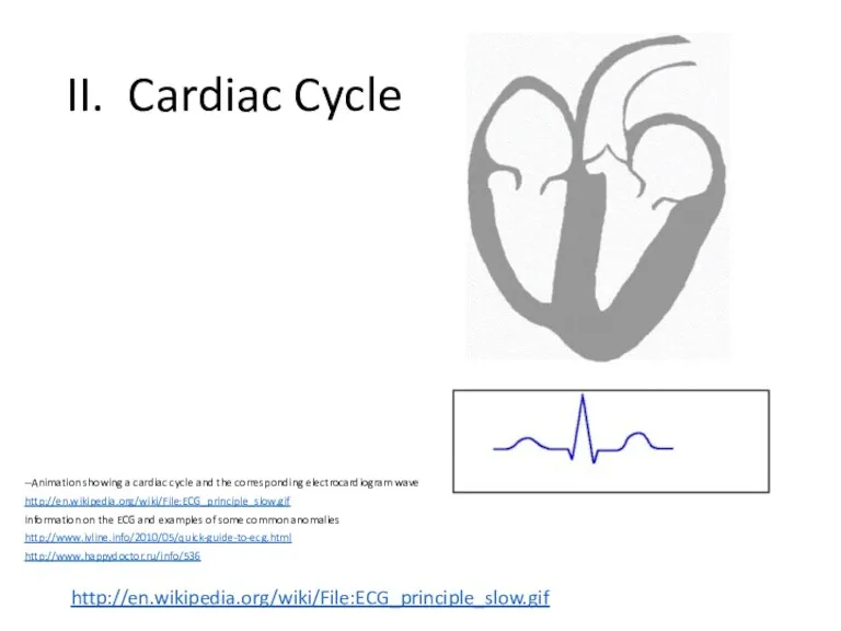
- 20. II. Cardiac Cycle http://en.wikipedia.org/wiki/File:ECG_principle_slow.gif --Animation showing a cardiac cycle and the corresponding electrocardiogram wave http://en.wikipedia.org/wiki/File:ECG_principle_slow.gif Information
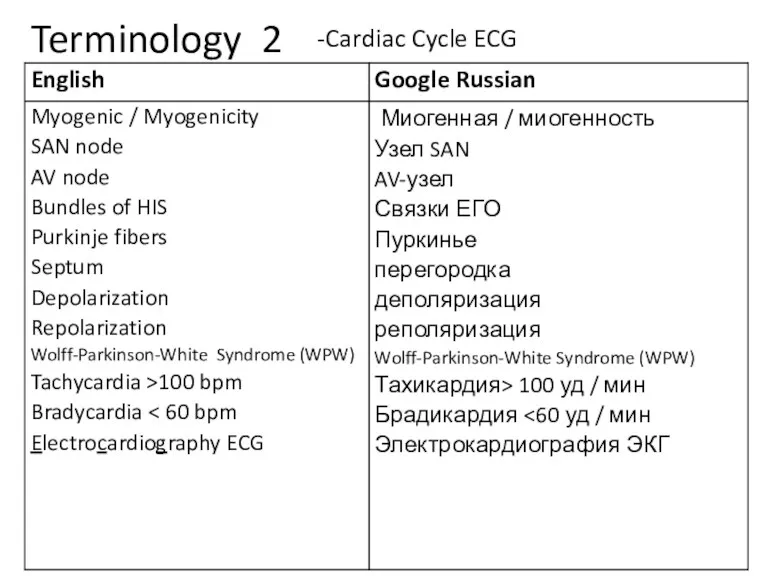
- 21. Terminology 2 -Cardiac Cycle ECG
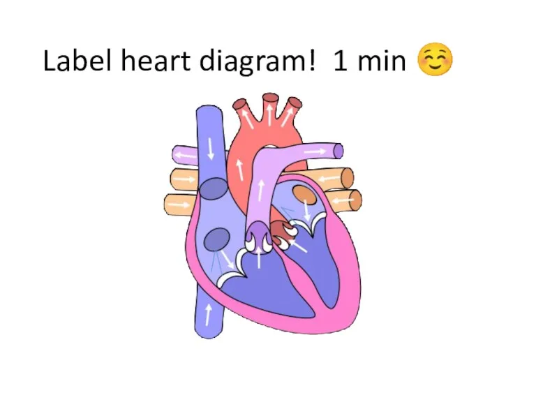
- 22. Label heart diagram! 1 min ☺
- 23. Aorta – Leaves the left ventricle and distributes oxygenated blood (through systemic circulation) to the body
- 24. What is myogenic? muscles or tissues that can contract on their own, without any external electrical
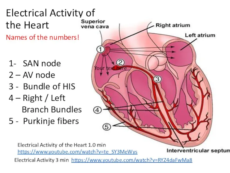
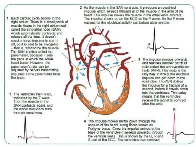
- 25. Electrical Activity of the Heart Electrical Activity of the Heart 1.0 min https://www.youtube.com/watch?v=te_SY3MeWys 1- SAN node
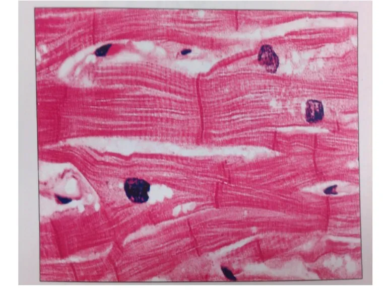
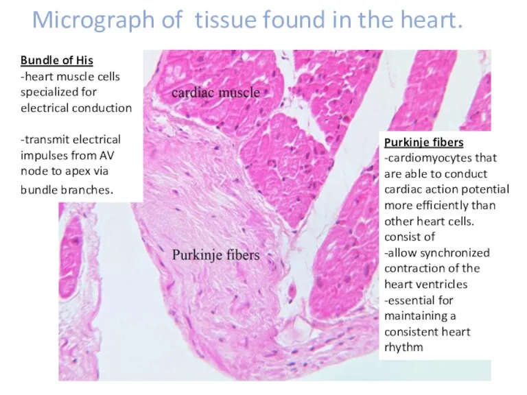
- 26. Micrograph of tissue found in the heart. Bundle of His -heart muscle cells specialized for electrical
- 27. Atrioventricular valves- link the atria to the ventricles. Semi-lunar- valves link the ventricles to the pulmonary
- 28. Rest: (=)+ outside - inside Depolarization: (=) - outside + inside Repolarization: Returns to: + outside
- 30. Describe the difference between polarisation, depolarisation and repolarisation. Sarcolemma –resting potential IONS INVOLVED - K+, Na+
- 31. depolarization….. Depolarization is when a cell membrane's charge becomes positive to generate an action potential. This
- 32. repolarization….. Repolarization is when a cell membrane's charge returns to negative after depolarization. This is caused
- 33. The QRS complex the combination of three of the graphical deflections seen on a typical deflections
- 34. EKG or ECG - Electrocardiogram
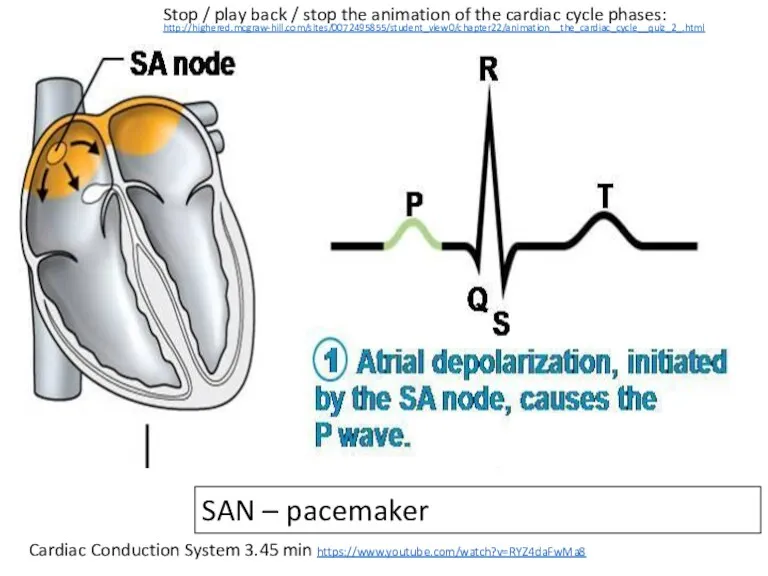
- 35. Cardiac Conduction System 3.45 min https://www.youtube.com/watch?v=RYZ4daFwMa8 SAN – pacemaker Stop / play back / stop the
- 36. 0.2 seconds -- time for the impulse to be conducted from the SAN to the ventricles
- 37. Ventricle contraction – wave of depolarization flows through the B. of HIS.
- 39. Relaxation phase
- 41. What are some ways that SAN and AVN control the heart beat?
- 42. -SAN initiates heartbeat -Beat of heart is myogenic – spontaneous not started by nervous system stimulus
- 43. EKG wave animation.. http://en.wikipedia.org/wiki/Electrocardiography#mediaviewer/File:ECG_principle_slow.gif
- 44. EKG or ECG - Electrocardiography
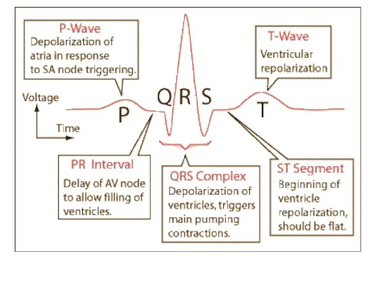
- 45. During the cardiac cycle (one contraction of the heart plus the relaxation period that follows), electrical
- 46. (1) P wave. A small upward (positive) wave that indicates atrial polarization (the spread of an
- 47. http://www.ivline.org/2010/05/quick-guide-to-ecg.html#at_pco=smlre-1.0&at_si=5420a7df299cca88&at_ab=per-2&at_pos=0&at_tot=4
- 48. Ventricular fibrillation What do you think is happening in the ECG? Heart Block
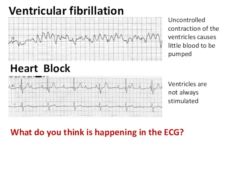
- 49. Ventricular fibrillation What do you think is happening in the ECG? Heart Block Uncontrolled contraction of
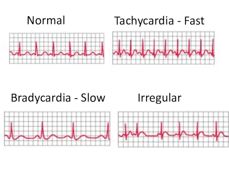
- 50. Match the beat with the ECG. Tachycardia Irregular Normal Bradycardia
- 51. Tachycardia - Fast Irregular Normal Bradycardia - Slow
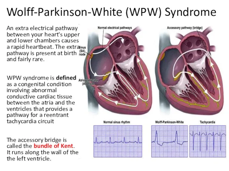
- 52. Wolff-Parkinson-White (WPW) Syndrome An extra electrical pathway between your heart's upper and lower chambers causes a
- 53. Wolff Parkinson Wright Syndrome (WPW) -Write information from the video and label the 2nd heart on
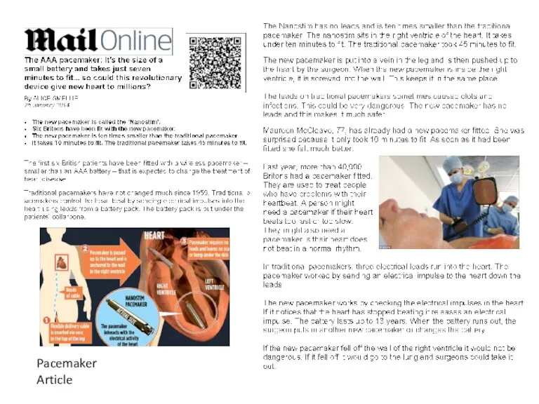
- 54. Pacemaker Article
- 56. Pacemaker
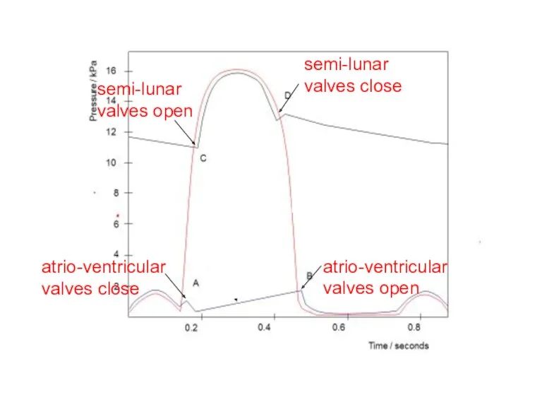
- 57. atrio-ventricular valves open atrio-ventricular valves close semi-lunar valves open semi-lunar valves close
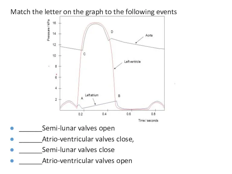
- 59. Match the letter on the graph to the following events ______Semi-lunar valves open ______Atrio-ventricular valves close,
- 60. atrio-ventricular valves open atrio-ventricular valves close semi-lunar valves open semi-lunar valves close
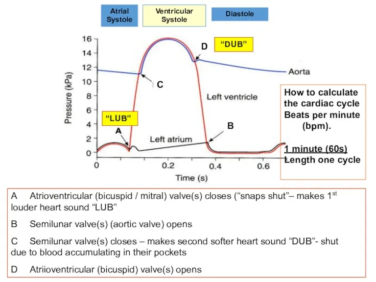
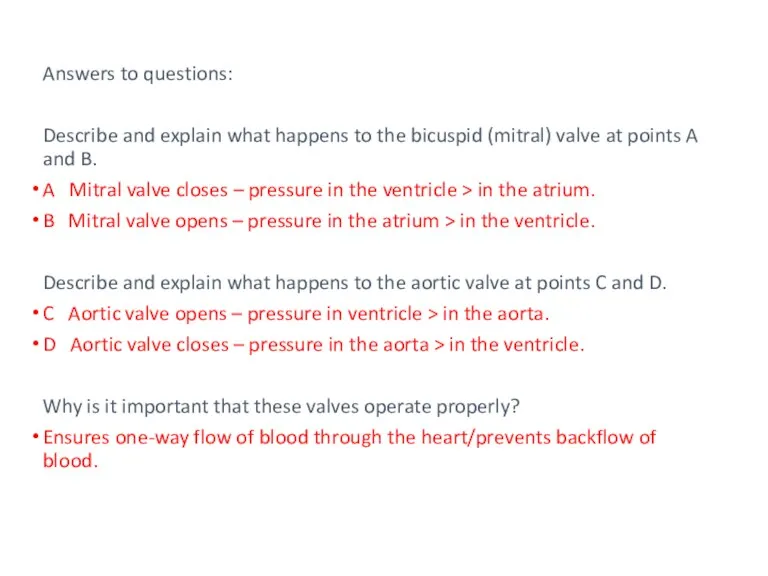
- 61. A Atrioventricular (bicuspid / mitral) valve(s) closes (“snaps shut”– makes 1st louder heart sound “LUB” B
- 62. https://www.youtube.com/watch?v=RYZ4daFwMa8 Electrical activity in heart https://www.twig-bilim.kz/film/heart-976/ - Revision of structure and function On your own electrical
- 63. Extra Information
- 64. Tachycardia Increased heart rate is a normal response to: exercise excitement stress drugs e.g. caffeine, nicotine,
- 65. Bradycardia Pattern of electrical activity is normal but slow. Reduced heart rate could indicate: good aerobic
- 66. Heart block There is separation of the P wave and the QRS complex. Pacemaker activity and
- 67. Fibrillation Contraction of cardiac muscle is normally coordinated. In VF the ventricles contract, but it is
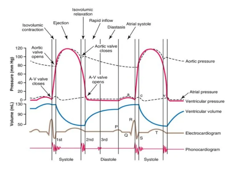
- 68. Cardiac Cycle General Principles. Contraction of the myocardium generates pressure changes which result in the orderly
- 69. Atrial systole The heart is full of blood and the ventricles are relaxed Both the atria
- 70. Ventricular systole The atria relax. The ventricle walls contract, forcing the blood out The pressure of
- 71. Ventricular systole The pressure of blood opens the semi-lunar valves. Blood passes into the aorta and
- 72. Diastole The ventricles relax Pressure in the ventricles falls below that in the arteries Blood under
- 73. Blood from the vena cava and pulmonary veins enter the atria. The whole cycle starts again.
- 74. Match the letter on the graph to the following events ______Semi-lunar valves open ______Atrio-ventricular valves close,
- 75. atrio-ventricular valves open atrio-ventricular valves close semi-lunar valves open semi-lunar valves close
- 76. A Atrioventricular (bicuspid / mitral) valve(s) closes (“snaps shut”– makes 1st louder heart sound “LUB” B
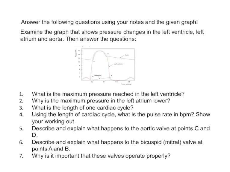
- 77. Examine the graph that shows pressure changes in the left ventricle, left atrium and aorta. Then
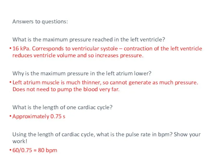
- 78. Answers to questions: What is the maximum pressure reached in the left ventricle? 16 kPa. Corresponds
- 79. Answers to questions: Describe and explain what happens to the bicuspid (mitral) valve at points A
- 81. Electrical Activity In The Heart. The heart's electrical activity begins in the sinoatrial (SA) node and
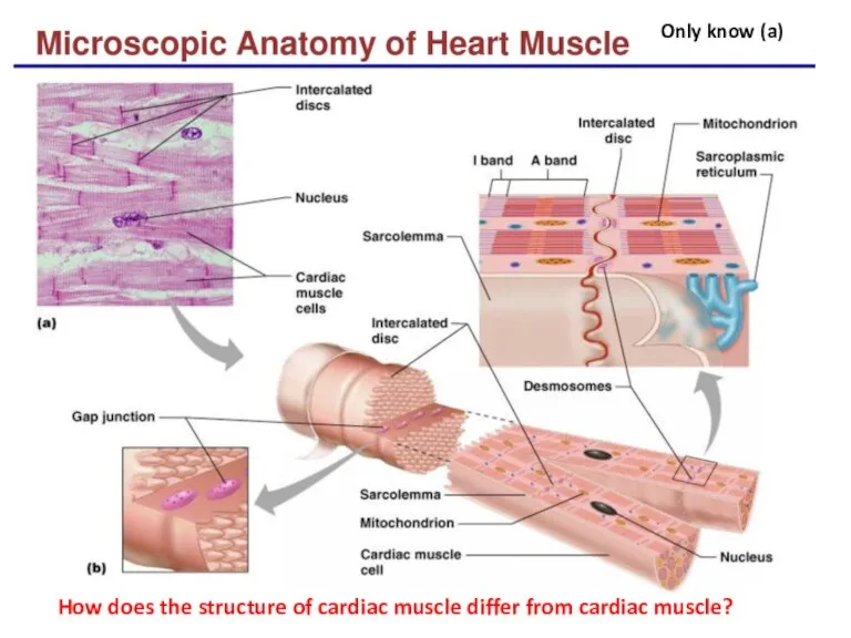
- 82. How does the structure of cardiac muscle differ from cardiac muscle? Only know (a)
- 84. Скачать презентацию
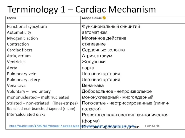
Terminology 1 – Cardiac Mechanism
https://quizlet.com/173937887/chapter-7-cardiac-cycle-conduction-system-of-the-heart-flash-cards/ Flash Cards
Terminology 1 – Cardiac Mechanism
https://quizlet.com/173937887/chapter-7-cardiac-cycle-conduction-system-of-the-heart-flash-cards/ Flash Cards
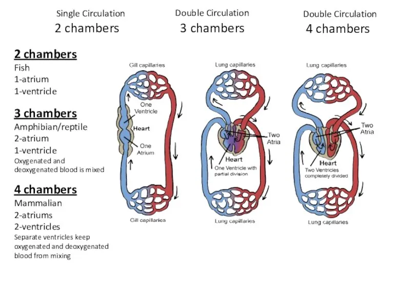
2 chambers
3 chambers
2 chambers
Fish
1-atrium
1-ventricle
4 chambers
3 chambers
Amphibian/reptile
2-atrium
1-ventricle
Oxygenated and
2 chambers
3 chambers
2 chambers
Fish
1-atrium
1-ventricle
4 chambers
3 chambers
Amphibian/reptile
2-atrium
1-ventricle
Oxygenated and
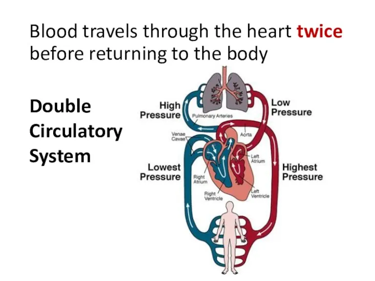
Blood travels through the heart twice before returning to the body
Double
Blood travels through the heart twice before returning to the body
Double

Heart Double Pump
Pulmonary – lungs
Systemic - body
Heart Double Pump
Pulmonary – lungs
Systemic - body
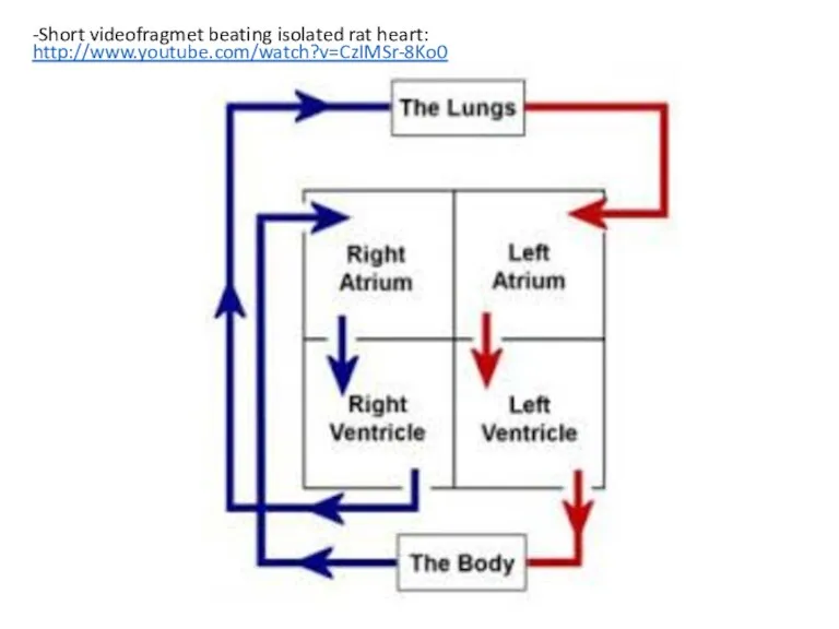
-Short videofragmet beating isolated rat heart:
http://www.youtube.com/watch?v=CzIMSr-8Ko0
-Short videofragmet beating isolated rat heart:
http://www.youtube.com/watch?v=CzIMSr-8Ko0
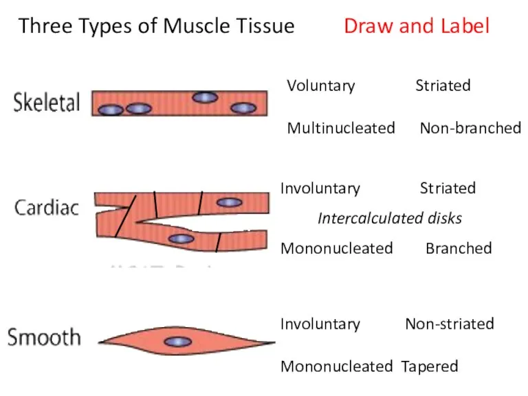
Voluntary Striated
Multinucleated Non-branched
Involuntary Non-striated
Mononucleated Tapered
Involuntary Striated
Intercalculated disks
Mononucleated Branched
Three Types of
Voluntary Striated
Multinucleated Non-branched
Involuntary Non-striated
Mononucleated Tapered
Involuntary Striated
Intercalculated disks
Mononucleated Branched
Three Types of
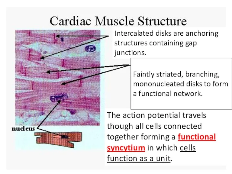
The action potential travels though all cells connected together forming a
The action potential travels though all cells connected together forming a
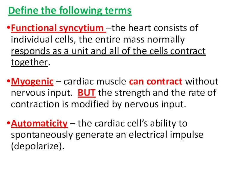
Define the following terms
Functional syncytium –the heart consists of individual cells,
Define the following terms
Functional syncytium –the heart consists of individual cells,
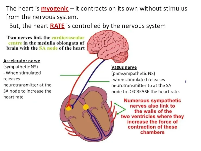
The heart is myogenic – it contracts on its own without
The heart is myogenic – it contracts on its own without
Heart Function- More definitions
Aorta-is connected to the left ventricle and carries
Heart Function- More definitions
Aorta-is connected to the left ventricle and carries
Label Heart - 1 min
Label Heart - 1 min
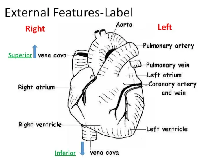
External Features-Label
Superior
Inferior
Left
Right
External Features-Label
Superior
Inferior
Left
Right
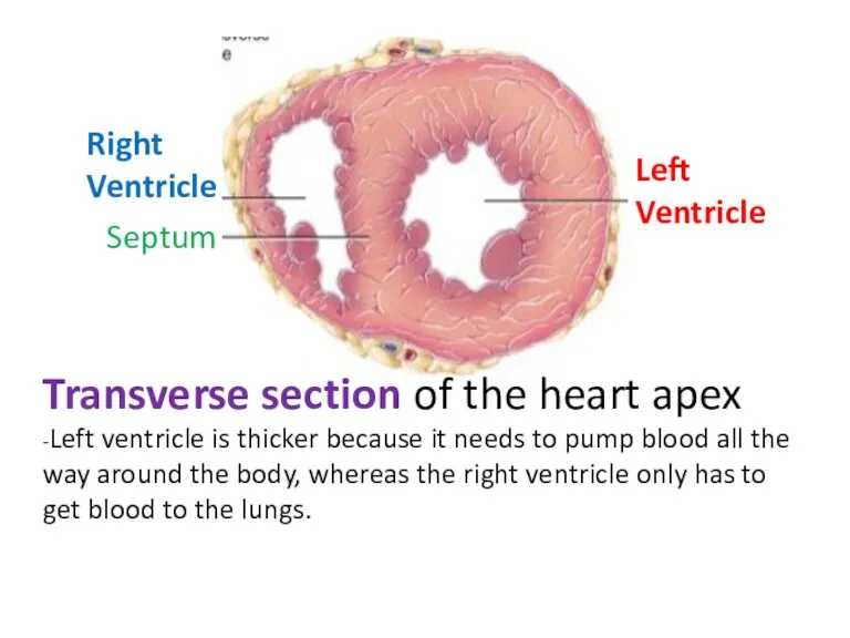
Transverse section of the heart apex
-Left ventricle is thicker because
Transverse section of the heart apex
-Left ventricle is thicker because
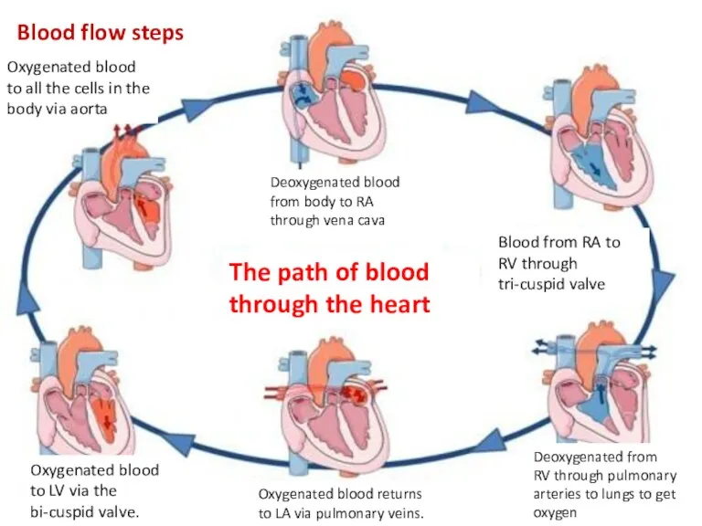
Deoxygenated blood from body to RA through vena cava
Blood from RA
Deoxygenated blood from body to RA through vena cava
Blood from RA
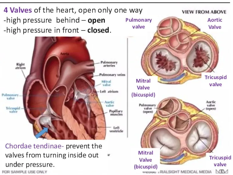
Chordae tendinae- prevent the valves from turning inside out under pressure.
Chordae tendinae- prevent the valves from turning inside out under pressure.
Chordae tendinae- prevent the valves from turning inside out under pressure
https://img-s3.onedio.com/id-586dc25e8af48d87160028dc/rev-0/raw/s-38552e457ef8be1d75f2e890341c6b74d906a440.gif
Animation
Chordae tendinae- prevent the valves from turning inside out under pressure
https://img-s3.onedio.com/id-586dc25e8af48d87160028dc/rev-0/raw/s-38552e457ef8be1d75f2e890341c6b74d906a440.gif
Animation
II. Cardiac Cycle
http://en.wikipedia.org/wiki/File:ECG_principle_slow.gif
--Animation showing a cardiac cycle and the corresponding electrocardiogram
II. Cardiac Cycle
http://en.wikipedia.org/wiki/File:ECG_principle_slow.gif
--Animation showing a cardiac cycle and the corresponding electrocardiogram
Terminology 2
-Cardiac Cycle ECG
Terminology 2
-Cardiac Cycle ECG
Label heart diagram! 1 min ☺
Label heart diagram! 1 min ☺
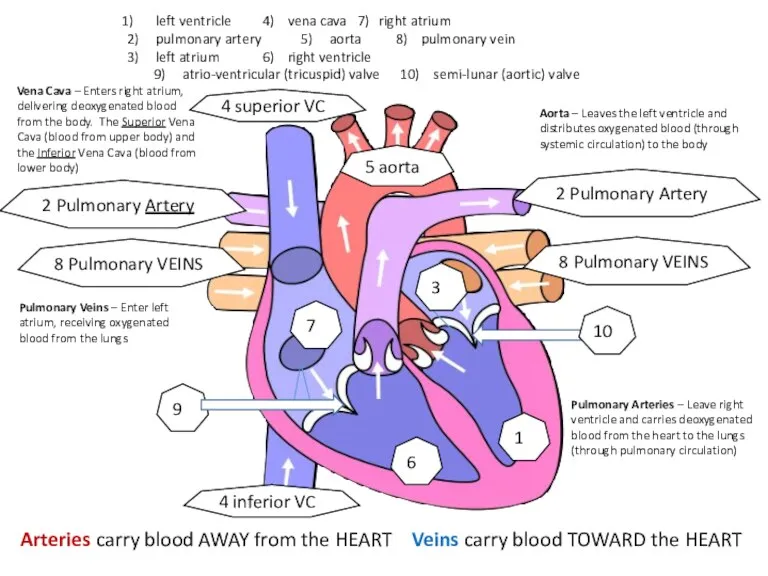
Aorta – Leaves the left ventricle and distributes oxygenated blood (through
Aorta – Leaves the left ventricle and distributes oxygenated blood (through
What is myogenic?
muscles or tissues that can contract on their own,
What is myogenic?
muscles or tissues that can contract on their own,
Electrical Activity of the Heart
Electrical Activity of the Heart 1.0 min
https://www.youtube.com/watch?v=te_SY3MeWys
1-
Electrical Activity of the Heart
Electrical Activity of the Heart 1.0 min
https://www.youtube.com/watch?v=te_SY3MeWys
1-
Micrograph of tissue found in the heart.
Bundle of His
-heart muscle cells
Micrograph of tissue found in the heart.
Bundle of His
-heart muscle cells
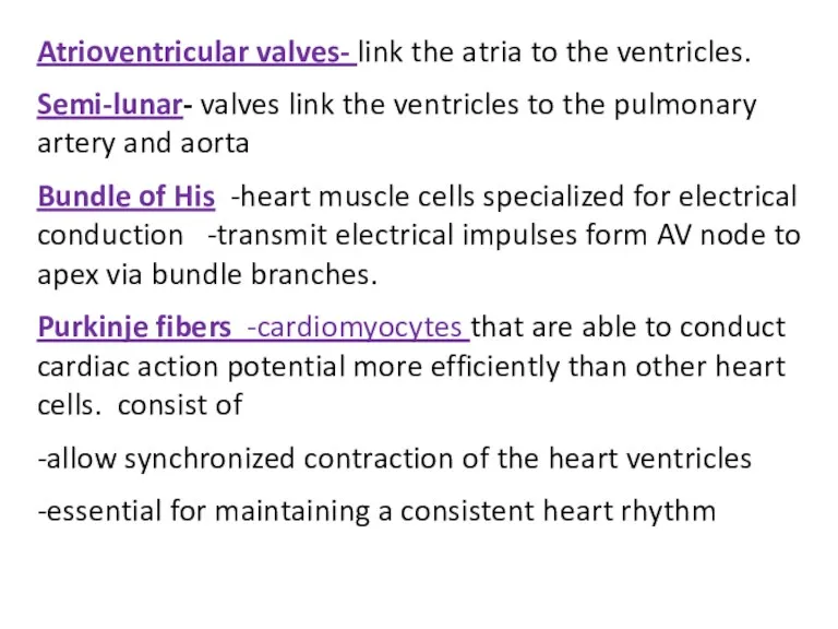
Atrioventricular valves- link the atria to the ventricles.
Semi-lunar- valves link the
Atrioventricular valves- link the atria to the ventricles.
Semi-lunar- valves link the
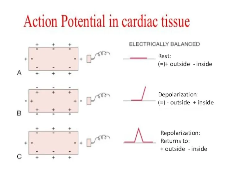
Rest:
(=)+ outside - inside
Depolarization:
(=) - outside + inside
Rest:
(=)+ outside - inside
Depolarization:
(=) - outside + inside
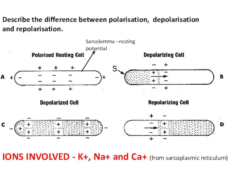
Describe the difference between polarisation, depolarisation and repolarisation.
Sarcolemma –resting potential
IONS INVOLVED
Describe the difference between polarisation, depolarisation and repolarisation.
Sarcolemma –resting potential
IONS INVOLVED
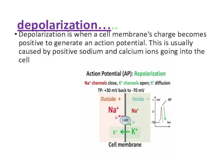
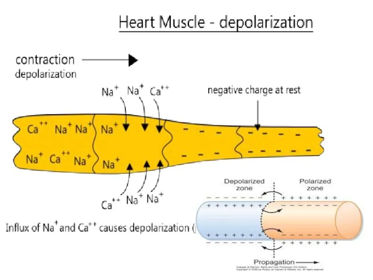
depolarization…..
Depolarization is when a cell membrane's charge becomes positive to
depolarization…..
Depolarization is when a cell membrane's charge becomes positive to
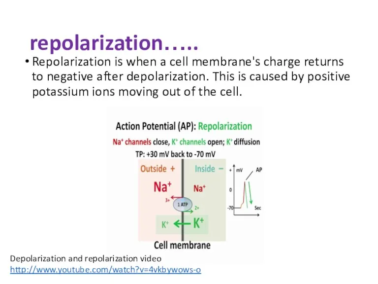
repolarization…..
Repolarization is when a cell membrane's charge returns to negative after
repolarization…..
Repolarization is when a cell membrane's charge returns to negative after
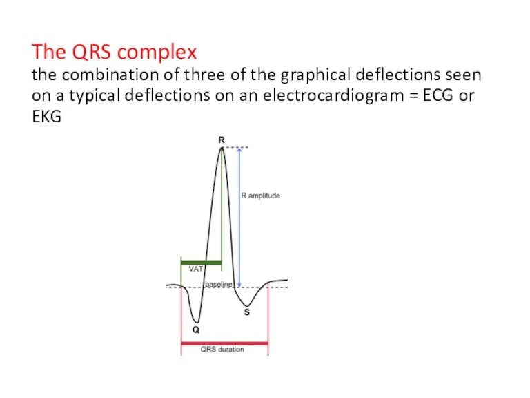
The QRS complex
the combination of three of the graphical deflections
The QRS complex the combination of three of the graphical deflections
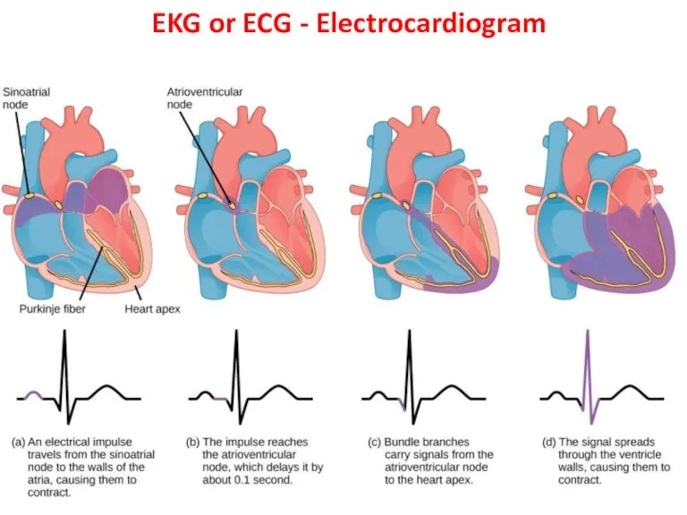
EKG or ECG - Electrocardiogram
EKG or ECG - Electrocardiogram
Cardiac Conduction System 3.45 min https://www.youtube.com/watch?v=RYZ4daFwMa8
SAN – pacemaker
Stop / play
Cardiac Conduction System 3.45 min https://www.youtube.com/watch?v=RYZ4daFwMa8
SAN – pacemaker
Stop / play
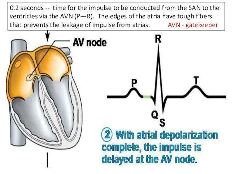
0.2 seconds -- time for the impulse to be conducted from
0.2 seconds -- time for the impulse to be conducted from
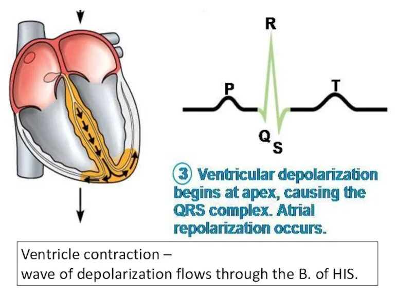
Ventricle contraction –
wave of depolarization flows through the B. of
Ventricle contraction –
wave of depolarization flows through the B. of
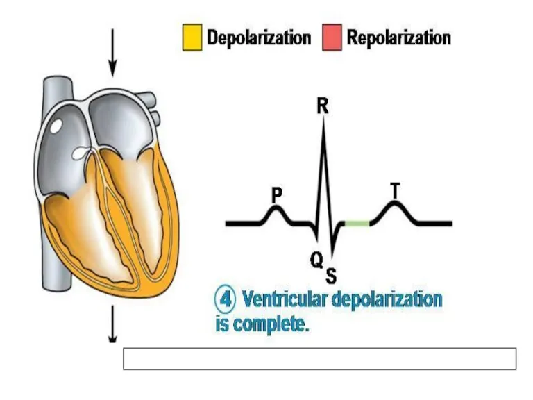
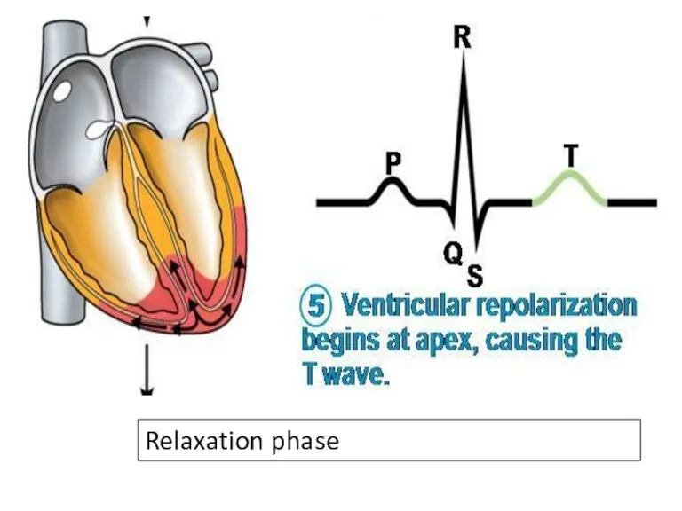
Relaxation phase
Relaxation phase
What are some ways that SAN and AVN control the heart
What are some ways that SAN and AVN control the heart
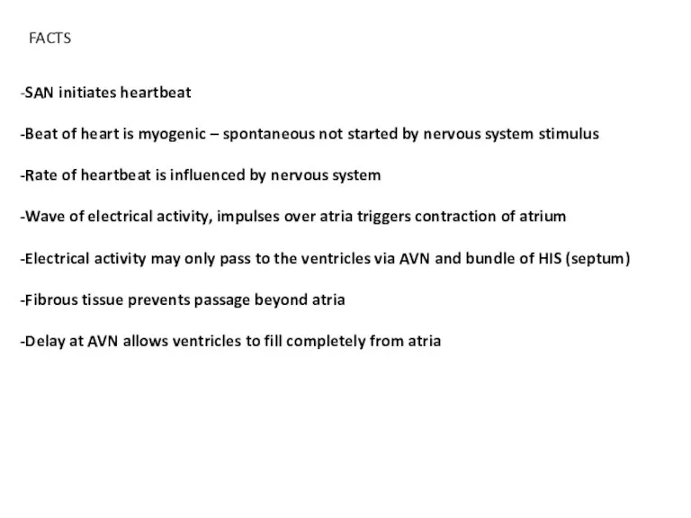
-SAN initiates heartbeat
-Beat of heart is myogenic – spontaneous not started
-SAN initiates heartbeat -Beat of heart is myogenic – spontaneous not started
EKG wave animation..
http://en.wikipedia.org/wiki/Electrocardiography#mediaviewer/File:ECG_principle_slow.gif
EKG wave animation..
http://en.wikipedia.org/wiki/Electrocardiography#mediaviewer/File:ECG_principle_slow.gif
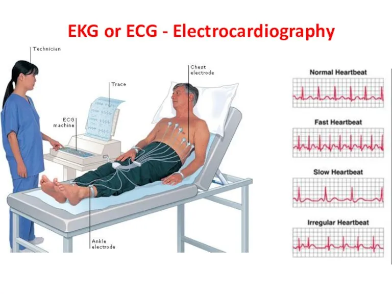
EKG or ECG - Electrocardiography
EKG or ECG - Electrocardiography
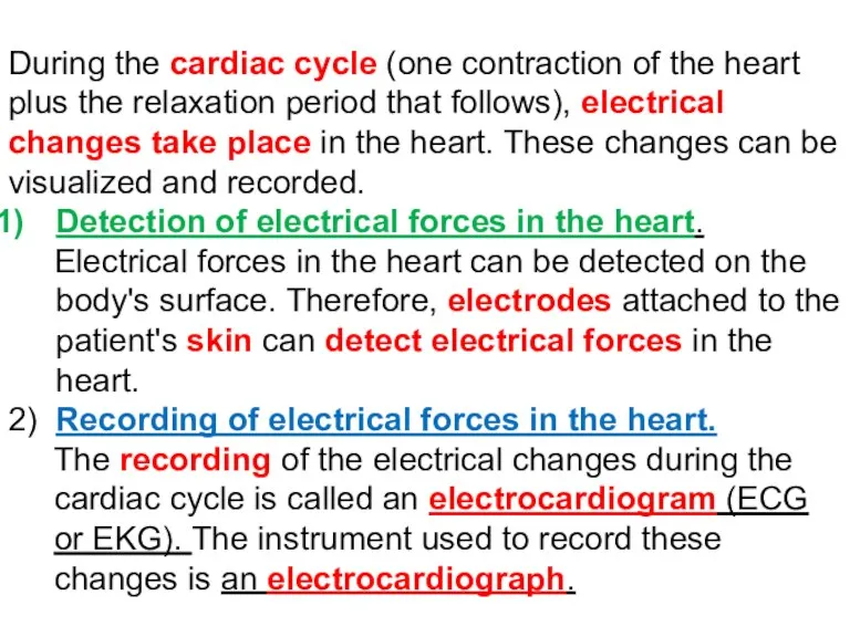
During the cardiac cycle (one contraction of the heart plus the
During the cardiac cycle (one contraction of the heart plus the
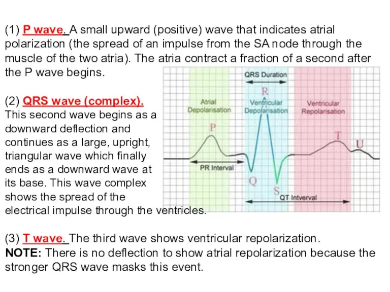
(1) P wave. A small upward (positive) wave that indicates atrial polarization (the
(1) P wave. A small upward (positive) wave that indicates atrial polarization (the
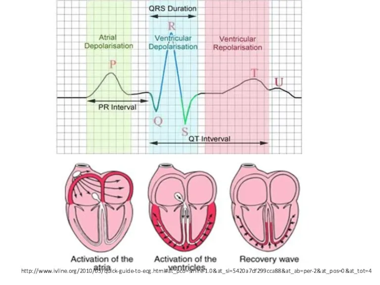
http://www.ivline.org/2010/05/quick-guide-to-ecg.html#at_pco=smlre-1.0&at_si=5420a7df299cca88&at_ab=per-2&at_pos=0&at_tot=4
http://www.ivline.org/2010/05/quick-guide-to-ecg.html#at_pco=smlre-1.0&at_si=5420a7df299cca88&at_ab=per-2&at_pos=0&at_tot=4
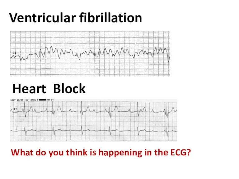
Ventricular fibrillation
What do you think is happening in the ECG?
Heart
Ventricular fibrillation
What do you think is happening in the ECG?
Heart
Ventricular fibrillation
What do you think is happening in the ECG?
Heart
Ventricular fibrillation
What do you think is happening in the ECG?
Heart
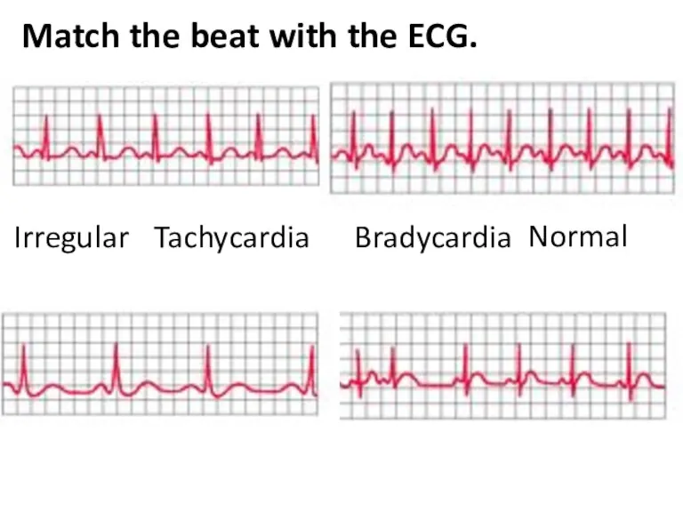
Match the beat with the ECG.
Tachycardia
Irregular
Normal
Bradycardia
Match the beat with the ECG.
Tachycardia
Irregular
Normal
Bradycardia
Tachycardia - Fast
Irregular
Normal
Bradycardia - Slow
Tachycardia - Fast
Irregular
Normal
Bradycardia - Slow
Wolff-Parkinson-White (WPW) Syndrome
An extra electrical pathway between your heart's upper and
Wolff-Parkinson-White (WPW) Syndrome
An extra electrical pathway between your heart's upper and
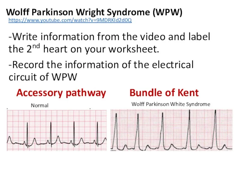
Wolff Parkinson Wright Syndrome (WPW)
-Write information from the video and label
Wolff Parkinson Wright Syndrome (WPW)
-Write information from the video and label
Pacemaker
Article
Pacemaker
Article
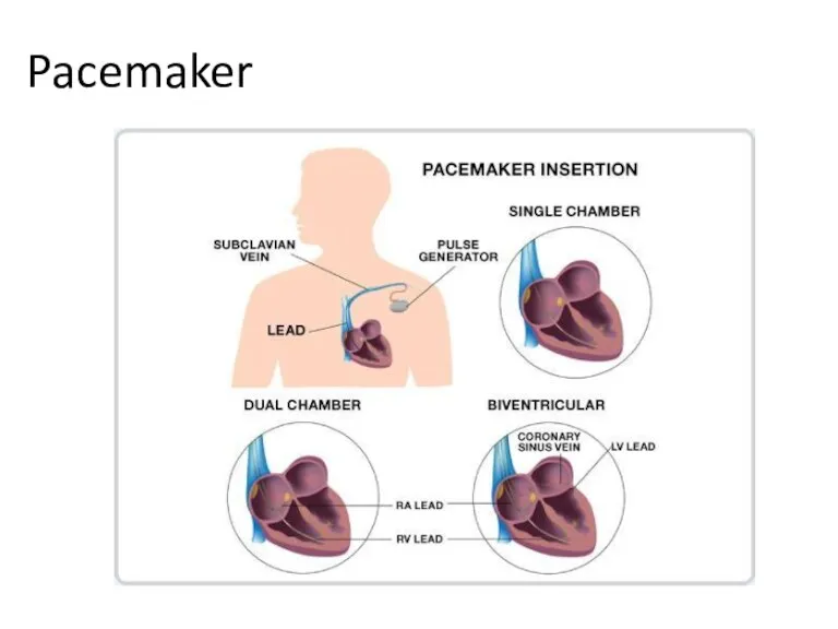
Pacemaker
Pacemaker
atrio-ventricular valves open
atrio-ventricular valves close
semi-lunar
valves open
semi-lunar
valves close
atrio-ventricular valves open
atrio-ventricular valves close
semi-lunar
valves open
semi-lunar
valves close

Match the letter on the graph to the following events
______Semi-lunar valves
Match the letter on the graph to the following events
______Semi-lunar valves

atrio-ventricular valves open
atrio-ventricular valves close
semi-lunar
valves open
semi-lunar
valves close
atrio-ventricular valves open
atrio-ventricular valves close
semi-lunar
valves open
semi-lunar
valves close

A Atrioventricular (bicuspid / mitral) valve(s) closes (“snaps shut”– makes 1st
A Atrioventricular (bicuspid / mitral) valve(s) closes (“snaps shut”– makes 1st
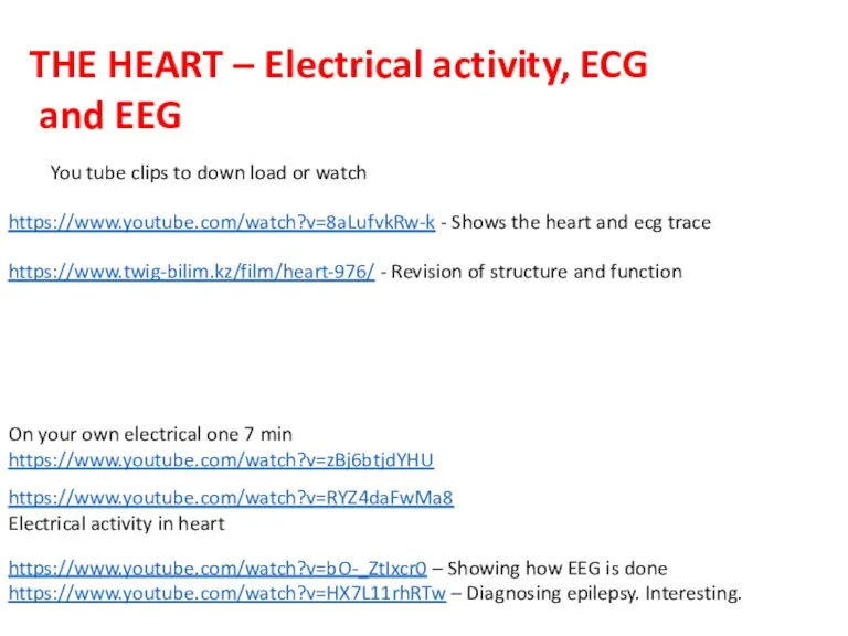
https://www.youtube.com/watch?v=RYZ4daFwMa8
Electrical activity in heart
https://www.twig-bilim.kz/film/heart-976/ - Revision of structure and function
On your
https://www.youtube.com/watch?v=RYZ4daFwMa8
Electrical activity in heart
https://www.twig-bilim.kz/film/heart-976/ - Revision of structure and function
On your
Extra Information
Extra Information
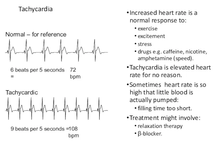
Tachycardia
Increased heart rate is a normal response to:
exercise
excitement
stress
drugs
Tachycardia
Increased heart rate is a normal response to:
exercise
excitement
stress
drugs
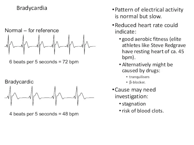
Bradycardia
Pattern of electrical activity is normal but slow.
Reduced heart rate could
Bradycardia
Pattern of electrical activity is normal but slow.
Reduced heart rate could
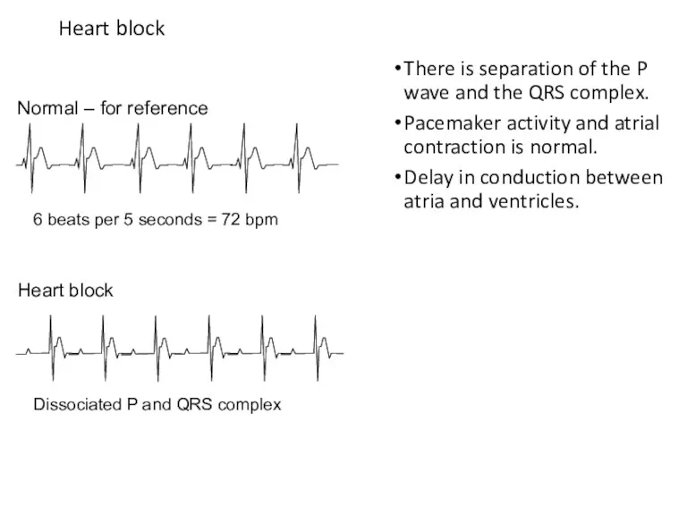
Heart block
There is separation of the P wave and the QRS
Heart block
There is separation of the P wave and the QRS
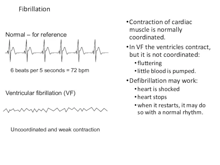
Fibrillation
Contraction of cardiac muscle is normally coordinated.
In VF the ventricles contract,
Fibrillation
Contraction of cardiac muscle is normally coordinated.
In VF the ventricles contract,
Cardiac Cycle
General Principles.
Contraction of the myocardium generates pressure changes which
Cardiac Cycle
General Principles.
Contraction of the myocardium generates pressure changes which
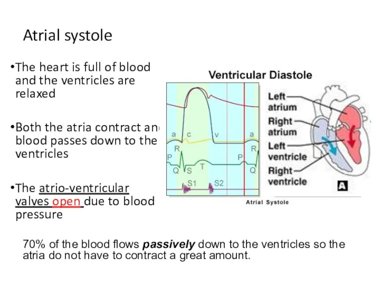
Atrial systole
The heart is full of blood and the ventricles are
Atrial systole
The heart is full of blood and the ventricles are
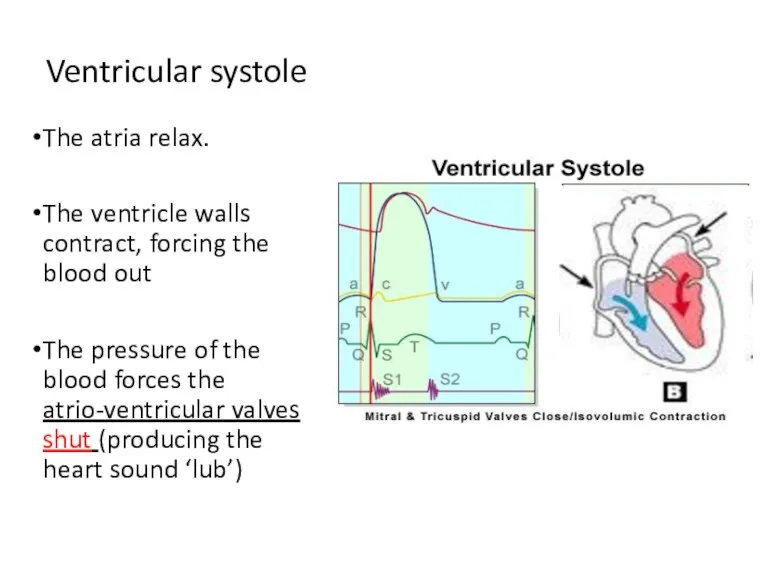
Ventricular systole
The atria relax.
The ventricle walls contract, forcing the blood out
The
Ventricular systole
The atria relax.
The ventricle walls contract, forcing the blood out
The
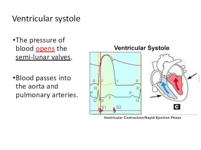
Ventricular systole
The pressure of blood opens the semi-lunar valves.
Blood passes into
Ventricular systole
The pressure of blood opens the semi-lunar valves.
Blood passes into
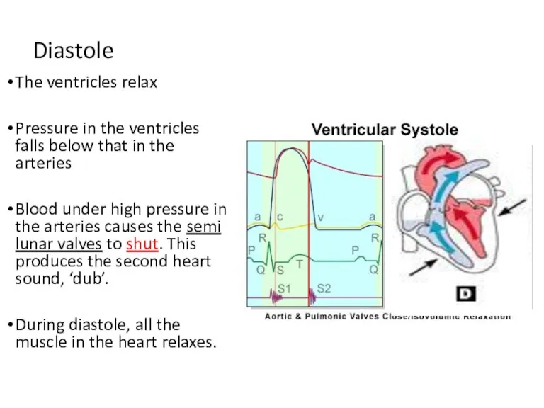
Diastole
The ventricles relax
Pressure in the ventricles falls below that in the
Diastole
The ventricles relax
Pressure in the ventricles falls below that in the
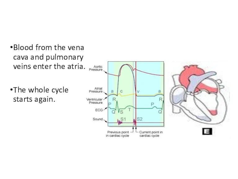
Blood from the vena cava and pulmonary veins enter the atria.
The
Blood from the vena cava and pulmonary veins enter the atria.
The
Match the letter on the graph to the following events
______Semi-lunar valves
Match the letter on the graph to the following events
______Semi-lunar valves

atrio-ventricular valves open
atrio-ventricular valves close
semi-lunar
valves open
semi-lunar
valves close
atrio-ventricular valves open
atrio-ventricular valves close
semi-lunar
valves open
semi-lunar
valves close
A Atrioventricular (bicuspid / mitral) valve(s) closes (“snaps shut”– makes 1st
A Atrioventricular (bicuspid / mitral) valve(s) closes (“snaps shut”– makes 1st
Examine the graph that shows pressure changes in the left ventricle,
Examine the graph that shows pressure changes in the left ventricle,
Answers to questions:
What is the maximum pressure reached in the left
Answers to questions:
What is the maximum pressure reached in the left
Answers to questions:
Describe and explain what happens to the bicuspid (mitral)
Answers to questions:
Describe and explain what happens to the bicuspid (mitral)
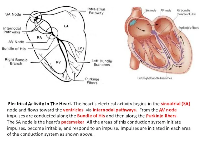
Electrical Activity In The Heart. The heart's electrical activity begins in the
Electrical Activity In The Heart. The heart's electrical activity begins in the
How does the structure of cardiac muscle differ from cardiac muscle?
Only
How does the structure of cardiac muscle differ from cardiac muscle?
Only
 Заболевания верхних дыхательных путей у детей
 Экстракорпоральное оплодотворение (ЭКО)
 Механизмы действия гомеопатических лекарственных средств
 Венгерский курорт Хевиз
 Ограниченные возможности здоровья
 Патология слуховой системы
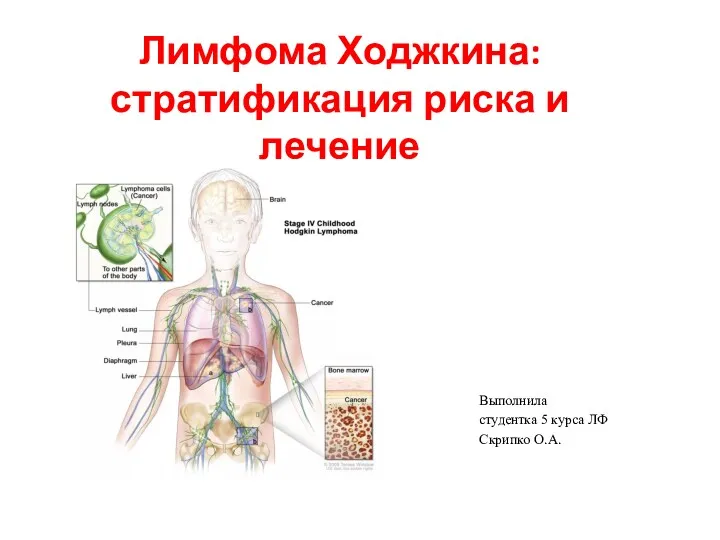 Лимфома Ходжкина. Стратификация риска и лечение
 Внезапная смерть: причины, клиника, диагностика, алгоритм неотложной помощи
 Геморрагическая лихорадка Эбола - современная мировая проблема
 Нейроэндокринные опухоли желудочно-кишечного тракта
 Патопсихология эмоционально-волевой сферы
 Косоглазие
 Ювенильді идиопатиялық артрит емінде адам иммуноглобулинін тағайындау
 Фиксация съемных ортопедических конструкций
 Заболевания мужской репродуктивной системы
 Гериатрические аспекты заболеваний опорно-двигательного аппарата
 Подари улыбку. Здоровое долголетие - образовательно-оздоровительная программа для пожилых
 Планирование ортодонтического лечения с учетом контакта больного с врачом. Показания к ортодонтическому лечению
 Первичный туберкулезный комплекс
 Ультрафиолетовое излучение и его применение в медицине
 Torch-инфекции: клиническая диагностика и этиологическая верификация
 Клиническая фармакология препаратов, влияющих на органы пищеварения
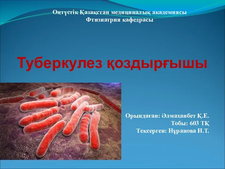 Туберкулез қоздырғышы
 Аномальді бүйректің гистоморфологиялық сипаттамасы
 Прийоми та правила надання першої домедичної допомоги постраждалим
 Регуляция менструального цикла
 Патогенное действие факторов внешней среды
 Системные васкулиты