Слайд 2
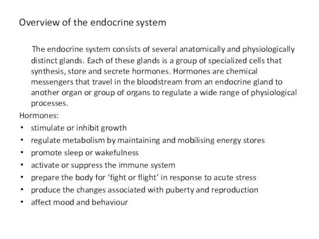
Overview of the endocrine system
The endocrine system consists of several
anatomically and physiologically distinct glands. Each of these glands is a group of specialized cells that synthesis, store and secrete hormones. Hormones are chemical messengers that travel in the bloodstream from an endocrine gland to another organ or group of organs to regulate a wide range of physiological processes.
Hormones:
stimulate or inhibit growth
regulate metabolism by maintaining and mobilising energy stores
promote sleep or wakefulness
activate or suppress the immune system
prepare the body for ‘fight or flight’ in response to acute stress
produce the changes associated with puberty and reproduction
affect mood and behaviour
Слайд 3
Слайд 4
Chemical classification of hormones
peptides
amines
lipids (mainly steroids)
Peptide hormones
The hormones in this class
are chains of amino acids (polypeptides). These chains range in length. They may be short and comprise only a few amino acids (e.g. Antidiuretic hormone), or they may be very long molecules (e.g. follicle-stimulating hormone, FSH). Peptide hormones have a large molecular weight.
Слайд 5
Chemical classification of hormones
Amine hormones
Amine hormones are derived from aromatic
amino acids such as tryptophan, phenylalanine and tyrosine. Aromatic amino acids have an aromatic side chain, i.e. one containing a stable, planar unsaturated ring of atoms.
Lipid hormones
Hormones in this class are derived from cholesterol and are either alcohols or ketones.
Alcohol lipid hormones have names ending in ‘-ol’ (e.g. oestradiol)
Ketone lipid hormones have names ending in ‘-one’ (e.g. aldosterone)
Слайд 6
Hormonal signalling pathway
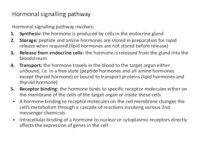
Hormonal signalling pathway involves:
Synthesis: the hormone is produced by
cells in the endocrine gland
Storage: peptide and amine hormones are stored in preparation for rapid release when required (lipid hormones are not stored before release)
Release from endocrine cells: the hormone is released from the gland into the bloodstream
Transport: the hormone travels in the blood to the target organ either unbound, i.e. in a free state (peptide hormones and all amine hormones except thyroid hormone) or bound to transport proteins (lipid hormones and thyroid hormone)
Receptor binding: the hormone binds to specific receptor molecules either on the membrane of the cells of the target organ or inside these cells
A hormone binding to receptor molecules on the cell membrane changes the cell’s metabolism through a cascade of reactions involving various 2nd messenger chemicals
Intracellular binding of a hormone to nuclear or cytoplasmic receptors directly affects the expression of genes in the cell
Слайд 7
Hormonal signalling pathway
6. Release from the cells of the target organ
The cells secrete the hormone unchanged
Alternatively, the cells metabolise the hormone to an inactive form
7. Further transport: the hormone or its breakdown products travel in the bloodstream to the liver or kidneys
8. Excretion: the hormone or its breakdown are excreted by the liver (in bile) or the kidneys (in urine)
Слайд 8
Hormone synthesis and storage
Endocrine cells synthesise peptide and amine hormones
from amino acids, and lipid hormones from cholesterol.
Peptide hormones
Hormones in this class are synthesised as precursor molecules. These prohormones undergo processing in the intracellular endoplasmic reticulum and Golgi apparatus. In the Golgi apparatus, the processed peptide hormones are packaged into secretory granules. They are stored in high concentration in these granules, ready for stimulated release from the endocrine cells into the bloodstream.
Amine hormones
These hormones are synthesised from aromatic amino acids. These amino acids are chemically altered by enzymes in the cells of endocrine glands to synthesise specific hormones. For example, in cells of the adrenal medulla, adrenaline (epinephrine) is synthesised from the amino acid tyrosine. Various enzymes catalyse the steps in adrenaline production; the final step is the conversion of noradrenaline (norepinephrine) to adrenaline by the enzyme phenylethanolamine-N-methyltransferase. Like peptide hormones, amine hormones are stored in secretory granules.
Слайд 9
Hormone synthesis and storage
Lipid hormones
These are synthesised from cholesterol. The
cholesterol is metabolised by enzymes in the cells of an endocrine gland to produce lipid hormones that are either alcohols or ketones.
The onset of action of lipid hormones is slower than that of amine hormones. Therefore, unlike amine and peptide hormones, lipid hormones are not stored in secretory granules for rapid release. Instead, they are synthesised as required, with the rate of synthesis directly determining blood concentration.
Слайд 10
Hormone release
When an endocrine cell is activated, secretory granules (containing
peptide or amine hormones) move to the cell surface. Here, the vesicular membranes of the granules fuse with the plasma membrane of the cell surface to release their contents to the exterior of the cell. This process is called exocytosis, which literally means ‘out of cell’.
Membrane transport of lipid hormones (such as testosterone) occurs in a passive manner across the cell membrane due to the non-polarised nature of the lipid-rich cell membrane. This form of hormone secretion depends upon the difference in concentration of the hormone in the intracellular space (high) to equalize with the hormone concentration in the extracellular space (low) by random motion of molecules (Brownian motion).
Слайд 11
Hormone transport
Peptide hormones are able to travel unbound (free) in the
bloodstream, because they are hydrophilic (‘water loving’). Amine hormones are also hydrophilic and also able to travel unbound in the blood. The hydrophobic thyroid hormones are the exception.
Peptide and amine hormones, other than thyroid hormones, are able to pass through capillary membranes to reach their target cells.
Lipid hormones are hydrophobic (‘water hating’), so they must be bound to transport proteins in plasma to enable them to travel in the bloodstream. Lipid hormones undergo continuous and spontaneous binding and unbinding from their carrier molecules. Because lipid hormones are bound to transport proteins, they have a longer half-life (the time taken for half of the hormone molecules to be excreted or metabolised) than amine hormones, which are transported unbound.
Only a small fraction of lipid hormones present in the bloodstream are in an unbound state. For example, 99% of cortisol in the blood is bound to proteins; the unbound remainder, the free cortisol, is biologically active. This is true of all lipid hormones.
Слайд 12
Hormone receptor binding
Hormones travel through the bloodstream and thus come into
contact with many cell types. However, a cellular response is initiated only in cells with the specific receptors for a hormone. These receptors may be on the cell membrane or in the cytoplasm.
Multiple types of cell may have receptors for a particular hormone. This allows a hormone, for example thyroxine (T4), to bind to receptors in the cells of many different tissues and thus have widespread effects on metabolism throughout the body.
The effects of a hormone binding to a receptor in one type of cell will differ from those of the same hormone binding to a receptor on another type of cell due to differing downstream processes associated with each receptor. For example, when adrenaline (epinephrine) binds to β adrenergic receptors in cardiac myocytes, it causes the heart muscle to contract more forcefully; however, the same hormone causes muscle relaxation when it binds to β receptors in the bronchioles.
Слайд 13
Hormone receptor binding
Peptide hormone receptors
Peptide hormones are lipophobic (‘lipid hating’), so
they are unable to diffuse freely through the cell membrane, which consists of two layers of lipid molecules. Therefore peptide hormone receptors composed of transmembrane proteins are necessary to communicate the hormonal message from outside the cell to the target molecules inside the cell.
The peptide hormone receptor is part of a signal transduction system. In this system, the hormone acts as the 1st messenger by binding to its receptor on the extracellular surface of the cell. This hormone−receptor binding activates 2nd messengers such as cyclic AMP (cAMP), which relay the signal within the cell.
Слайд 14
Hormone transport
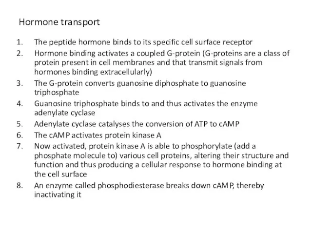
The peptide hormone binds to its specific cell surface receptor
Hormone
binding activates a coupled G-protein (G-proteins are a class of protein present in cell membranes and that transmit signals from hormones binding extracellularly)
The G-protein converts guanosine diphosphate to guanosine triphosphate
Guanosine triphosphate binds to and thus activates the enzyme adenylate cyclase
Adenylate cyclase catalyses the conversion of ATP to cAMP
The cAMP activates protein kinase A
Now activated, protein kinase A is able to phosphorylate (add a phosphate molecule to) various cell proteins, altering their structure and function and thus producing a cellular response to hormone binding at the cell surface
An enzyme called phosphodiesterase breaks down cAMP, thereby inactivating it
Слайд 15
Hormone transport
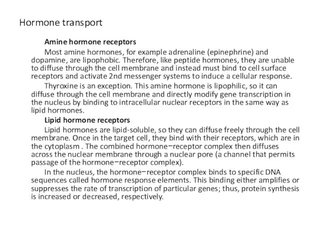
Amine hormone receptors
Most amine hormones, for example adrenaline (epinephrine) and
dopamine, are lipophobic. Therefore, like peptide hormones, they are unable to diffuse through the cell membrane and instead must bind to cell surface receptors and activate 2nd messenger systems to induce a cellular response.
Thyroxine is an exception. This amine hormone is lipophilic, so it can diffuse through the cell membrane and directly modify gene transcription in the nucleus by binding to intracellular nuclear receptors in the same way as lipid hormones.
Lipid hormone receptors
Lipid hormones are lipid-soluble, so they can diffuse freely through the cell membrane. Once in the target cell, they bind with their receptors, which are in the cytoplasm . The combined hormone−receptor complex then diffuses across the nuclear membrane through a nuclear pore (a channel that permits passage of the hormone−receptor complex).
In the nucleus, the hormone−receptor complex binds to specific DNA sequences called hormone response elements. This binding either amplifies or suppresses the rate of transcription of particular genes; thus, protein synthesis is increased or decreased, respectively.
Слайд 16
Hormone degradation and clearance
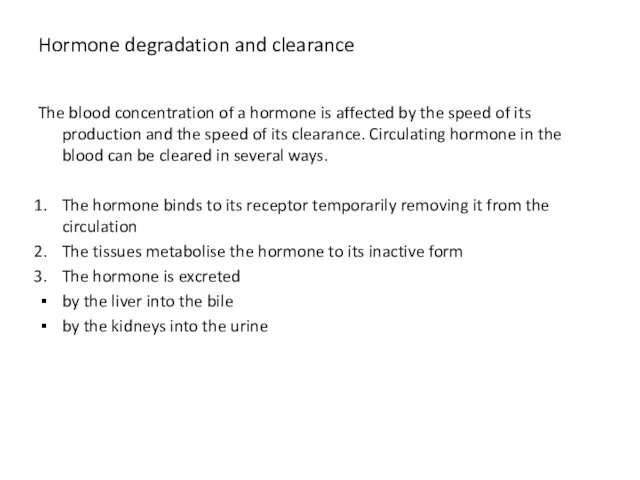
The blood concentration of a hormone is affected
by the speed of its production and the speed of its clearance. Circulating hormone in the blood can be cleared in several ways.
The hormone binds to its receptor temporarily removing it from the circulation
The tissues metabolise the hormone to its inactive form
The hormone is excreted
by the liver into the bile
by the kidneys into the urine
Слайд 17
Hormonal regulation
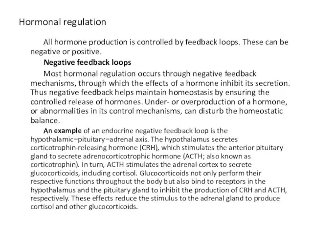
All hormone production is controlled by feedback loops. These can
be negative or positive.
Negative feedback loops
Most hormonal regulation occurs through negative feedback mechanisms, through which the effects of a hormone inhibit its secretion. Thus negative feedback helps maintain homeostasis by ensuring the controlled release of hormones. Under- or overproduction of a hormone, or abnormalities in its control mechanisms, can disturb the homeostatic balance.
An example of an endocrine negative feedback loop is the hypothalamic−pituitary−adrenal axis. The hypothalamus secretes corticotrophin-releasing hormone (CRH), which stimulates the anterior pituitary gland to secrete adrenocorticotrophic hormone (ACTH; also known as corticotrophin). In turn, ACTH stimulates the adrenal cortex to secrete glucocorticoids, including cortisol. Glucocorticoids not only perform their respective functions throughout the body but also bind to receptors in the hypothalamus and the pituitary gland to inhibit the production of CRH and ACTH, respectively. These effects reduce the stimulus to the adrenal gland to produce cortisol and other glucocorticoids.
Слайд 18
Hormonal regulation
Positive feedback loops
In positive feedback, a hormone’s effects stimulate its
secretion. An example occurs in the female reproductive cycle. When luteinising hormone causes a surge in the production of oestrogen by the ovary, the released oestrogen stimulates the anterior pituitary gland to produce more luteinising hormone. This positive feedback mechanism results in the luteinising hormone surge that stimulates ovulation.
Слайд 19
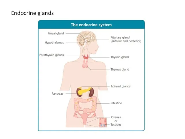
The hypothalamus
The hypothalamus is an almond-sized symmetrical structure in the brain.
It is below and anterior to the thalamus, superior to the pituitary gland and either side of the 3rd ventricle.
As an endocrine gland, the hypothalamus is responsible for control of the pituitary gland. It also has major effects on other, non-endocrine physiological processes, such as regulation of body temperature.
Two different types of neuroendocrine cell in the hypothalamus separately regulate the anterior and posterior pituitary glands:
The neuroendocrine cells that make up the tuberoinfundibular tract project into the capillary bed, where they form synapses (the end terminal of a nerve cell that transmits the cells electrical or chemical signal to another cell) and release hormones that stimulate or inhibit the anterior pituitary
The neuroendocrine cells that make up the agnocellular−neurohypophysial tract synapse directly in the posterior pituitary gland and release the hormones antidiuretic hormone and oxytocin.
Слайд 20
Antidiuretic hormone
Antidiuretic hormone (also known as vasopressin) is a polypeptide
hormone. It is synthesised by hypothalamic neuroendocrine cells that project into the posterior pituitary gland, from which the hormone is secreted. Antidiuretic hormone is stored in granules at the terminal ends of magnocellular neuroendocrine cells in the posterior pituitary gland.
Actions
The overall action of antidiuretic hormone is to increase the water content of the body. is is primarily achieved through its e ect on the collecting ducts of the kidney, where antidiuretic hormone causes aquaporin 2 channel proteins to migrate to the luminal membrane. Aquaporin 2 channels permit water absorption from the urine into the kidney. us water transport across the impermeable membrane is increased, and more water is reabsorbed from the collecting duct.
Antidiuretic hormone also binds to receptors on the vascular smooth muscle in blood vessel walls; it causes vasoconstriction and thus increases blood pressure.
Слайд 21
Oxytocin
This peptide hormone is synthesised in magnocellular cells of the
hypothalamus and secreted from their terminal ends in the posterior pituitary.
Actions
The main roles of oxytocin are to stimulate:
uterine contraction during labour
milk let-down to facilitate lactation
Oxytocin also has roles in the menstrual cycle in women and in erectile function in men.
Слайд 22
Growth hormone− releasing hormone and somatostatin
Growth hormone−releasing hormone and somatostatin
are peptide hormones produced in the tuberoinfundibular tract of the hypothalamus that bind to G-protein−coupled receptors in somatotrophs (cells in the anterior pituitary gland that produce growth hormone).
Actions
Growth hormone−releasing hormone and somatostatin have antagonistic actions on the somatotrophs:
GHRH stimulates the production and release of growth hormone
Somatostatin inhibits growth hormone production
Growth hormone−releasing hormone also promotes somatotroph replication.
As well as reducing the production of growth hormone in the pituitary, somatostatin inhibits the production of GHRH in the hypothalamus.
Somatostatin also has other endocrine effects; for example, it is produced in the pancreas and inhibits insulin secretion.
Слайд 23
Corticotrophin-releasing hormone
Corticotrophin-releasing hormone is a pep- tide hormone produced in
the hypothalamus. It binds to a G-protein−coupled receptor on corticotrophs (ACTH-producing cells in the anterior pituitary gland).
Actions
CRH stimulates corticotrophs to produce ACTH. ACTH, in turn, increases the production of cortisol in the adrenal glands.
Слайд 24
Thyrotrophin-releasing hormone
Thyrotrophin-releasing hormone is a peptide hormone produced in the
paraventricular nucleus of the hypothalamus. It binds to G-protein−coupled receptors on thyrotrophs (cells in the anterior pituitary gland that produce TSH) to stimulate the production and release of TSH.
Secretion
The release of TSH is controlled by negative feedback from the thyroid hormones, T3 and T4. In the physiological states of fasting and illness, TRH production is down- regulated by inhibitory neuronal input of multiple cells of the peripheral and central nervous system. This effect conserves energy by producing a state of low thyroid hormone levels and a reduced metabolic rate.
Transport
Thyrotrophin-releasing hormone is transported unbound in the blood. Once secreted into the median eminence from granules at the distal end of the hypothalamic neurons it travels to the anterior pituitary gland in the blood via the hypothalamic-pituitary portal system the hypothalamic-pituitary portal system.
Слайд 25
Gonadotrophin-releasing hormone
Gonadotrophin-releasing hormone is a peptide hormone produced by cells
widely distributed throughout the medial hypothalamus. GnRH binds to a G-protein−coupled receptor on cells of the anterior pituitary gland.
This hormone promotes sexual develop- ment, sex hormone production and reproduc- tion by stimulating production of the gonado- trophins (hormones that stimulate gonadal function, e.g. luteinising hormone and FSH).
Actions
Gonadotrophin-releasing hormone stimulates gonadotrophin production by gonadotrophs (cells in the anterior pituitary that produce gonadotrophins). GnRH activ- ity is low in childhood but is activated around puberty when genetic triggers are activated by hypothalamic hormones called Kisspeptins.
Secretion
Gonadotrophin-releasing hormone is released in pulses. The pulsatile nature of GnRH is refected in its effects on the gonadotrophs. For example, changes in the frequency of GnRH pulses determine the frequency of pulses of gonadotrophins from the pituitary.
In women and girls, such changes enable progression through the phases of the menstrual cycle, as when a surge of luteinising hormone precipitates ovulation
In men, the pulsatility of GnRH and thus gonadotrophins is less variable
Слайд 26
The pituitary gland
The pituitary gland lies in the pituitary fossa
at the base of the skull. Although small (about 0.5 cm in diameter), the gland controls many of the body’s endocrine systems.
The gland is anatomically and functionally separated into two parts:
the anterior pituitary gland (also known as the adenohypophysis)
the posterior pituitary gland (also known as the neurohypophysis)
A wide variety of hormones are secreted from multiple cell types in the anterior pituitary gland. Hormones of the posterior pituitary gland (antidiuretic hormone and oxytocin) are released in the posterior pituitary having been synthesised in the hypothalamus.
Слайд 27
Adrenocorticotrophic hormone
This peptide hormone is derived from a large precursor
molecule called pro-opiomelanocortin. Cleavage of this polypeptide produces several peptides, including ACTH. ACTH is synthesised by the corticotrophs of the anterior pituitary gland. These cells make up about a quarter of the anterior pituitary gland.
Actions
Adrenocorticotrophic hormone binds to ACTH receptors, which are transmembrane receptors in the zona fasciculata and zona reticularis of the adrenal cortex. Binding of ACTH to its receptor stimulates the production of cortisol.
In addition, ACTH binds to cells in the zona glomerulosa and zona reticularis to stimulate synthesis of aldosterone and adrenal androgens. However, these hormones have other, more potent stimuli for secretion. For example, aldosterone secretion is primarily stimulated by angiotensin II.
Aside from its role in hormone production, ACTH also stimulates proliferation of adrenal cortex cells to maintain the adrenal cortex at a size sufficient to produce adequate amounts of cortisol.
Слайд 28
Adrenocorticotrophic hormone
Secretion
The secretion of ACTH is stimulated by CRH
from the hypothalamus. e hormone is transported unbound in the systemic circulation.
There are many other physiological stimuli for ACTH secretion. These include antidiuretic hormone, catecholamines and growth hormone (i.e. other hormones that control the stress response and affect metabolism). ACTH secretion has physiological characteristics that directly inuence the reactive production of cortisol:
ACTH secretion is pulsatile
ACTH secretion has a circadian rhythm
ACTH release is stimulated by stress
The circadian rhythm is a pattern of secretion that follows a 24-h cycle set by a hypothalamic pacemaker. ACTH is not unique in having such a rhythm; other hormones, such as testosterone and growth hormone, also follow a circadian pattern.
The frequency of ACTH pulses remains constant, but circadian rhythm occurs by changes in the quantity of ACTH released with each pulse. e highest peaks are early in the morning, and the lowest troughs are in the middle of the night.
Stress leads to cytokine, hormone and neurotransmitter release, which stimulates the release of CRH. CRH, in turn, increases overall ACTH secretion and cortisol production. For example, ACTH release can be caused by hypotension, pain, emotional strain and metabolic stressors such as hypoglycaemia.
Слайд 29
Growth hormone
Growth hormone is a polypeptide hormone released from somatotrophs
in the anterior pituitary gland. It is the most abundant anterior pituitary hormone.
In childhood, growth hormone has a key role in promoting growth. It also has a role in adulthood in increasing muscle growth and increasing blood glucose, even after the body has reached its final height.
Actions
The biological effects of growth hormone are varied and complex, but almost all its actions are through its effector hormone, insulin-like growth factor-1. Growth hormone stimulates the production of insulin-like growth factor-1 (a peptide hormone with a similar chemical structure to insulin) in the liver via activation of growth hormone receptors.
Insulin-like growth factor-1 has effects on multiple tissues, including promotion of cellular proliferation and stimulation of metabolism. However, the key target organ of IGF-1 in childhood is the epiphyseal growth plates in long bones; therefore ICF-1 stimulates long bone growth children.
Growth hormone also affects tissues directly to cause growth by cellular proliferation.
Слайд 30
Growth hormone
Secretion
The transcription and release of growth hormone are
stimulated by GHRH. Growth hormone secretion is also stimulated by ghrelin produced by the stomach, because ghrelin augments GHRH and inhibits somatostatin (an inhibitor of growth hormone release).
The release of growth hormone is pulsatile. Pulses occur less than a dozen times per day. During the intervening times, growth hormone levels are low. Growth hormone secretion has a circadian pattern; pulses occur in greater frequency and amplitude during sleep, when peak growth occurs in children. Growth hormone is present from birth. However, the onset of puberty causes a marked increase in the amplitude of growth hormone pulses as a result of genetic stimuli. In adulthood, growth hormone secretion declines with age but the pulse frequency remains constant.
Growth hormone is not the only hormone to promote growth. Growth velocity decreases during periods of illness in childhood, and multiple factors produce the ‘catch-up growth’ that occurs after a period of prolonged illness; this type of growth is the result of a complex interaction of hormones and physical state (e.g. nutritional status).
Growth hormone is transported bound to proteins in the blood.
Слайд 31
Thyroid-stimulating hormone
Thyroid-stimulating hormone is a glycoprotein synthesised in the anterior
pituitary. Through its actions on TSH receptors in the thyroid, it is a major stimulus for thyroid cell growth, differentiation and function.
TSH has two subunits.
The α subunit is nearly identical to that of human chorionic gonadotrophin, luteinising hormone and FSH
The β subunit is unique to TSH and is responsible for binding to the TSH receptor
Actions
Thyroid-stimulating hormone promotes production of the thyroid hormones, T3 and T4, by the follicular cells of the thyroid gland. TSH also stimulates thyroid cell growth and differentiation.
Secretion
Thyroid-stimulating hormone secretion is pulsatile and circadian, peaking in the evening. Secretion from the anterior pituitary gland is stimulated by TRH from the hypothalamus.
The production and secretion of TSH by the anterior pituitary gland are directly inhibited by negative feedback from T3 and T4.
The hormone is transported unbound in the serum.
Слайд 32
Luteinising hormone and follicle-stimulating hormone
Luteinising hormone and FSH are called
gonadotrophins, because they stimulate the gonads (the testes in males and the ovaries in females).They are glycoproteins produced by the gonadotrophs of the pituitary gland.
Similar to TSH and human chorionic gonadotrophin, they comprise homologous α chains and unique β chains. Theβ chains bind to activate the receptors.
The gonadotrophins are not essential to life (as individuals with genetic disorders of gonadotrophin deficiency, e.g. Kallman’s syndrome, have only partially reduced life-expectancy). However, they are essential for pubertal development and fertility.
Actions
Luteinising hormone and FSH stimulate sex hormone (see pages 51 and 56) and gamete production in both males and females. FSH is responsible for stimulating:
development of the ovarian follicles in women
spermatogenesis (sperm-production) in the Sertoli cells in men
Luteinising hormone stimulates production of the sex steroids. In response to luteinising hormone, testosterone is secreted from the Leydig cells in men and from theca cells in women. In women, the testosterone is then converted into oestrogen in ovarian granulosa cells, adjacent to the theca cells, during the follicular phase of the menstrual cycle (in which the developing follicle produces oestrogen in response to stimulation by LH and FSH)
Слайд 33
Luteinising hormone and follicle-stimulating hormone
Secretion
Luteinising hormone and FSH are
produced in response to pulses of GnRH from the hypothalamus. e increased frequency and amplitude of these pulses are detected by activation of the GnRH receptors on the gonadotrophs and increase the amount of luteinising hormone and FSH produced.
In women, there is a surge in gonadotrophins, predominantly luteinising hormone, just before ovulation. The luteinising hormone helps turn the remaining follicle into the corpus luteum. The corpus luteum secretes progesterone, which helps prepare the endometrium for possible implantation. The cyclicity of gonadotrophin secretion in women is not present in men as semen production and fertility are continuous rather than cyclical as in the female.

 Жас балалардағы тырнақ және шаш ерекшіліктері
 Профилактика стоматологических заболеваний в раннем детском возрасте
 Геморрагический васкули
 Болевые синдромы. Сенсорные расстройства
 Патологическая стираемость генерализованного типа
 Медицина Московского государства (XV – XVII вв.)
 Туберкулез почек, мочевыводящей системы и мужских половых органов
 Воспалительные заболевания глазного яблока и зрительного нерва
 Язвенная болезнь желудка и 12-перстной кишки у детей
 Шкода алкоголю, куріння та наркотиків
 Первая медицинская помощь при травмах опорнодвигательного аппарата
 Организация здравоохранения в зарубежных странах. Страховая, бюджетная и частная системы здравоохранения
 Гипоксия и дыхательная недостаточность
 Медицинский отряд специального назначения
 Сенсорные и гностические нарушения при поражении зрительного и слухового анализаторов
 Современные методы исследования в офтальмологии
 Стоматологическое просвещение населения. (Лекция 17)
 Цитомегаловирусты инфекция
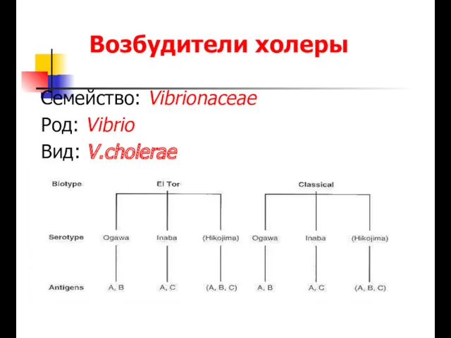 Возбудители холеры
 Профилактика ВИЧ-инфекции
 Зәр шығару жүйесі
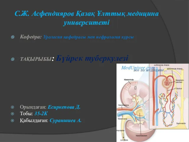 Бүйрек туберкулезі
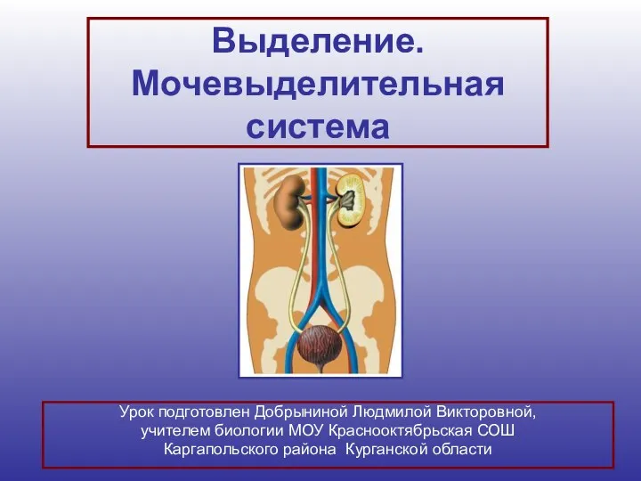 Выделение. Мочевыделительная система
 Амбулаториялық-емханалық көмек көрсететін денсаулық сақтау ұйымдарының қызметі туралы ережені бекіту туралы
 ЭХО-КГ в норме у детей
 Работники медицинских учреждений
 Балалардың іш қатуы
 Лекция №23. Острые гнойные заболевания мягких тканей