- Esophagus stomach

Содержание
- 2. Alimentary Canal Same basic construction end to end Four distinct layers: Mucosa Epithelium lamina propria, MUSCULARIS
- 3. Mucosa Mucosa has three functions: Barrier separates the lumen (which is in contact with the environment)
- 4. Mucosa: Epithelium Epithelium secretes: Digestive enzymes into lumen onto apical plasma membrane Hormones Mucous Antibodies which
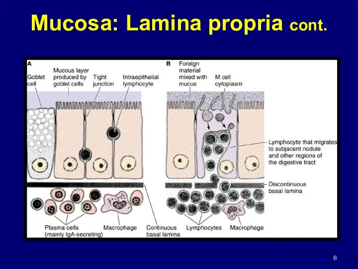
- 5. Mucosa: Lamina Propria Areolar (loose) connective tissue under epithelium Contains: - glands - vessels to receive
- 6. Mucosa: Lamina propria cont.
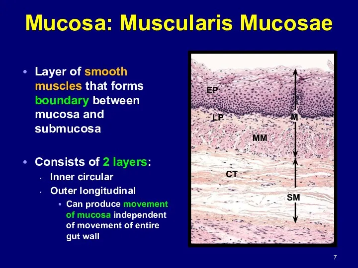
- 7. Mucosa: Muscularis Mucosae Layer of smooth muscles that forms boundary between mucosa and submucosa Consists of
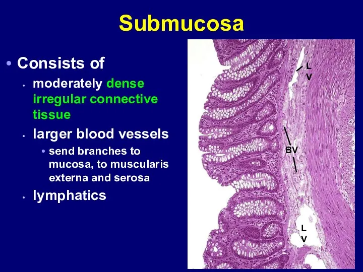
- 8. Submucosa Consists of moderately dense irregular connective tissue larger blood vessels send branches to mucosa, to
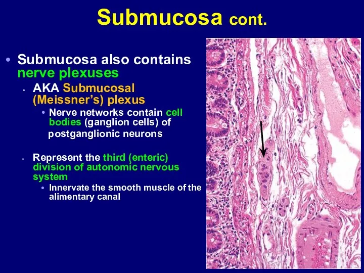
- 9. Submucosa cont. Submucosa also contains nerve plexuses AKA Submucosal (Meissner’s) plexus Nerve networks contain cell bodies
- 10. Neurons of the enteric division show the same pathologic changes that can occur in neurons of
- 11. Glands occur in submucosa of esophagus and initial part of duodenum Presence of these glands aids
- 12. Muscularis Externa Also called the MUSCULARIS Usually consists of two concentric thick layers of smooth muscle
- 13. Muscularis cont. Located between the 2 muscle layers is a thin connective tissue layer contains the
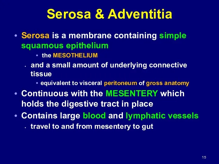
- 15. Serosa & Adventitia Serosa is a membrane containing simple squamous epithelium the MESOTHELIUM and a small
- 16. Serosa & Adventitia cont. Large amounts of fat can accumulate in serosa Where gut has no
- 17. Esophagus Mucosa: Epithelium non-keratinized stratified squamous Lamina propria has diffuse lymphatic tissue and lymphatic nodules Muscularis
- 18. Muscularis externa IC OL
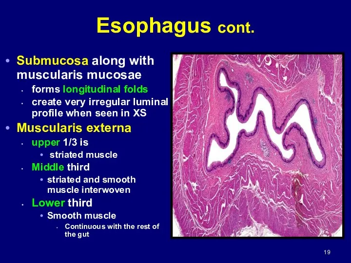
- 19. Esophagus cont. Submucosa along with muscularis mucosae forms longitudinal folds create very irregular luminal profile when
- 21. Esophagus has adventitia until it enters abdominal cavity where it is covered by SEROSA Esophagus cont.
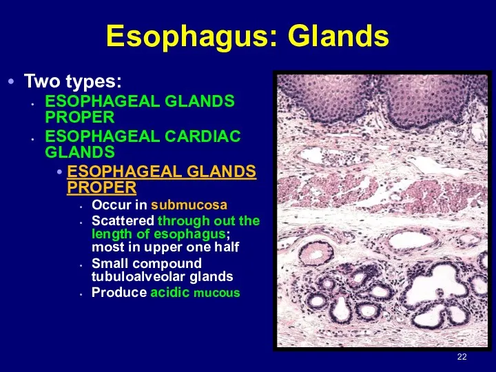
- 22. Esophagus: Glands Two types: ESOPHAGEAL GLANDS PROPER ESOPHAGEAL CARDIAC GLANDS ESOPHAGEAL GLANDS PROPER Occur in submucosa
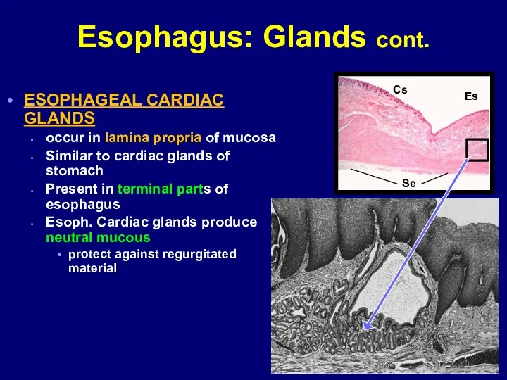
- 24. Esophagus: Glands cont. ESOPHAGEAL CARDIAC GLANDS occur in lamina propria of mucosa Similar to cardiac glands
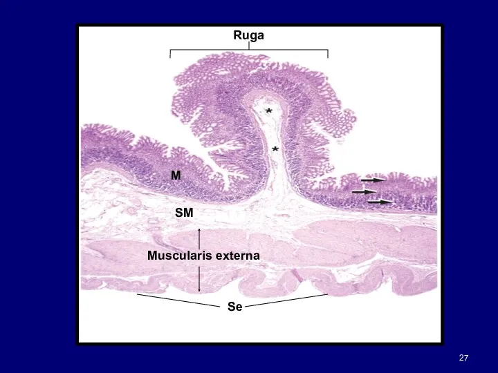
- 26. Stomach Expanded part of alimentary canal Same organization as remaining gut Mucosa submucosa muscularis serosa Inner
- 28. Stomach cont. Numerous openings seen in mucosal surface GASTRIC PITS or FOVEOLAE Easily seen in SEM
- 29. Histologically divided into 3 regions: Cardia pylorus Fundus Stomach cont.
- 30. CARDIA (cardiac region) part near esophagus contains cardiac glands PYLORUS (pyloric region) part proximal to pyloric
- 31. Stomach: Gastric Secretion 2 liters of fluid/day Gastric secretions include: Pepsinogen inactive precursor of proteolytic enzyme
- 32. Stomach: Absorption Stomach lining absorbs some water salts lipid-soluble drugs certain drugs Asprin enters by damaging
- 33. Stomach: Gastric Mucosa Simple columnar epithelium Called SURFACE MUCOUS CELLS Cells have a large apical deposit
- 34. Fundic Glands Fundic glands; also called gastric glands Produce digestive juice of stomach Present throughout gastric
- 35. Fundic Glands cont. Several glands open into one gastric pit Each gland has long NECK SEGMENT
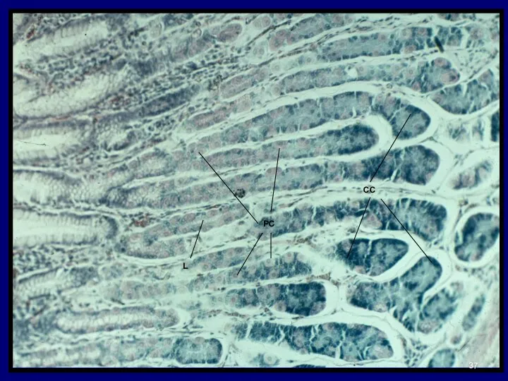
- 36. Composed of 4 functional cell types: MUCOUS NECK CELLS CHIEF CELLS PARIETAL CELLS (Oxyntic cells) ENTEROENDOCRINE
- 37. PC CC L
- 40. Fundic Glands: Mucous Neck Cells Located in neck region Shorter than surface mucous cell Nucleus tends
- 41. Fundic Glands: Chief Cells Typical protein-secreting cells Occupy the basal portion of the gland Cells easily
- 42. Fundic Glands: Parietal Cells Called OXYNTIC CELLS Secrete HCl and intrinsic factor Most numerous in upper
- 44. Fundic Glands: Parietal Cell cont. Nucleus is spherical Cytoplasm intensely eosinophilic easily recognized by size and
- 45. EM shows extensive INTRACELLULAR CANALICULAR SYSTEM communicates with lumen of fundic gland Numerous surface microvilli project
- 46. TUBULOVESICULAR MEMBRANE SYSTEM located in cytoplasm adjacent to canalicular system represent reserve membrane for insertion into
- 47. Fundic Glands: Enteroendocrines Open and closed types Open are chemoreceptors Located at any level of gland
- 51. Cardiac Glands Limited to narrow region of stomach near esophageal orifice Secretion, along with that of
- 52. Pyloric Glands Located between fundus and pylorus Branched tubular glands Coiled Cells Similar to surface mucous
- 55. Скачать презентацию
Alimentary Canal
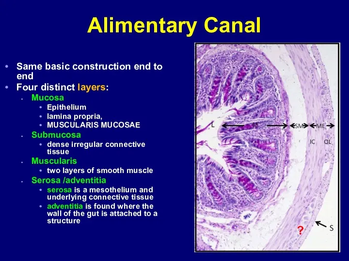
Same basic construction end to end
Four distinct layers:
Mucosa
Epithelium
lamina propria,
MUSCULARIS MUCOSAE
Submucosa
dense
Alimentary Canal
Same basic construction end to end
Four distinct layers:
Mucosa
Epithelium
lamina propria,
MUSCULARIS MUCOSAE
Submucosa
dense
Mucosa
Mucosa has three functions:
Barrier
separates the lumen (which is in contact with
Mucosa
Mucosa has three functions:
Barrier
separates the lumen (which is in contact with
Mucosa: Epithelium
Epithelium secretes:
Digestive enzymes
into lumen
onto apical plasma membrane
Hormones
Mucous
Antibodies
which
Mucosa: Epithelium
Epithelium secretes:
Digestive enzymes
into lumen
onto apical plasma membrane
Hormones
Mucous
Antibodies
which
Mucosa: Lamina Propria
Areolar (loose) connective tissue under epithelium
Contains:
- glands
-
Mucosa: Lamina Propria
Areolar (loose) connective tissue under epithelium
Contains:
- glands
-
Mucosa: Lamina propria cont.
Mucosa: Lamina propria cont.
Mucosa: Muscularis Mucosae
Layer of smooth muscles that forms boundary between mucosa
Mucosa: Muscularis Mucosae
Layer of smooth muscles that forms boundary between mucosa
Submucosa
Consists of
moderately dense irregular connective tissue
larger blood vessels
send branches
Submucosa
Consists of
moderately dense irregular connective tissue
larger blood vessels
send branches
Submucosa cont.
Submucosa also contains nerve plexuses
AKA Submucosal (Meissner’s) plexus
Nerve networks contain
Submucosa cont.
Submucosa also contains nerve plexuses
AKA Submucosal (Meissner’s) plexus
Nerve networks contain
Neurons of the enteric division show the same pathologic changes that
Neurons of the enteric division show the same pathologic changes that
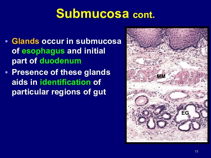
Glands occur in submucosa of esophagus and initial part of duodenum
Presence
Glands occur in submucosa of esophagus and initial part of duodenum
Presence
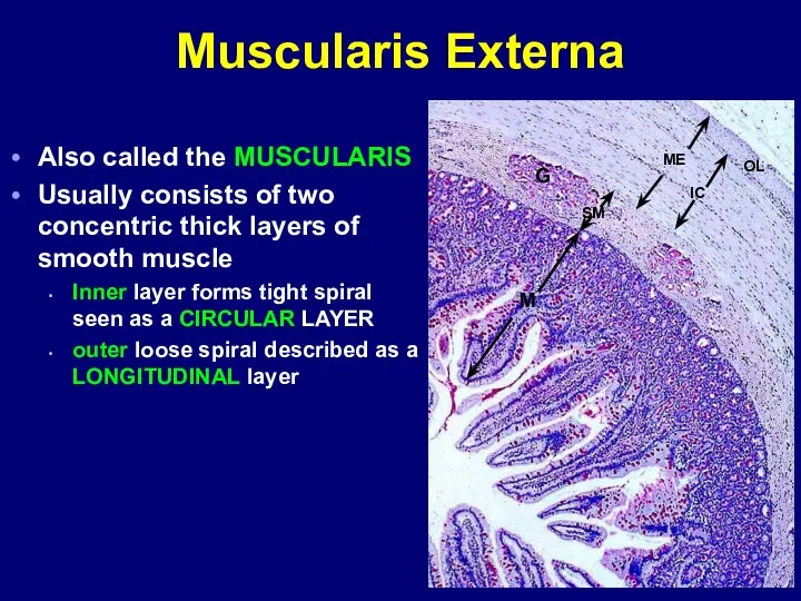
Muscularis Externa
Also called the MUSCULARIS
Usually consists of two concentric thick layers
Muscularis Externa
Also called the MUSCULARIS
Usually consists of two concentric thick layers
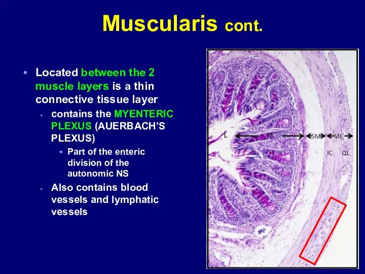
Muscularis cont.
Located between the 2 muscle layers is a thin connective
Muscularis cont.
Located between the 2 muscle layers is a thin connective

Serosa & Adventitia
Serosa is a membrane containing simple squamous epithelium
the
Serosa & Adventitia
Serosa is a membrane containing simple squamous epithelium
the
Serosa & Adventitia cont.
Large amounts of fat can accumulate in serosa
Where
Serosa & Adventitia cont.
Large amounts of fat can accumulate in serosa
Where
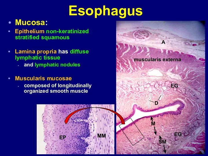
Esophagus
Mucosa:
Epithelium non-keratinized stratified squamous
Lamina propria has diffuse lymphatic tissue
and lymphatic
Esophagus
Mucosa:
Epithelium non-keratinized stratified squamous
Lamina propria has diffuse lymphatic tissue
and lymphatic
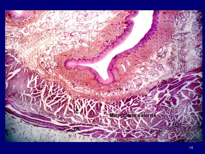
Muscularis externa
IC
OL
Muscularis externa
IC
OL
Esophagus cont.
Submucosa along with muscularis mucosae
forms longitudinal folds
create very irregular
Esophagus cont.
Submucosa along with muscularis mucosae
forms longitudinal folds
create very irregular

Esophagus has adventitia until it enters abdominal cavity
where it is
where it is
Esophagus: Glands
Two types:
ESOPHAGEAL GLANDS PROPER
ESOPHAGEAL CARDIAC GLANDS
ESOPHAGEAL GLANDS PROPER
Esophagus: Glands
Two types:
ESOPHAGEAL GLANDS PROPER
ESOPHAGEAL CARDIAC GLANDS
ESOPHAGEAL GLANDS PROPER

Esophagus: Glands cont.
ESOPHAGEAL CARDIAC GLANDS
occur in lamina propria of mucosa
Similar
Esophagus: Glands cont.
ESOPHAGEAL CARDIAC GLANDS
occur in lamina propria of mucosa
Similar
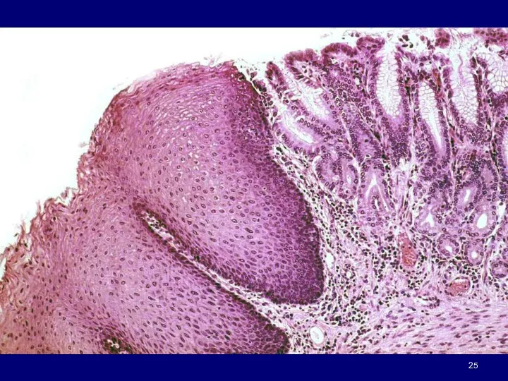
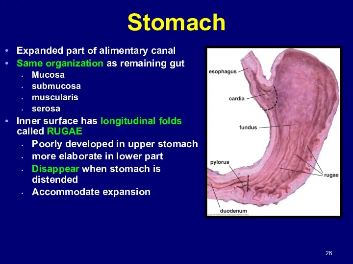
Stomach
Expanded part of alimentary canal
Same organization as remaining gut
Mucosa
submucosa
muscularis
serosa
Inner surface has
Stomach
Expanded part of alimentary canal
Same organization as remaining gut
Mucosa
submucosa
muscularis
serosa
Inner surface has
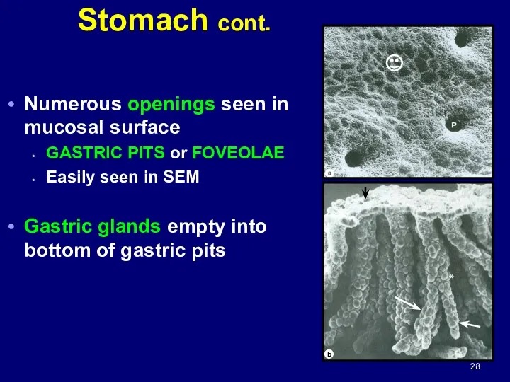
Stomach cont.
Numerous openings seen in mucosal surface
GASTRIC PITS or FOVEOLAE
Easily seen
Stomach cont.
Numerous openings seen in mucosal surface
GASTRIC PITS or FOVEOLAE
Easily seen
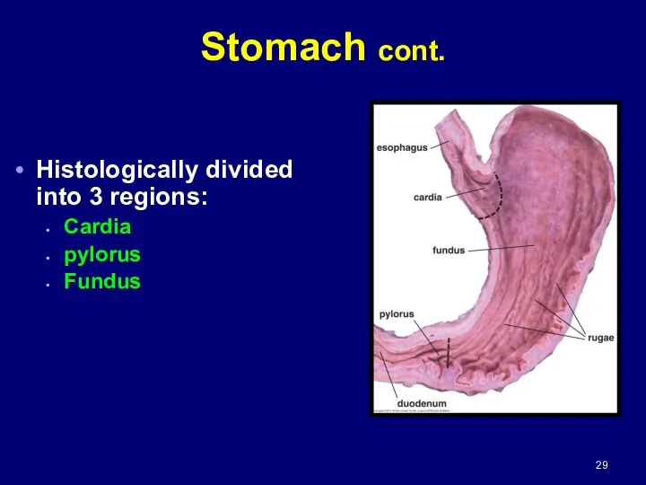
Histologically divided into 3 regions:
Cardia
pylorus
Fundus
Stomach cont.
Histologically divided into 3 regions:
Cardia
pylorus
Fundus
Stomach cont.
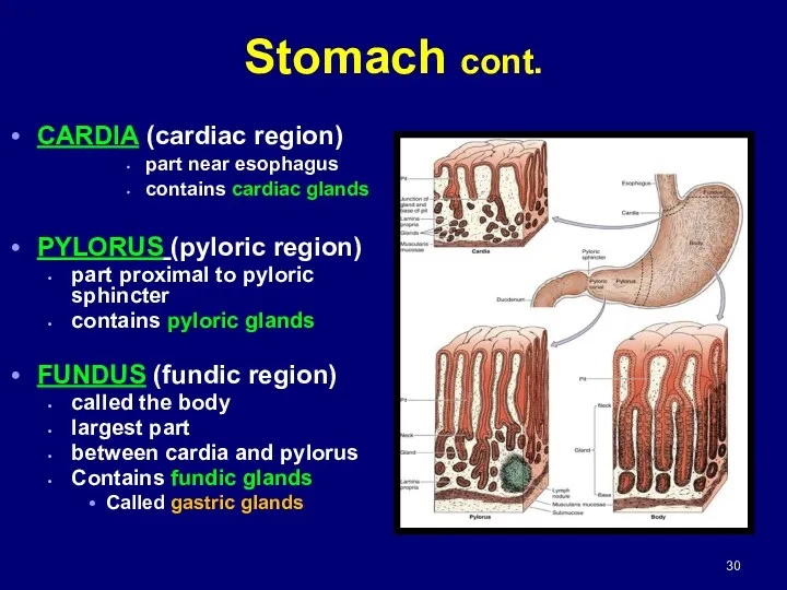
CARDIA (cardiac region)
part near esophagus
contains cardiac glands
PYLORUS (pyloric region)
part proximal
CARDIA (cardiac region)
part near esophagus
contains cardiac glands
PYLORUS (pyloric region)
part proximal
Stomach: Gastric Secretion
2 liters of fluid/day
Gastric secretions include:
Pepsinogen
inactive precursor of proteolytic
Stomach: Gastric Secretion
2 liters of fluid/day
Gastric secretions include:
Pepsinogen
inactive precursor of proteolytic
Stomach: Absorption
Stomach lining absorbs
some water
salts
lipid-soluble drugs
certain drugs
Asprin enters
Stomach: Absorption
Stomach lining absorbs
some water
salts
lipid-soluble drugs
certain drugs
Asprin enters
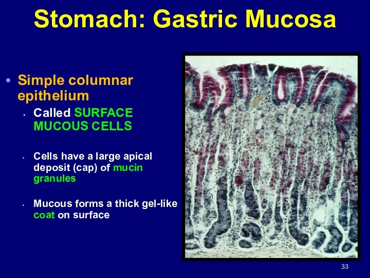
Stomach: Gastric Mucosa
Simple columnar epithelium
Called SURFACE MUCOUS CELLS
Cells have a
Stomach: Gastric Mucosa
Simple columnar epithelium
Called SURFACE MUCOUS CELLS
Cells have a
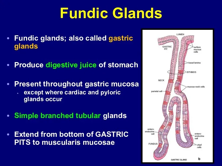
Fundic Glands
Fundic glands; also called gastric glands
Produce digestive juice of stomach
Present
Fundic Glands
Fundic glands; also called gastric glands
Produce digestive juice of stomach
Present
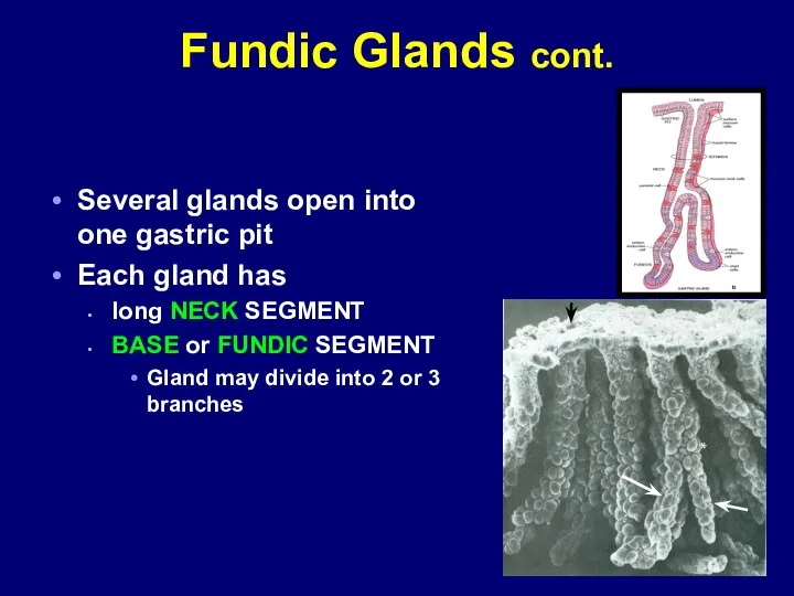
Fundic Glands cont.
Several glands open into one gastric pit
Each gland has
long
Fundic Glands cont.
Several glands open into one gastric pit
Each gland has
long
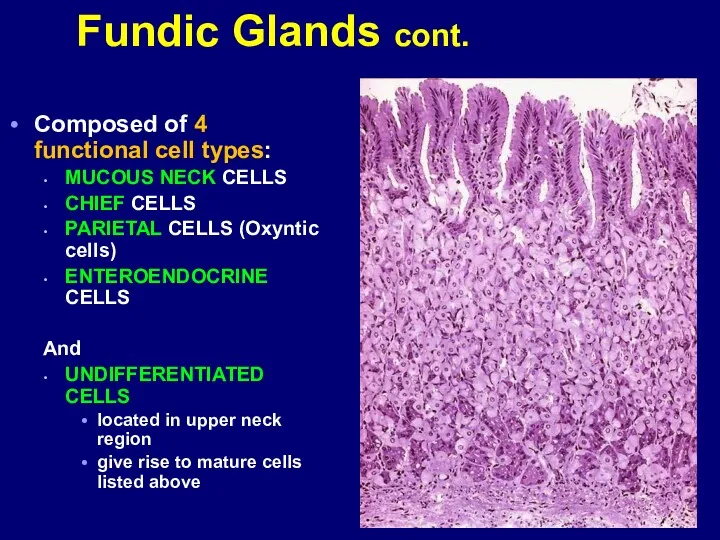
Composed of 4 functional cell types:
MUCOUS NECK CELLS
CHIEF CELLS
PARIETAL CELLS (Oxyntic
Composed of 4 functional cell types:
MUCOUS NECK CELLS
CHIEF CELLS
PARIETAL CELLS (Oxyntic
PC
CC
L
PC
CC
L


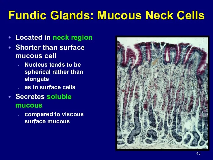
Fundic Glands: Mucous Neck Cells
Located in neck region
Shorter than surface mucous
Fundic Glands: Mucous Neck Cells
Located in neck region
Shorter than surface mucous
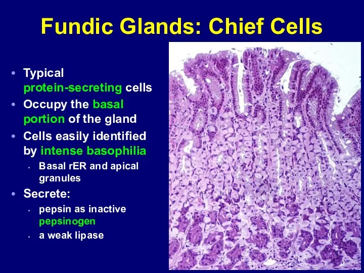
Fundic Glands: Chief Cells
Typical protein-secreting cells
Occupy the basal portion of the
Fundic Glands: Chief Cells
Typical protein-secreting cells
Occupy the basal portion of the
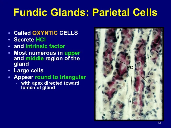
Fundic Glands: Parietal Cells
Called OXYNTIC CELLS
Secrete HCl
and intrinsic factor
Most numerous
Fundic Glands: Parietal Cells
Called OXYNTIC CELLS
Secrete HCl
and intrinsic factor
Most numerous

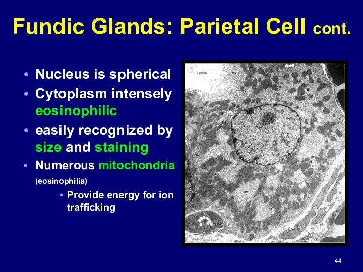
Fundic Glands: Parietal Cell cont.
Nucleus is spherical
Cytoplasm intensely eosinophilic
easily recognized by
Fundic Glands: Parietal Cell cont.
Nucleus is spherical
Cytoplasm intensely eosinophilic
easily recognized by
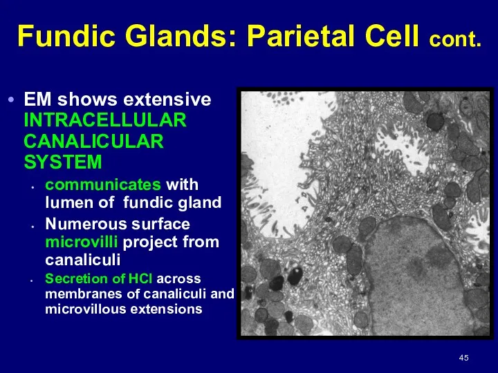
EM shows extensive INTRACELLULAR CANALICULAR SYSTEM
communicates with lumen of fundic gland
Numerous
EM shows extensive INTRACELLULAR CANALICULAR SYSTEM
communicates with lumen of fundic gland
Numerous
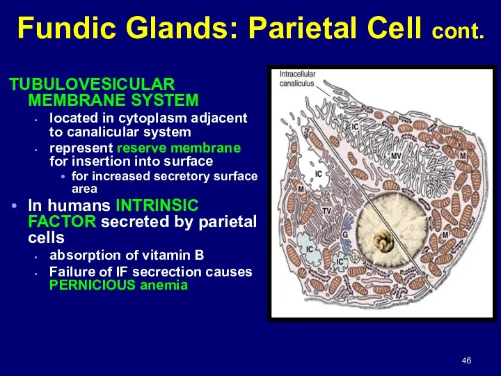
TUBULOVESICULAR MEMBRANE SYSTEM
located in cytoplasm adjacent to canalicular system
represent reserve membrane
TUBULOVESICULAR MEMBRANE SYSTEM
located in cytoplasm adjacent to canalicular system
represent reserve membrane
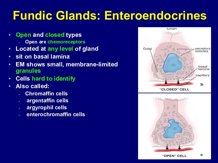
Fundic Glands: Enteroendocrines
Open and closed types
Open are chemoreceptors
Located at any level
Fundic Glands: Enteroendocrines
Open and closed types
Open are chemoreceptors
Located at any level


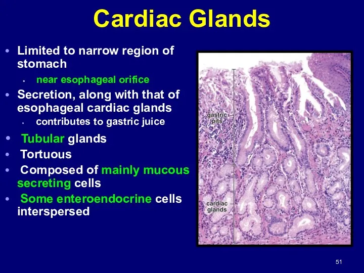
Cardiac Glands
Limited to narrow region of stomach
near esophageal orifice
Secretion, along
Cardiac Glands
Limited to narrow region of stomach
near esophageal orifice
Secretion, along
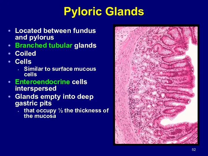
Pyloric Glands
Located between fundus and pylorus
Branched tubular glands
Coiled
Cells
Similar to surface
Pyloric Glands
Located between fundus and pylorus
Branched tubular glands
Coiled
Cells
Similar to surface

 Державна санітарно-епідеміологічна експертиза, як елемент соціально-гігієнічного моніторингу. Основні положення та організація
 Операции на органах шеи
 Физиология паращитовидных желёз
 Повреждения и заболевания мочеполовых органов
 Хирург Н.Н. Бурденко
 Арбовирусты инфекциялар. Кенелік энцефалит вирусы
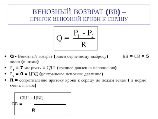 Венозный возврат (ВВ) – приток венозной крови к сердцу
 Шум и вибрация
 Алкогольный цирроз
 Возрастные особенности системы крови и иммунитета
 Неврозы
 Противоаритмические лекарственные средства
 Здоровье на работе. Что должен знать о ВИЧ/СПИДе каждый?
 Гигиена аптечных заведений
 Гиперчувствительность. Иммунодефициты. Аутоиммунные процессы
 Послеродовые депрессии
 Аллергия. Стоматология
 84-я Всероссийская научная конференция студентов и молодых ученых. Отчет. Секция: Общая хирургия
 Клинико-экономические исследования
 Химиотерапевтические лекарственные препараты, макролиды и азалиды
 Пороки сердца
 Асқорыту жолдарының қатерлі және қатерсіз ісіктері
 Мировые демографические показатели рождаемость, смертность в развитых и развивающихся странах. Демографическая ситуация в Росси
 Классификация геморрагического васкулита
 Белки
Белки ЦМК СД в акушерстве и гинекологии ,
 Medical Education in Japan
 Заболевания органов пищеварения у пожилых людей