- Face department of the head part 1

Содержание
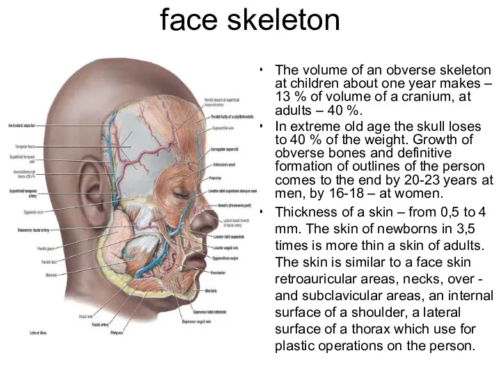
- 2. face skeleton The volume of an obverse skeleton at children about one year makes – 13
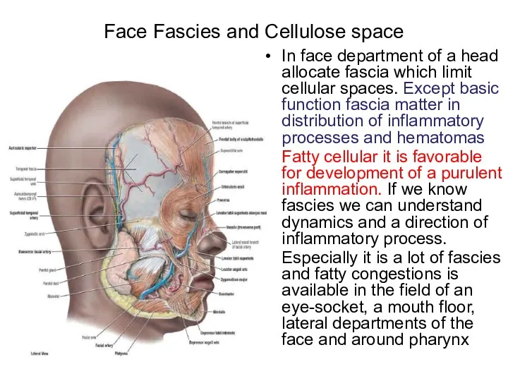
- 3. Face Fascies and Cellulose space In face department of a head allocate fascia which limit cellular
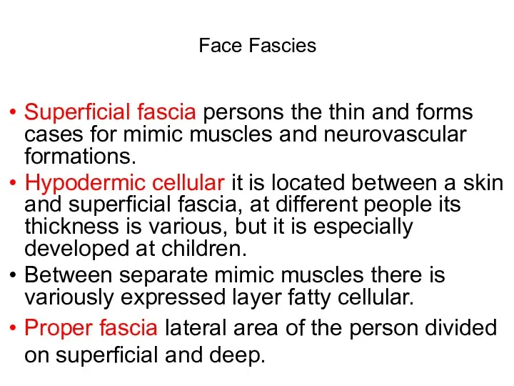
- 4. Face Fascies Superficial fascia persons the thin and forms cases for mimic muscles and neurovascular formations.
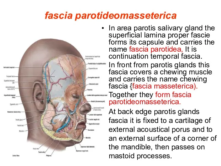
- 5. fascia parotideomasseterica In area parotis salivary gland the superficial lamina proper fascie forms its capsule and
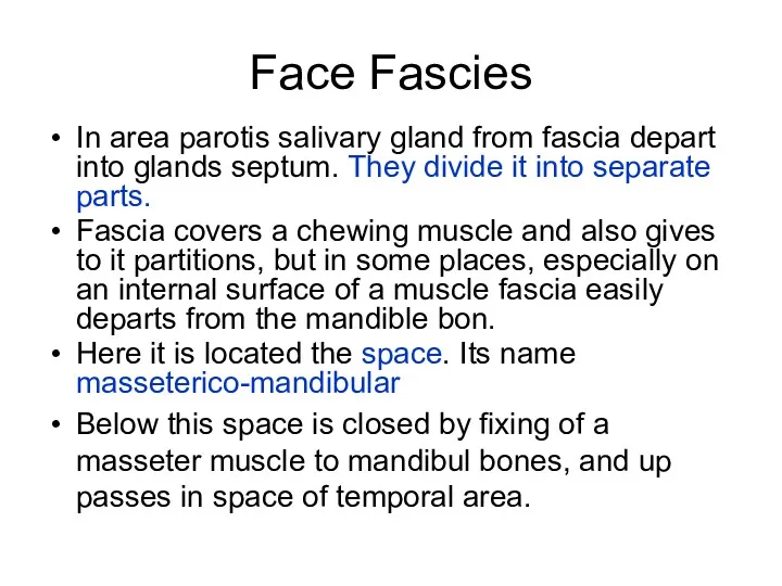
- 6. Face Fascies In area parotis salivary gland from fascia depart into glands septum. They divide it
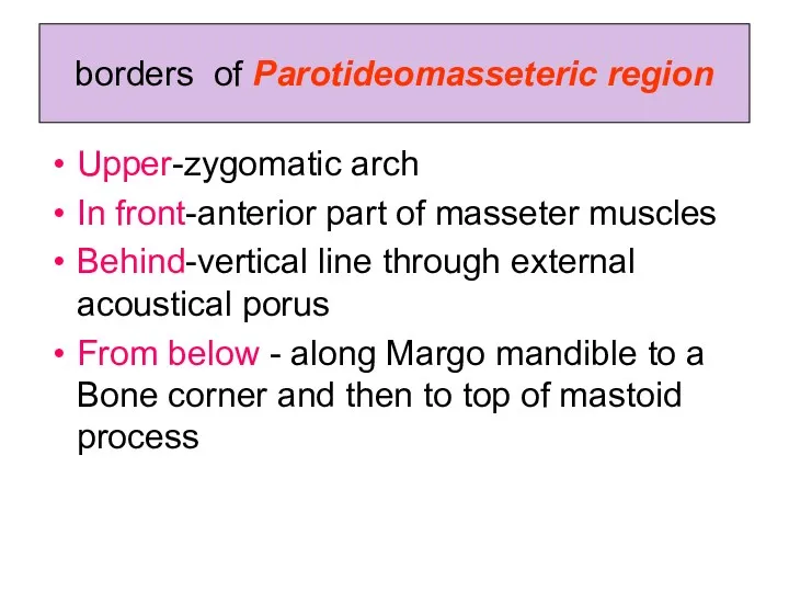
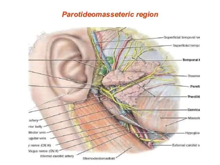
- 7. borders of Parotideomasseteric region Upper-zygomatic arch In front-anterior part of masseter muscles Behind-vertical line through external
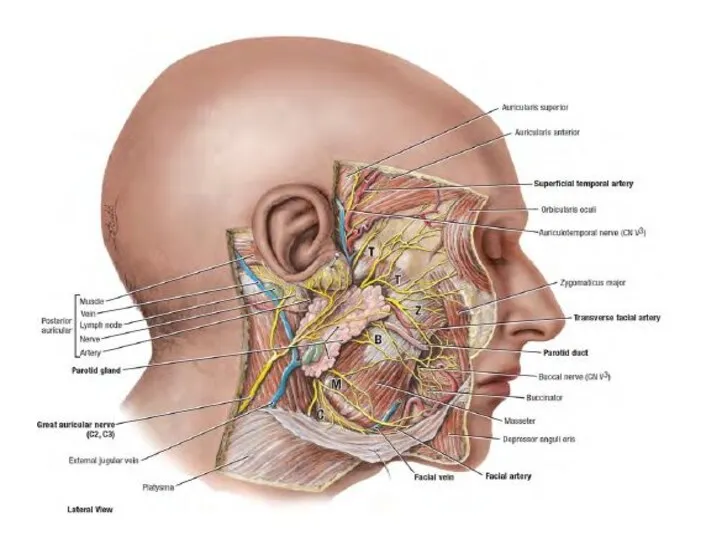
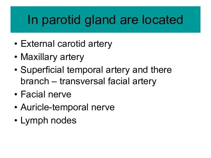
- 9. In parotid gland are located External carotid artery Maxillary artery Superficial temporal artery and there branch
- 10. Parotideomasseteric region
- 11. weak places of a capsule of parotid gland Internal surface (there is not capsule) The top
- 12. Transverse cut through parotid gland. (Internal pharyngeal part)
- 13. Your conclusions about inflammatory process in parotid gland
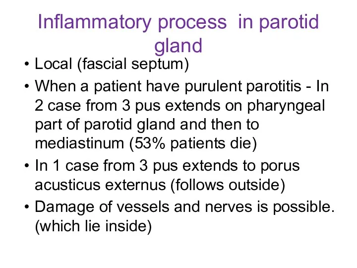
- 14. Inflammatory process in parotid gland Local (fascial septum) When a patient have purulent parotitis - In
- 15. Face department of the head part 2
- 16. Borders of buccal areas Above – Margo inferior orbital Below – Margo inferior mandibule In front
- 17. Superficial fascia Covers mimic muscles Forms a case for a lump of Bisha (being split on
- 18. Fascia proper In front from a masseter muscle fascia proper covered buccal muscle. This part carries
- 19. Corpus adipose buccal It is well expressed at small children. A capsule for it forms superficial
- 20. Superficial vessels buccal areas Facial artery and vеin. Artery has many bends Vein – forward stroke
- 21. Facial artery and vien
- 22. Face department of the head part 3
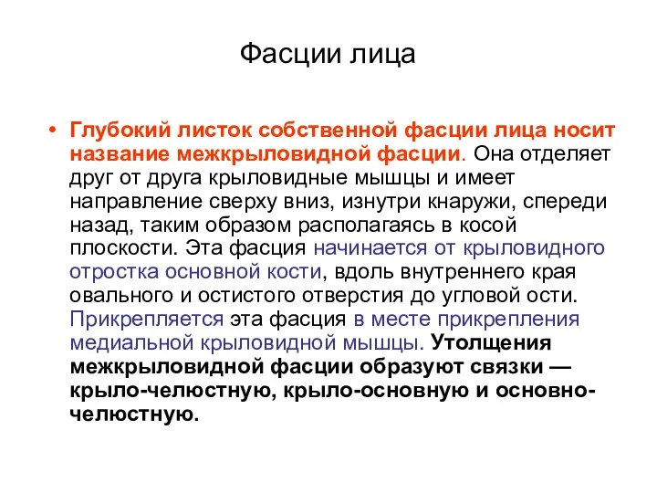
- 23. Фасции лица Глубокий листок собственной фасции лица носит название межкрыловидной фасции. Она отделяет друг от друга
- 24. Нижнечелюстной нерв и верхнечелюстная артерия
- 25. Глубокая область лица Занимает подвисочную ямку. Ограничена: Вверху-большое крыло клиновидной кости Спереди-бугор верхней челюсти Сзади-околоушная железа
- 26. Содержит Латеральную и медиальную крыловидные мышцы и конечный (нижний) участок височной мышцы Верхнечелюстную артерию Крыловидное венозное
- 27. жевательные мышцы
- 28. Глубокие жевательные мышцы
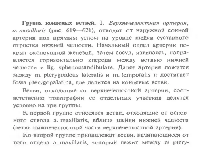
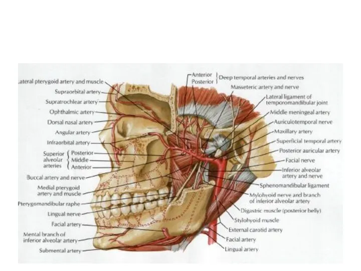
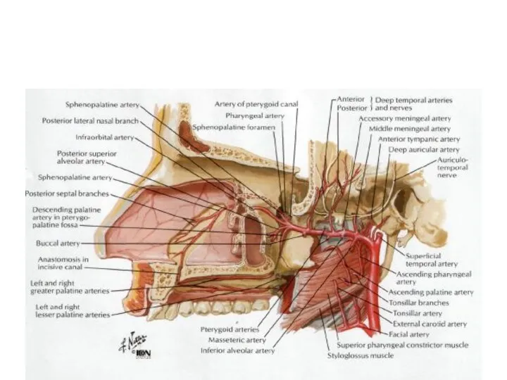
- 29. Артерия maxillaris
- 30. Вены лица
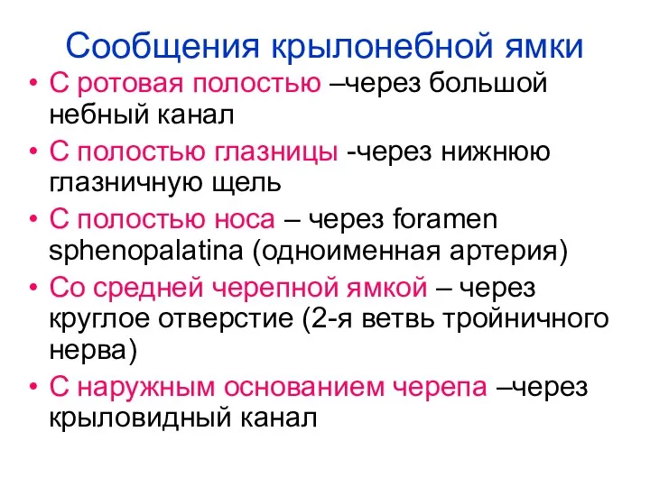
- 31. Сообщения крылонебной ямки С ротовая полостью –через большой небный канал С полостью глазницы -через нижнюю глазничную
- 32. Клетчаточные пространства глотки В окологлоточном пространстве различают передний и задний отделы. Границу между ними образует шиловидный
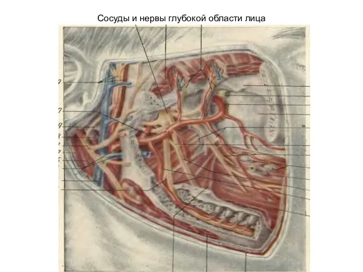
- 33. Сосуды и нервы глубокой области лица
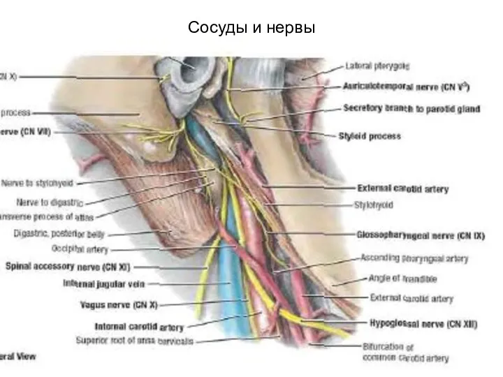
- 34. Сосуды и нервы
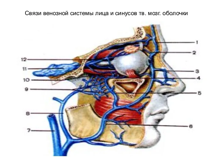
- 44. Связи венозной системы лица и синусов тв. мозг. оболочки
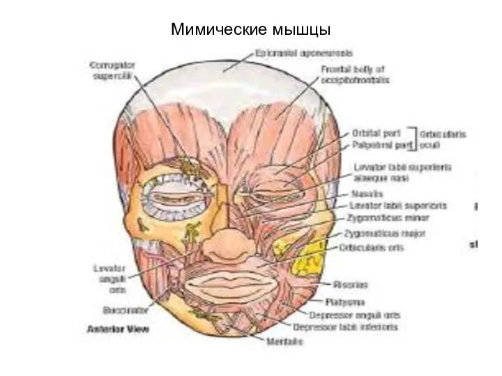
- 45. Мимические мышцы
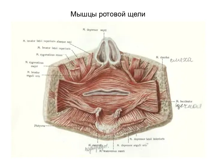
- 46. Мышцы ротовой щели
- 47. Ветви лицевого нерва Височная Скуловая Щечная Краевая нижней челюсти Шейная Задняя ушная
- 48. Мимические мышцы и лицевой нерв
- 49. occipitofrontalis
- 50. Orbicularis oculi
- 51. Buccinator +orbicularis oris
- 52. Zigomaticus major + minor
- 53. Depressor labii+levator labii superior
- 54. Rizorius (смеха)
- 55. Зоны иннервации тройничного нерва
- 56. Тройничный нерв и шейное сплетение
- 57. сосуды
- 58. вены
- 59. Мышцы глазного яблока
- 60. Мышцы глазного яблока
- 61. Глубокая область лица
- 62. Ветви артерии maxillaris
- 63. n.Maxillaris и mandibularis
- 64. Проекция ветвей тройничного нерва
- 67. Лимфатическая система лица и шеи
- 68. Твердое и мягкое небо
- 69. Мягкое небо
- 70. Мышцы мягкого неба
- 71. Латеральная стенка полости носа
- 72. раковины
- 73. Придаточные пазухи
- 74. Придаточные пазухи, вид со стороны глазницы
- 75. Медиальная стенка полости носа (перегородка)
- 76. Иннервация латеральной стенки носа
- 78. Скачать презентацию
face skeleton
The volume of an obverse skeleton at children about one
face skeleton
The volume of an obverse skeleton at children about one
Face Fascies and Cellulose space
In face department of a head allocate
Face Fascies and Cellulose space
In face department of a head allocate
Face Fascies
Superficial fascia persons the thin and forms cases for mimic
Face Fascies
Superficial fascia persons the thin and forms cases for mimic
fascia parotideomasseterica
In area parotis salivary gland the superficial lamina proper fascie
fascia parotideomasseterica
In area parotis salivary gland the superficial lamina proper fascie
Face Fascies
In area parotis salivary gland from fascia depart into glands
Face Fascies
In area parotis salivary gland from fascia depart into glands
borders of Parotideomasseteric region
Upper-zygomatic arch
In front-anterior part of masseter muscles
Behind-vertical line
borders of Parotideomasseteric region
Upper-zygomatic arch
In front-anterior part of masseter muscles
Behind-vertical line
In parotid gland are located
External carotid artery
Maxillary artery
Superficial temporal artery and
In parotid gland are located
External carotid artery
Maxillary artery
Superficial temporal artery and
Parotideomasseteric region
Parotideomasseteric region
weak places of a capsule of parotid gland
Internal surface (there is
weak places of a capsule of parotid gland
Internal surface (there is
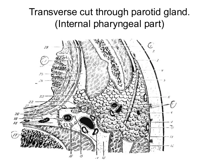
Transverse cut through parotid gland. (Internal pharyngeal part)
Transverse cut through parotid gland. (Internal pharyngeal part)
Your conclusions about inflammatory process in parotid gland
Your conclusions about inflammatory process in parotid gland
Inflammatory process in parotid gland
Local (fascial septum)
When a patient have purulent
Inflammatory process in parotid gland
Local (fascial septum)
When a patient have purulent
Face department of the head
part 2
Face department of the head
part 2
Borders of buccal areas
Above – Margo inferior orbital
Below – Margo inferior
Borders of buccal areas
Above – Margo inferior orbital
Below – Margo inferior
Superficial fascia
Covers mimic muscles
Forms a case for a lump of Bisha
Superficial fascia
Covers mimic muscles
Forms a case for a lump of Bisha
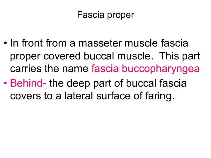
Fascia proper
In front from a masseter muscle fascia proper covered buccal
Fascia proper
In front from a masseter muscle fascia proper covered buccal
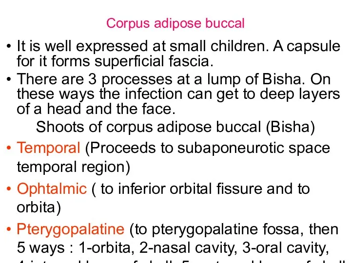
Corpus adipose buccal
It is well expressed at small children. A capsule
Corpus adipose buccal
It is well expressed at small children. A capsule
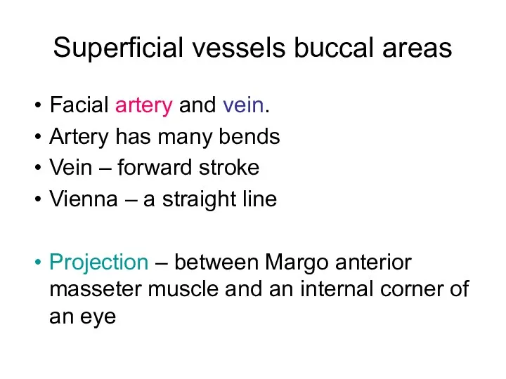
Superficial vessels buccal areas
Facial artery and vеin.
Artery has many bends
Vein
Superficial vessels buccal areas
Facial artery and vеin.
Artery has many bends
Vein

Facial artery and vien
Facial artery and vien
Face department of the head
part 3
Face department of the head
part 3
Фасции лица
Глубокий листок собственной фасции лица носит название межкрыловидной фасции. Она
Фасции лица
Глубокий листок собственной фасции лица носит название межкрыловидной фасции. Она
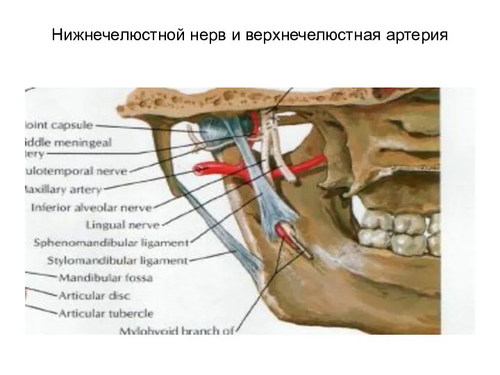
Нижнечелюстной нерв и верхнечелюстная артерия
Нижнечелюстной нерв и верхнечелюстная артерия
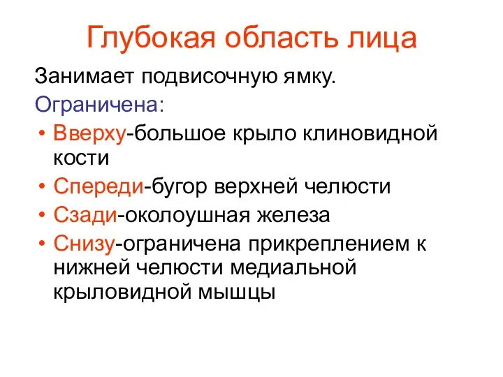
Глубокая область лица
Занимает подвисочную ямку.
Ограничена:
Вверху-большое крыло клиновидной кости
Спереди-бугор верхней челюсти
Сзади-околоушная
Глубокая область лица
Занимает подвисочную ямку.
Ограничена:
Вверху-большое крыло клиновидной кости
Спереди-бугор верхней челюсти
Сзади-околоушная
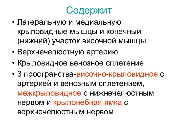
Содержит
Латеральную и медиальную крыловидные мышцы и конечный (нижний) участок височной мышцы
Верхнечелюстную
Содержит
Латеральную и медиальную крыловидные мышцы и конечный (нижний) участок височной мышцы
Верхнечелюстную
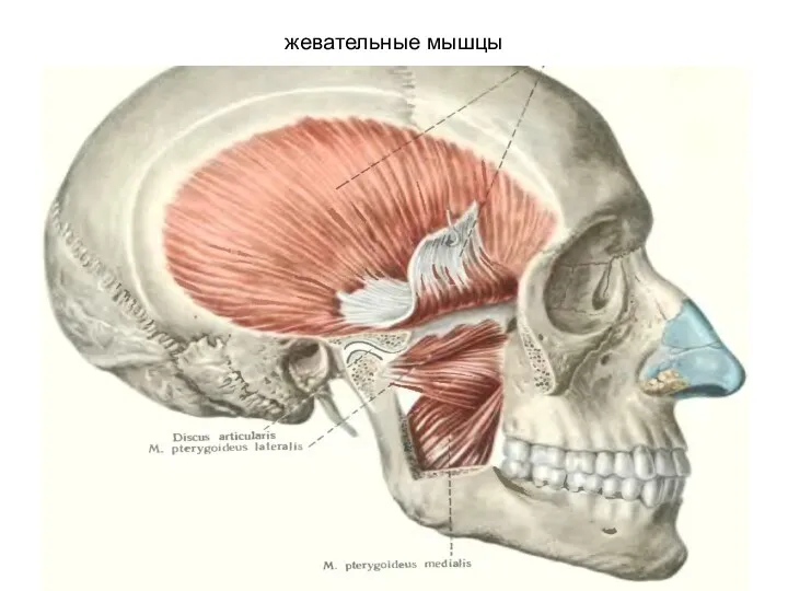
жевательные мышцы
жевательные мышцы
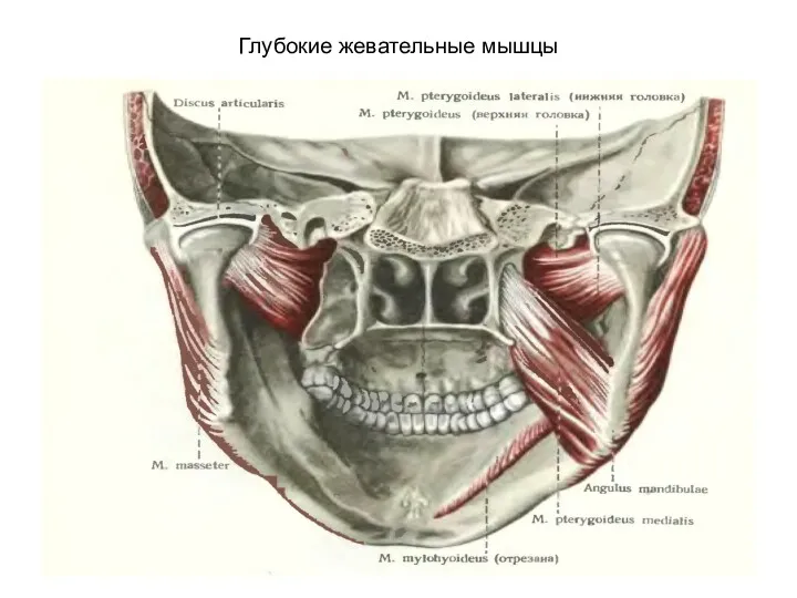
Глубокие жевательные мышцы
Глубокие жевательные мышцы
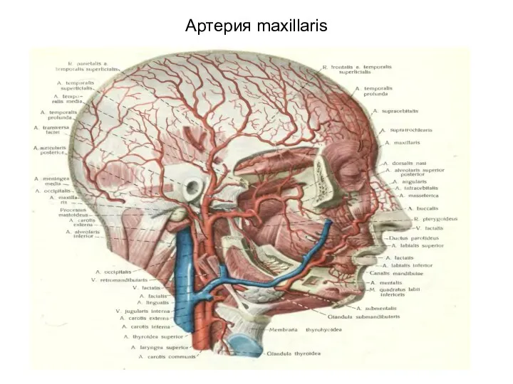
Артерия maxillaris
Артерия maxillaris
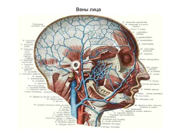
Вены лица
Вены лица
Сообщения крылонебной ямки
С ротовая полостью –через большой небный канал
С полостью глазницы
Сообщения крылонебной ямки
С ротовая полостью –через большой небный канал
С полостью глазницы

Клетчаточные пространства глотки
В окологлоточном пространстве различают передний и задний отделы. Границу
Клетчаточные пространства глотки
В окологлоточном пространстве различают передний и задний отделы. Границу
Сосуды и нервы глубокой области лица
Сосуды и нервы глубокой области лица
Сосуды и нервы
Сосуды и нервы
Связи венозной системы лица и синусов тв. мозг. оболочки
Связи венозной системы лица и синусов тв. мозг. оболочки
Мимические мышцы
Мимические мышцы
Мышцы ротовой щели
Мышцы ротовой щели
Ветви лицевого нерва
Височная
Скуловая
Щечная
Краевая нижней челюсти
Шейная
Задняя ушная
Ветви лицевого нерва
Височная
Скуловая
Щечная
Краевая нижней челюсти
Шейная
Задняя ушная
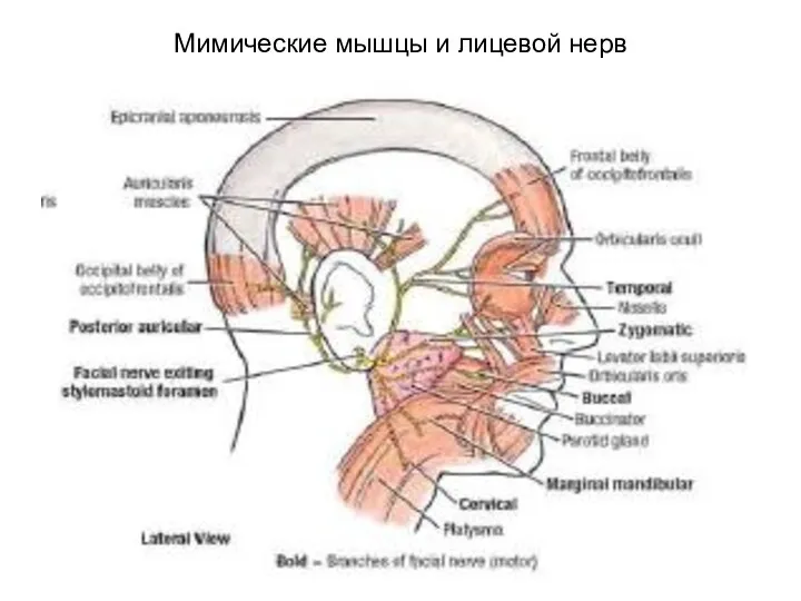
Мимические мышцы и лицевой нерв
Мимические мышцы и лицевой нерв
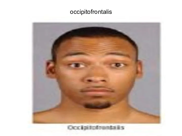
occipitofrontalis
occipitofrontalis
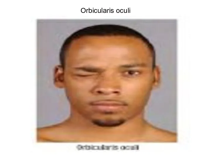
Orbicularis oculi
Orbicularis oculi
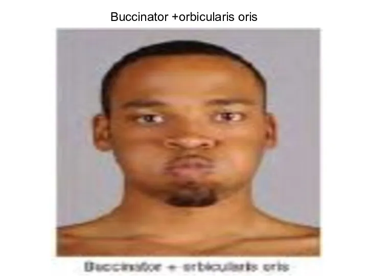
Buccinator +orbicularis oris
Buccinator +orbicularis oris
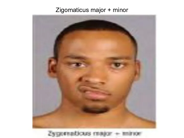
Zigomaticus major + minor
Zigomaticus major + minor

Depressor labii+levator labii superior
Depressor labii+levator labii superior
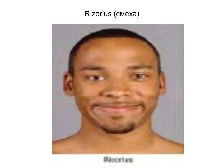
Rizorius (смеха)
Rizorius (смеха)
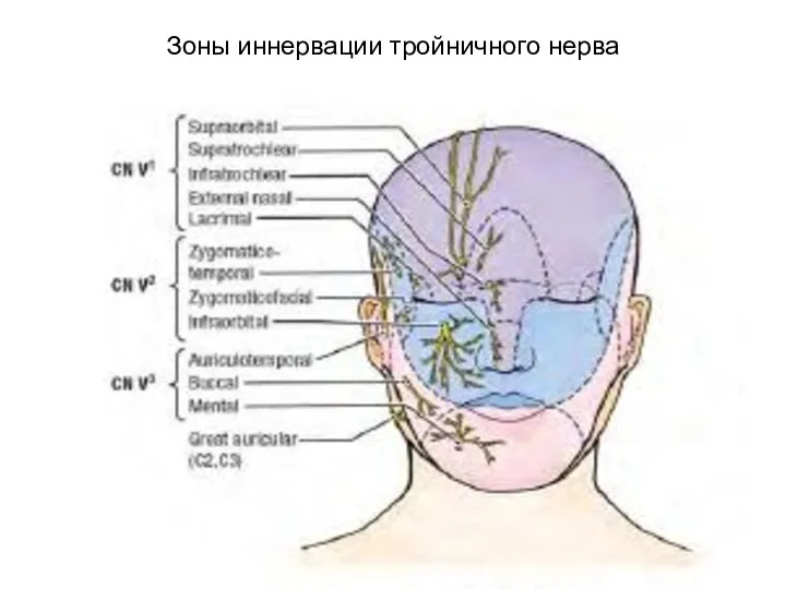
Зоны иннервации тройничного нерва
Зоны иннервации тройничного нерва
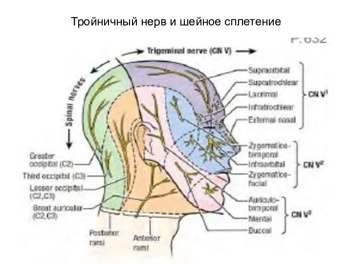
Тройничный нерв и шейное сплетение
Тройничный нерв и шейное сплетение
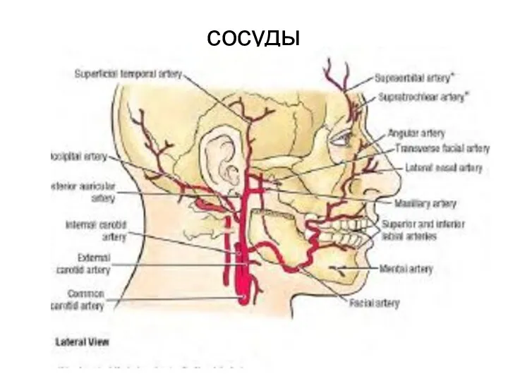
сосуды
сосуды
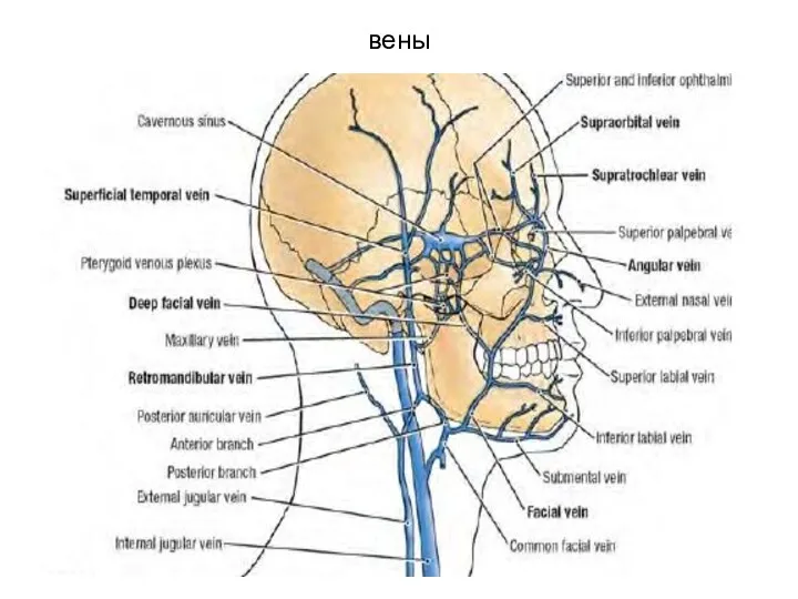
вены
вены
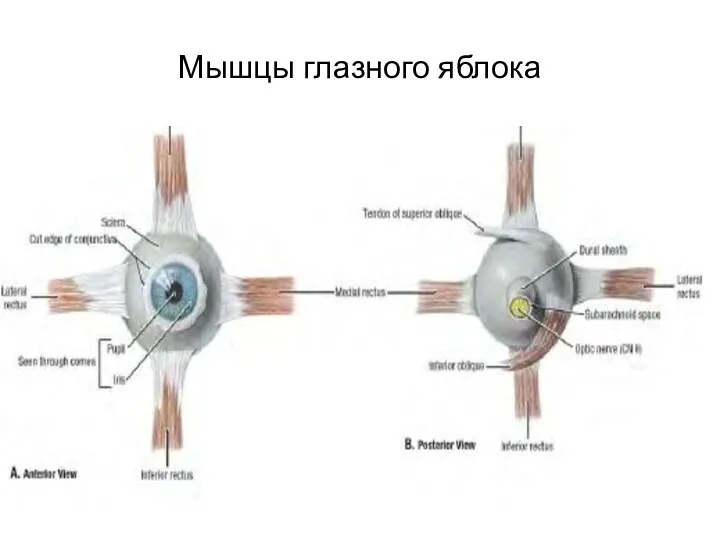
Мышцы глазного яблока
Мышцы глазного яблока
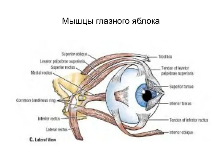
Мышцы глазного яблока
Мышцы глазного яблока
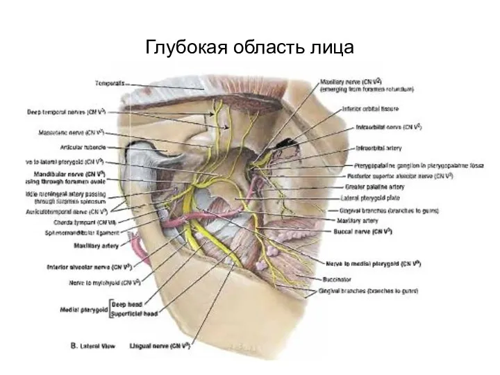
Глубокая область лица
Глубокая область лица
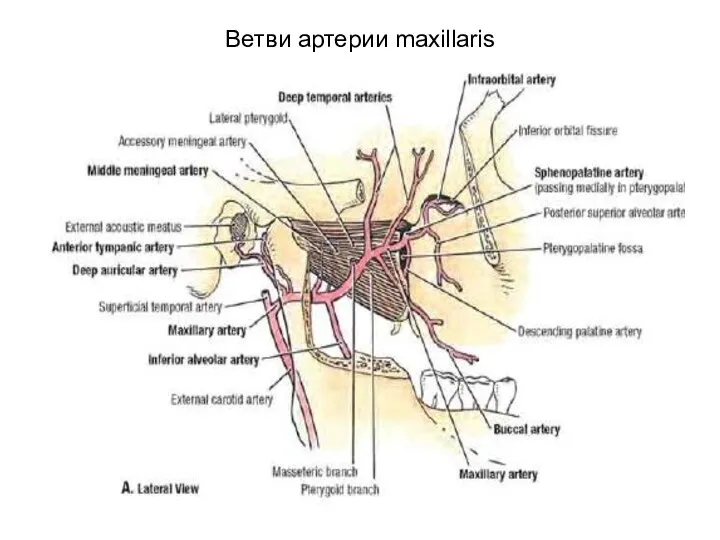
Ветви артерии maxillaris
Ветви артерии maxillaris
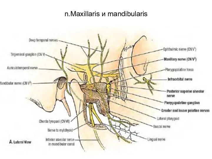
n.Maxillaris и mandibularis
n.Maxillaris и mandibularis
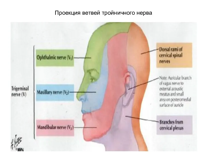
Проекция ветвей тройничного нерва
Проекция ветвей тройничного нерва
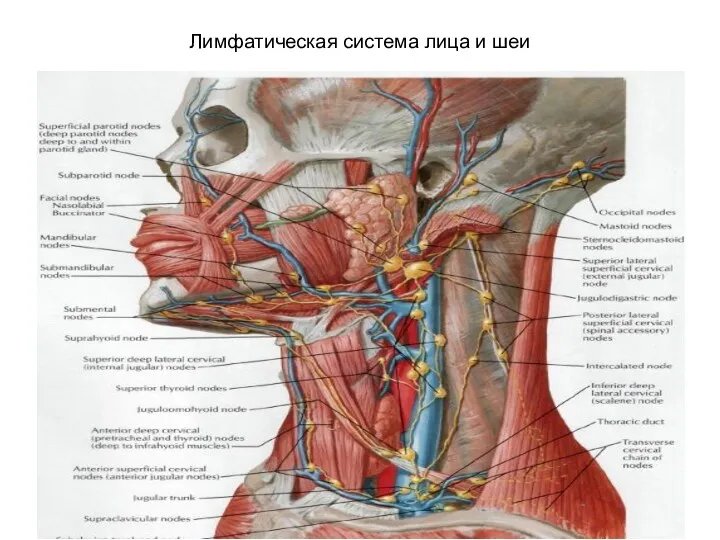
Лимфатическая система лица и шеи
Лимфатическая система лица и шеи
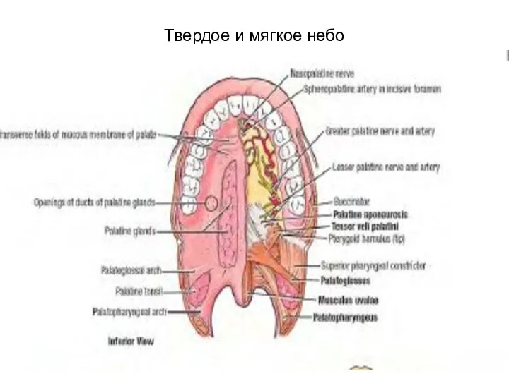
Твердое и мягкое небо
Твердое и мягкое небо
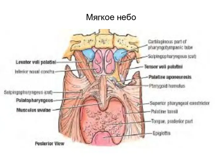
Мягкое небо
Мягкое небо
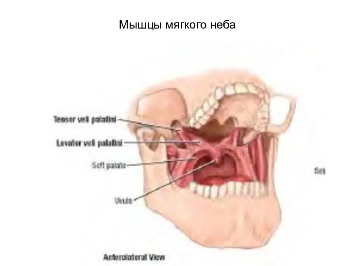
Мышцы мягкого неба
Мышцы мягкого неба
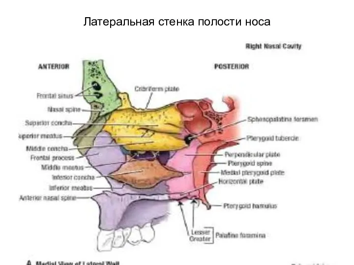
Латеральная стенка полости носа
Латеральная стенка полости носа
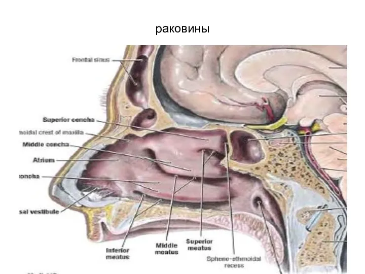
раковины
раковины
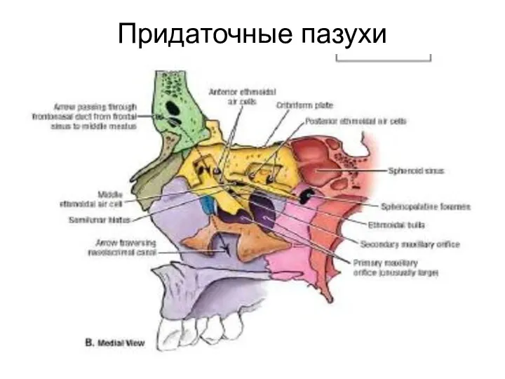
Придаточные пазухи
Придаточные пазухи
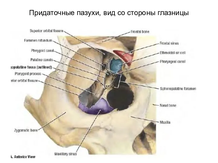
Придаточные пазухи, вид со стороны глазницы
Придаточные пазухи, вид со стороны глазницы
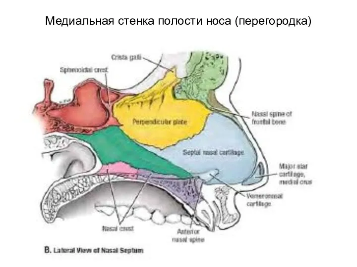
Медиальная стенка полости носа (перегородка)
Медиальная стенка полости носа (перегородка)
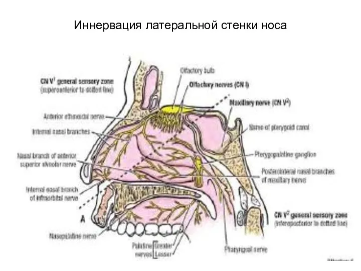
Иннервация латеральной стенки носа
Иннервация латеральной стенки носа
 Физиология и биохимия бактерий. Лекция 2
 Рак губы
 Коронавирус Covid-19
 Анализ электрокардиограммы (ЭКГ)
 Преэклампсияның ауыр дәрежесі
 Противоаритмические лекарственные средства
 Typhoid
 Ісікке қарсы препараттардың клиникалық фармакологиясы
 Сульфаниламидные средства
 Сосудистая хирургия
 Ожоги органа зрения
 Учение об иммунитете
 Роль медицинской сестры в реабилитации ожоговых больных
 Медицинская служба гражданской обороны
 Опухоли кишечника
 Техника транспортной иммобилизации верхней конечности
 Нормализация речевой моторики у детей с алалией
 Мази. Средства аппликационной терапии
 Патология кровообращения и лимфообращения
 Парапроктит. Этиологиясы, клиникасы, емдеу тактикасы
 Семиотика и диагностика заболеваний женской половой системы
 Физические методы антисептики
 Острый гломерулонефрит
 Современные подходы к антибактериальной терапии
 Рак желудка
 История сестриского дела. Этика в сестринской деятельности
 Понятие об общем уходе в хирургии и его элементах
 Хронический гепатит